These will also appear in metamorphic rocks
Quartz
Plagioclase
Potassium feldspars
Myrmeckite
Micas
Amphiboles
Pyroxenes
Other major primary minerals
Accessory (minor primary) minerals
Secondary (subsolidus) minerals
Quartz
|
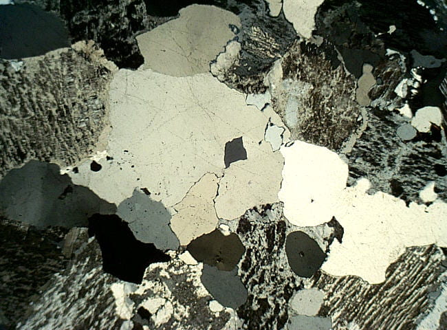
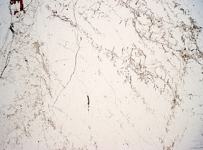
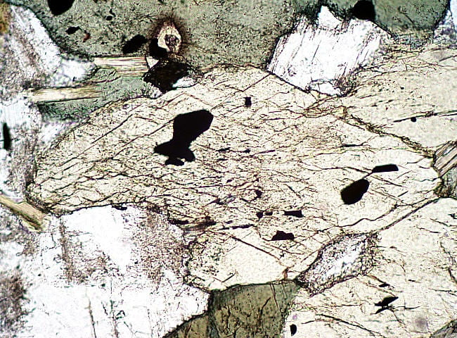
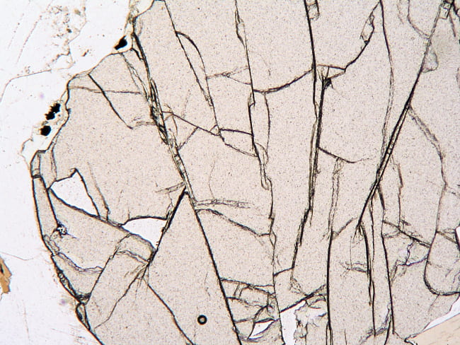
Quartz crystals in alkali granite. Quartz is typically the most transparent mineral in rocks because it is not very susceptible to alteration to fine-grained minerals, and it has no cleavages. Birefringence in the low to middle first order. Plane- and cross-polarized light views. Field width is 6 mm. NEIGC86-B2-7 |
|
 |
Strained quartz crystal in a metaluminous granite. Strain has caused the quartz crystal to deform into domains with slightly different extinction angles. Typical for quartz, but not feldspars. Cross-polarized light view only. Field width is 6 mm. DIG-D |
 |
Fluid inclusions in quartz in alkali granite. The center inclusion has an irregular outer boundary, inside of which is a layer of liquid water, a layer of liquid CO2, and a central bubble of vapor (mostly CO2). At the time the fluid was trapped, it was a homogeneous H2O-CO2 fluid. Plane-polarized light view only. Field width is 0.12 mm. NEIGC86-B2-7 |
 |
Fluid inclusions in quartz in alkali granite. Several inclusions containing (probably) water and a vapor bubble. Plane-polarized light view only. Field width is 0.12 mm. NEIGC86-B2-7 |

Plagioclase
|
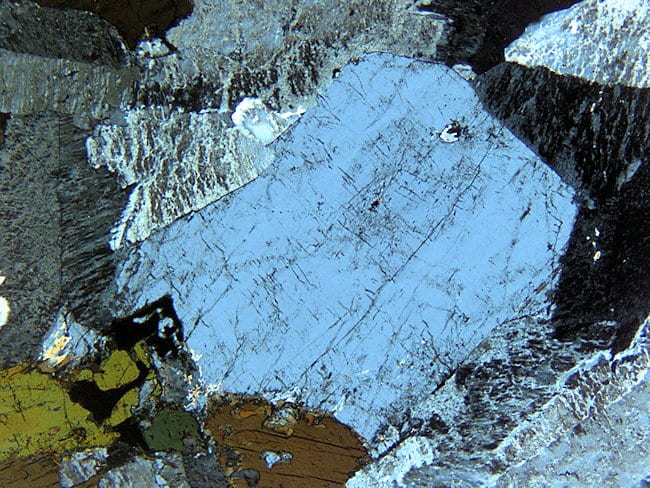
Plagioclase, unzoned, in a hornblende diorite. Ateration to fine-grained material typically occurs along fractures, twin planes, and cleavages. In the solid solution series, the plagioclase refractive index varies from slightly lower than quartz to somewhat above it. Here, albite twins are prominent, and some small pericline twins can also be seen, which is typical. Plane- and cross-polarized light views. Field width is 3 mm. NEIGC84-A5-5C |
|
|
Plagioclase, zoned, in a dacite porphyry. This grain appears quite homogeneous in plane light, without concentric zones of inclusions that are commonly seen elsewhere. In cross-polarized light, fine oscillatory zoning is visible, in which the composition varies back and forth between more and less anorthite-rich. Internal unconformities followed by euhedral overgrowths are also visible. Plane- and cross-polarized light views. Field width is 3 mm. Rhyolite-2 |
|
|
Plagioclase, zoned, in a dacite porphyry. Notice the concentric layers of inclusions. These probably formed during faster, skeletal crystal growth than the clear layers. In cross-polarized light you can see that the layers also have different birefringence, indicating they have different anorthite content, and so have different orientations of the indicatrix. The center of this crystal has patchy zoning rather than concentric, indicating skeletal early growth. Albite, carlsbad, and pericline twins cut across the layers. Plane- and cross-polarized light views. Field width is 1.2 mm. Rhyolite-2 |


Potassium feldspars
|
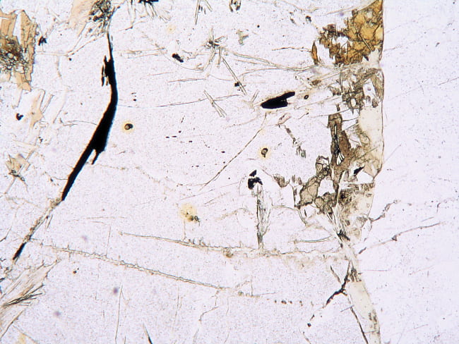
Orthoclase in a dacite shallow intrusive. In plane-polarized light K-feldspars may be difficult to tell from plagioclase, though plagioclase has higher refractive indexes. Hint: to highlight K-feldspar, switch to a medium magnification objective (usually 10X works well), mostly close the substage iris, and raise the stage slightly (yes, raise). Though the image will become somewhat out of focus, the low index K-feldspar grains will be surrounded by bright Becke lines just inside the grain boundaries. This is especially handy for tonalites, in which K-feldspar may be hard to find by other methods. Plane- and cross-polarized light views. Field width is 3 mm. Py-28 |
|
|
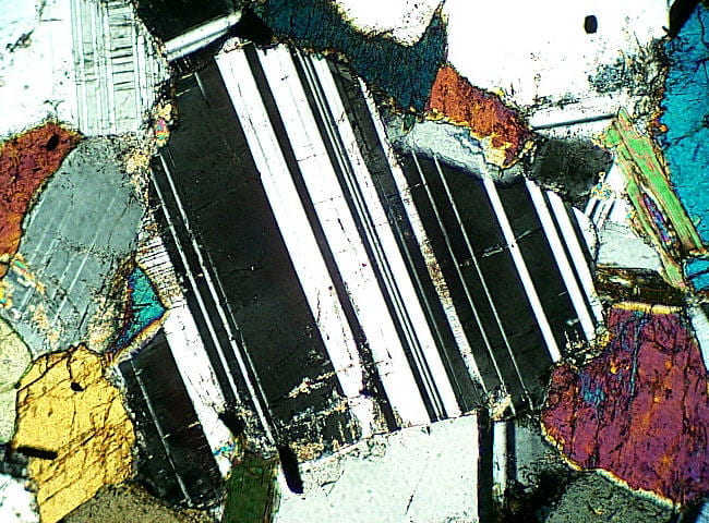
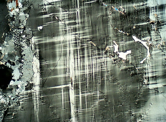
Microcline from a peraluminous granite. Characteristically featureless in plane light, but the inverse Becke line highlighting technique described above works just as well for microcline. In cross-polarized light you can see the striking “grid” or “tartan plaid” twinning pattern that results from crossing albite and pericline twin domains. These form during cooling, as the grain changes from monoclinic orthoclase to triclinic microcline. This transformation is associated with progressive ordering of aluminum and silicon. Albite and pericline twins are impossible in monoclinic orthoclase and sanidine, and develop like this only during inversion to triclinic microcline. The triclinic twin domains can nucleate with the triclinic angles leaning one way or another in both the albite and pericline twin directions. However, once they start, that is how the twin continues to grow. Because many twin domains nucleate throughout the crystal, there are large numbers of thin, spindly, and discontinuous domains in the final microcline product. Plane- cross-polarized light views. Field width is 6 mm. Kinsman |
|
 |
Microcline from a peraluminous granite. Close-up of the grid twinning. Notice how the twin domains are spindly and somewhat wispy. This is in contrast to the straight, and generally continuous twins in plagioclase. Note the variable spacing of the twin domains, indicating that different volumes of the crystal nucleated different numbers of twin domains during inversion. Cross-polarized light view only. Field width is 1.2 mm. Kinsman |
|
Perthite from a metaluminous biotite granite. Perthite is an unmixing texture of an originally homogeneous alkali feldspar grain. Microcline and albite both exsolved (unmixed) from the homogeneous solid solution during cooling. In this case, the exsolved albite is less altered (clearer) than adjacent microcline, which is grayish because of lots of minute alteration minerals. In cross-polarized light you can see the lighter, irregular stripes and patches of bright yellowish-white birefringent albite, between gray microcline. The birefringent color difference is mostly caused by the different optical orientations of the two different minerals. In this photo, the thin section was rotated to obscure twinning. Plane- and cross-polarized light views. Field width is 6 mm. 4.7.84H |
|
|
Perthite from a metaluminous biotite granite. The somewhat less-altered and narrower albite exsolution lamellae are in sharp contact with larger microcline domains. A Becke line test tells you which phase is which: the Becke line goes into the higher index phase, albite, when you lower the stage. Plane- and cross-polarized light views. Field width is 1.2 mm. 4.7.84H |
|
 |
Perthite from a metaluminous biotite granite. Close-up showing the characteristic grid twinning in the microcline host (upper center and upper left) and albite twinning in the lamellae (center to center right). Cross-polarized light view only. Field width is 0.6 mm. 4.7.84H |


Myrmeckite
|
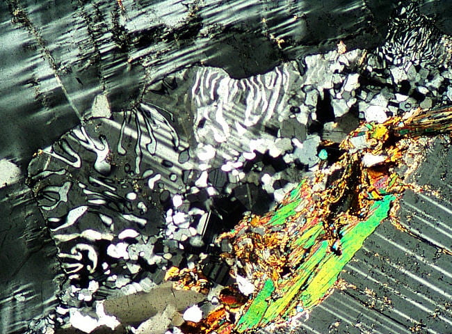
Myrmekite patch that appears to be replacing microcline. Though its presence is obscure in plane-polarized light, it is obvious in cross-polarized light as quartz worms in plagioclase. The plagioclase is twinned. Myrmekite is a subsolidus reaction texture that generally results from fluid flow. Here, K-feldpar was removed and quartz and plagioclase were deposited in its place. Plane- and cross-polarized light views. Field width is 3 mm. Kinsman |

Micas
|
Muscovite in a peraluminous granite. Igneous muscovite is generally colorless, with good cleavage. Pale brown radiation halos can sometimes be visible around radioactive inclusions (but not really visible here). Birefringence is up to the high second or lower third order. Plane- and cross-polarized light views. Field width is 3 mm. NEIGC84-A5-6 |
|
|
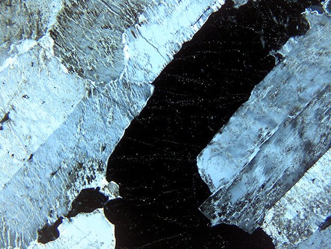
Biotite, metaluminous granite, showing several grains in different orientations. The grain on the far right is oriented with cleavages N-S, and is almost opaque. The largest grain is inclined and is lighter in color. Grains with the cleavages E-W have the least absorption (not shown here). In cross-polarized light the birefringent colors of the biotite are muted by the color of the biotite itself. Plane- and cross-polarized light views. Field width is 3 mm. 4.7.84G |
|
 |
Biotite, metaluminous granite, showing a close-up of one crystal. Damage produced during thin section grinding causes speckles of light in the biotite, where the crystal lattice has been deformed. This means that biotite in standard thin sections rarely goes completely extinct. This is called “incomplete extinction” or sometimes “birds eye maple extinction.” Cross-polarized light view only. Field width is 6 mm. 4.7.84G |
 |
Biotite, peraluminous granite. The red-brown biotite in this graphite- and garnet-bearing granite has high titanium and low Fe3+ content. This patch of biotite shows a range of pleochroic colors caused by different crystal orientations. Plane-polarized light view only. Ffield width is 0.6 mm Kinsman |

Amphiboles
|
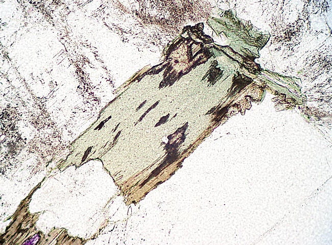
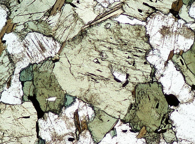
Green hornblende in a diorite. Pleochroism in this sample ranges from light-yellow-green to bluish-green to brownish-green. Birefringent colors are typically up to middle second order. Plane- and cross-polarized light views. Field width is 3 mm. NEIGC84-A5-5C |
|
 |
Green hornblende in a diorite. The ~120° and ~60° cleavage intersections are clearly visible in this crystal end section. Plane-polarized light view only. Field width is 1.2 mm. NEIGC84-A5-5C |
|
Brown hornblende in hornblende gabbro. The extensive dark and light brown areas are different hornblende crystals in different orientations. Note the numerous inclusions of opaques and plagioclase. Plane- and cross-polarized light views. Field width is 6 mm. 4.8.84Q |

Pyroxenes
|
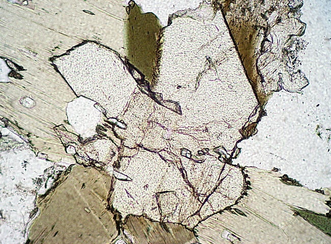
Enstatite (orthopyroxene, Opx) in norite. The large Opx in the center is oriented with its c crystallographic axis N-S. In cross-polarized light that crystal is at extinction, in keeping with the orthorhombic symmetry of this mineral. Birefringence ranges up to middle first order. Plane- and cross-polarized light views. Field width is 1.2 mm. NEIGC83-C1-14 |
|
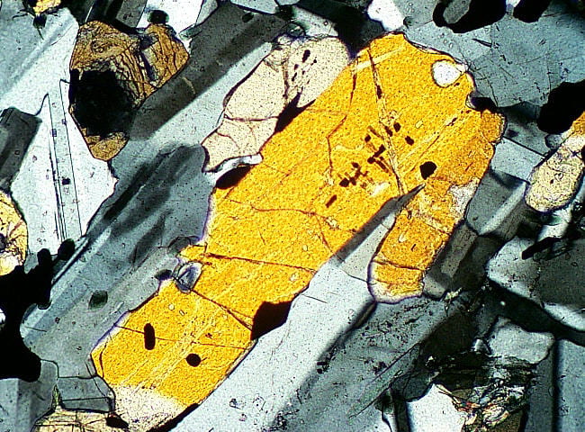 |
Enstatite (orthopyroxene, Opx) in norite. Here the thin section shown above has been rotated clockwise ~45° to show the birefringence of the large grain. Cross-polarized light view only. Field width is 1.2 mm. NEIGC83-C1-14 |
|
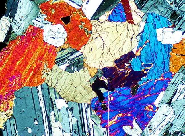
Augite (clinopyroxene, Cpx), in gabbro. This augite is slightly brownish, and has tiny exsolved rods and plates of Fe-Ti oxides. These form the darkish, cloudy regions, especially in the grain to the lower right. Augite usually has birefringence up to second order blue. Birefringence tends to be somewhat irregular in single grains because of compositional variations. Plane- and cross-polarized light views. Field width is 6 mm. 4.8.84A |
|
|
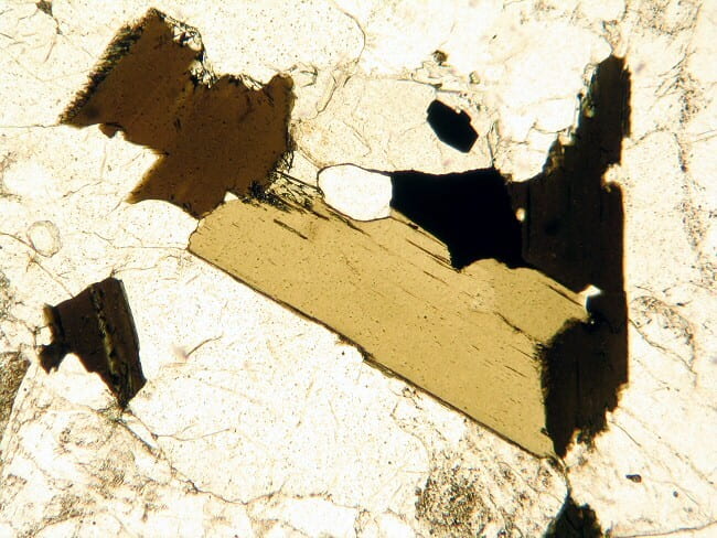
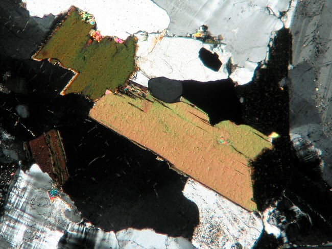
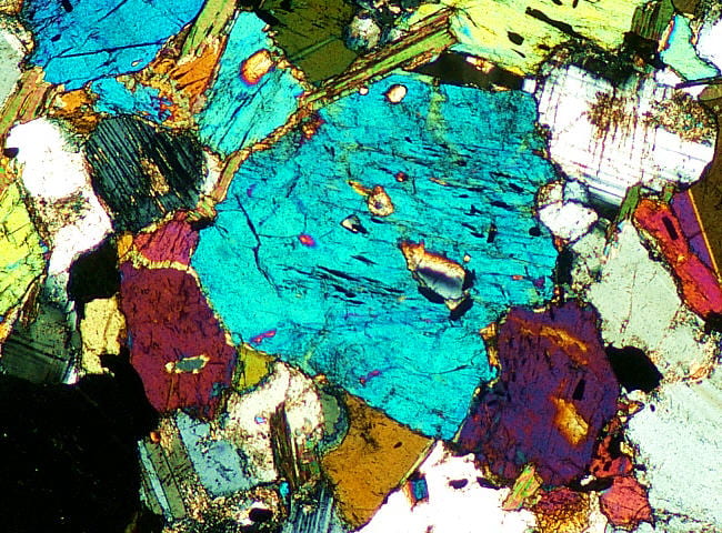
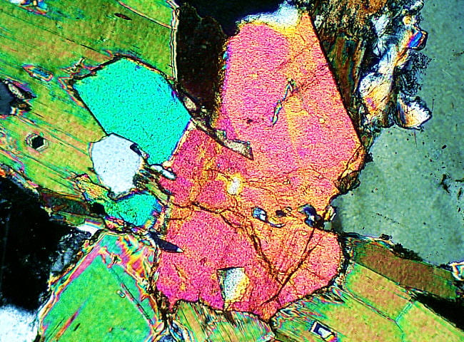
Aegirine (Na–Fe3+ clinopyroxene), alkaline granite. These crystals have a lot of inclusions of quartz and feldspar. The pleochroic colors are similar to hornblende, but it has approximately right-angle cleavage intersections like all pyroxenes. In cross-polarized light, aegerine can be seen to have a smaller extinction angle than most hornblende, and negative sign of elongation. Aegirine birefringence tends to be up to upper second to lower third order, a bit higher than amphiboles and most other clinopyroxenes. Plane- and cross-polarized light views. Field width is 3 mm. 4.8.84F |


Other major primary minerals
|
Olivine phenocryst in an Iceland basalt. Notice the fractures concentric with the crystal margin. In cross-polarized light, smaller “microphenocrysts” of brightly birefringent olivine and gray to white plagioclase can be seen. Augite occurs in the matrix as small, brownish crystals with brirefringence up to second order blue. Plane- and cross-polarized light views. Field width is 3 mm. I-1 |
|
|
Garnet in a peraluminous granite. The high refractive index of garnet causes fractures and the grain margin to stand out as dark lines, because of total internal reflection. The lack of birefringence distinguishes garnet from all other common high-index minerals. The bright cracks seen in cross-polarized light have thin films of calcite. Plane- and cross-polarized light views. Field width is 3 mm. NHM-9 |
|
|
Cordierite in a peraluminous granite. In plane- and cross-polarized light cordierite looks much like quartz and feldspar, and it can be twinned or untwinned. It has three distinguishing characteristics. 1) It tends to look dustier than quartz and feldspar, minute black specks all over the surface, such as can be seen here. 2) It commonly alters to brownish or orange material (top right), or to an intergrowth of Mg-rich chlorite and muscovite (some of the right margin and lower left). 3) Radiation halos are yellow and pleochroic, unless it is very Mg-rich. Interference colors are first order gray to white, like quartz and feldspar. It is more commonly euhedral than quartz in plutonic rocks. The muscovite alteration products are easily visible here, but the Mg-rich chlorite is not so visible because of its low birefringence and pale color. Plane- and cross-polarized light views. Field width is 3 mm. NHM-9 |
|
 |
Cordierite in a peraluminous granite. This shows three radioactive inclusions with their yellow radiation halos, caused by alpha particle radiation damage. Plane-polarized light view only. Field width is 1.2 mm. NHM-9 |
|
Epidote in a calc-alkaline granodiorite. In general, epidote in igneous rocks has rather high Fe3+ content, which may color it pale yellow-green. Birefringence and color may define zoning, and both color and birefringence increase with Fe3+ content. The birefringence is usually 2nd and 3rd order, so epidote, with its high relief and common lack of obvious color, can resemble olivine. It’s commonly associated with other Ca-rich minerals like hornblende, plagioclase, and titanite. Epidote may have simple twinning, and can occur with quartz and various hydrous minerals like chlorite and sericite. Olivine is usually not associated with quartz (except in highly Fe-rich, Mg-poor rocks), and is likely to be partly altered. Plane- and cross-polarized light views. Field width is 1.2 mm. UN30A |
|
|
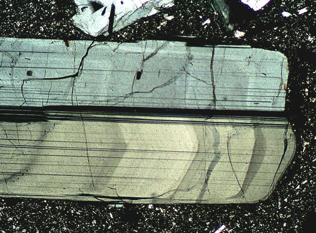
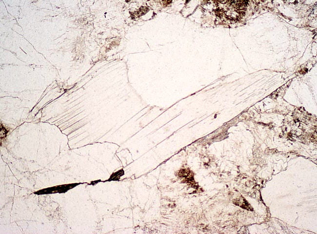
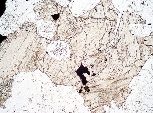
Nepheline in a nepheline syenite. It is distinguished from alkali feldspar by lack of perthitic intergrowths, from plagioclase by lack of polysynthetic twinning, from both by its parallel extinction on cleavages and uniaxial negative interference figure, from apatite by its much lower relief, and from quartz by its cleavage, common alteration, and uniaxial negative interference figure. Its birefringence is also somewhat lower than feldspars or quartz. Plane- and cross-polarized light views. Field width is 3 mm. RH-1 |
|
|
Sodalite in a nepheline syenite. This isotropic mineral has much lower relief than both garnet and fluorite, and its refractive index is closer to alkali feldspars and epoxy. In this view, healed fractures are highlighted by minute birefringent grains, probably carbonates inside fluid inclusions. Plane- and cross-polarized light views. Field width is 3 mm. RH-1 |


Accessory minerals
|
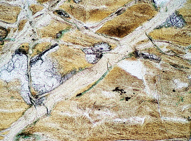
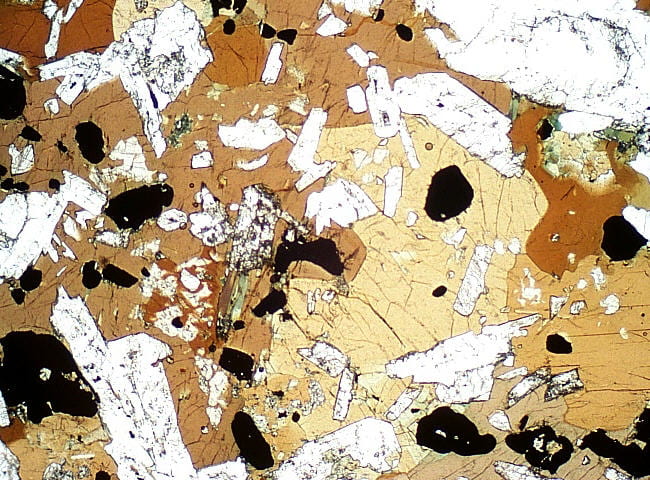
Opaques inside olivine in an olivine gabbro. The large crystals are titanomagnetite, which crystallized from a relatively evolved gabbroic magma. There are also peculiar paralellogram- and trapezoid-shaped patches of wormy opaque material in the olivine. These are magnetite that formed during subsolidus oxidation of iron in olivine. Oxidation turned some Fe2+ to Fe3+, precipitating magnetite by the reaction: olivine + O2 = magnetite + enstatite. Common opaque minerals include the oxides magnetite and ilmenite, sulfides pyrite, pyrrhotite, and chalcopyrite, and graphite, among others. Yellow sulfides can be distinguished from gray oxides by using oblique illumination from above the section, with the substage light off. The different sulfide and oxide colors become visible from light reflecting off the rough mineral surfaces. Plane- and cross-polarized light views. Field width is 1.2 mm. 4.8.84D |
|
|
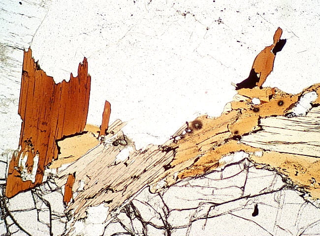
Chromite crystals in olivine in a primitive basalt. Chromite is a spinel-type mineral, like magnetite. Cr-Mg-Al-rich chromites are somewhat transparent, and can be dark-brown at full thin section thickness. As the magma evolves toward more Fe-rich compositions, chromite also becomes more Fe-rich and less transparent. Fe-rich chromite is typically opaque at full thin section thickness, but is transparent dark-brown along thin edges. The chromite shown here is relatively Fe-poor. Chromite is isotropic, like all spinels, with a very high refractive index. Plane- and cross-polarized light views. Field width is 0.6 mm. ICE-24B |
|
|
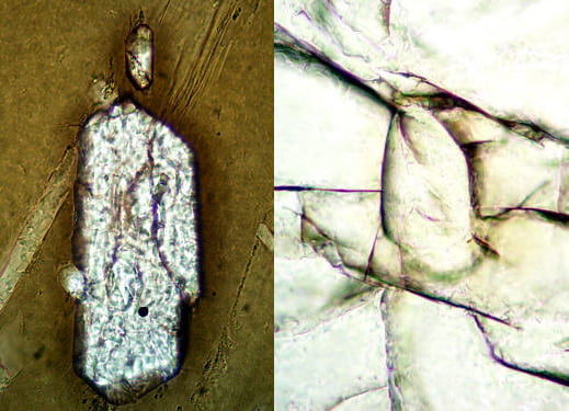
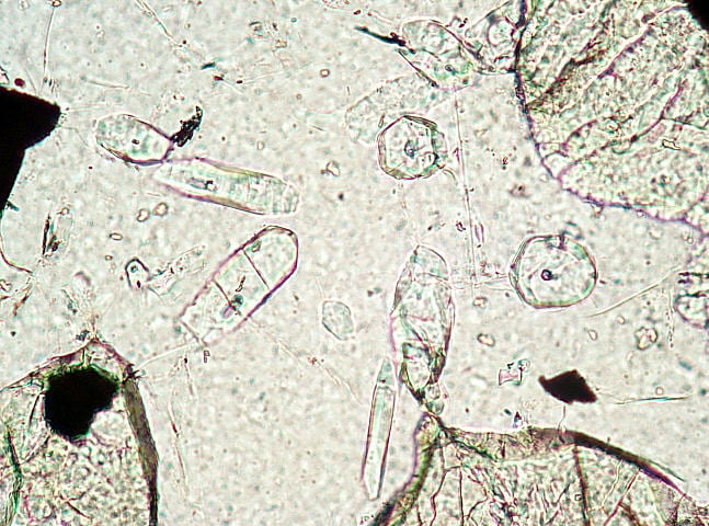
Apatite crystals in norite. This view has several apatite crystals, mostly in plagioclase with surrounding Opx. Apatite is colorless, commonly elongate, and typically with hexagonal end sections. Its relief is less than the pyroxenes but higher than any feldspar. The very low 1st order birefringence is obvious, and it has negative sign of elongation. Apatite is extremely common in small amounts in igneous rocks. Indeed, it is rare to find a terrestrial plutonic rock without at least some apatite. Plane- and cross-polarized light views. Field width is 1.2 mm. NEIGC83-C1-14 |
|
|
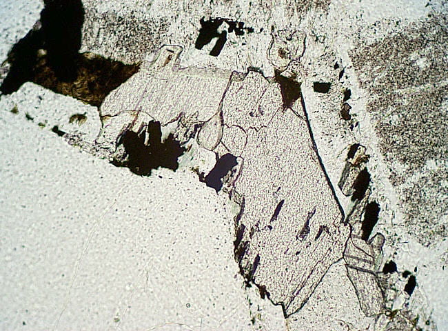
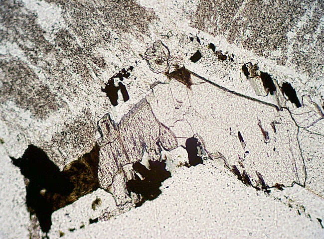
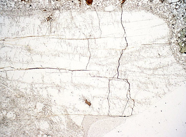
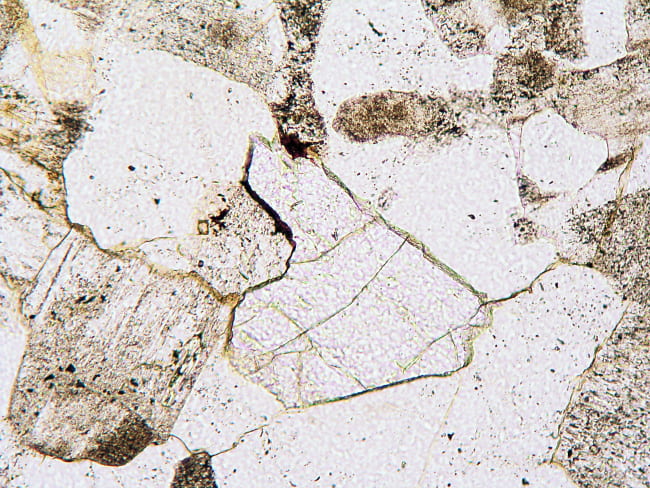
Fluorite in a metaluminous granite. The fluorite here is primary, as it occurs as euhedral and subhedral single crystals in a relatively fine-grained matrix. It has high negative relief, its refractive index being considerably lower than adjacent quartz, feldspar, and epoxy. Sodalite has much less pronounced relief and poorer cleavage. Fluorite, of course, is isotropic. Plane- and cross-polarized light views. Field width is 1.2 mm. NEIGC86-B2-3B |
|
|
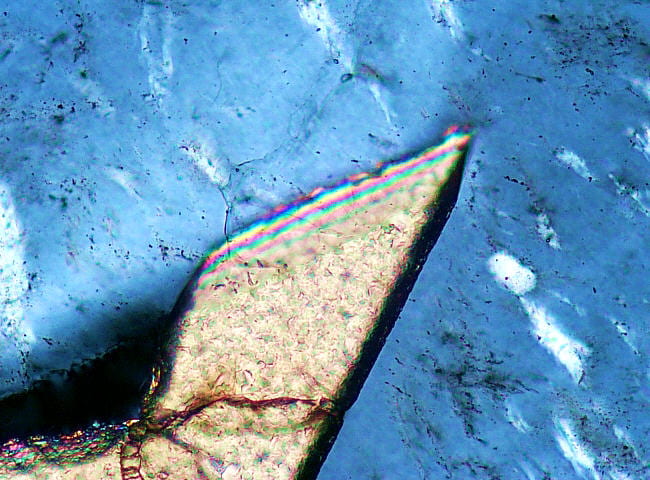
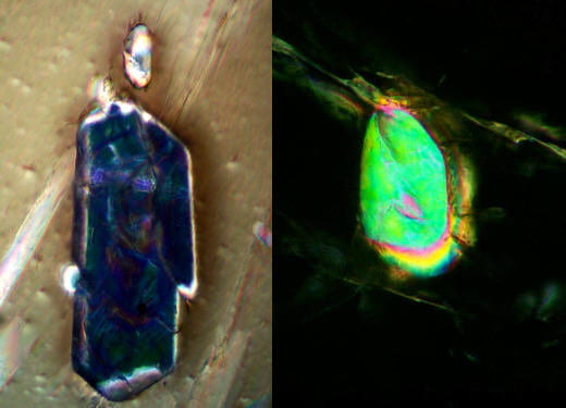
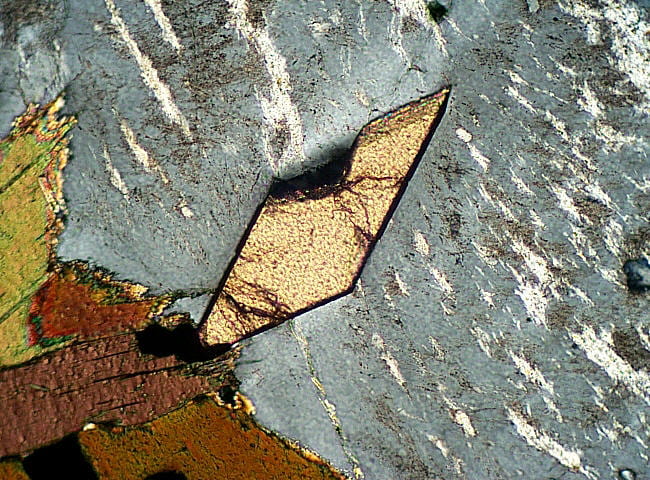
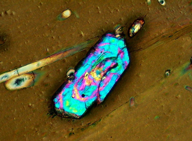
Titanite in a metaluminous granite. Titanite has a typical double-wedge or diamond shape, is typically light-brown, and has very high relief, higher than the pyroxenes or garnet. Titanite birefringence is very high, making it difficult to determine interference color order from the high-order pastel interference colors normally seen. Plane- and cross-polarized light views. Field width is 1.2 mm. DIG-D |
|
 |
Titanite in a metaluminous granite. At high magnification, the magenta bands can be counted up a thin edge to get a better idea of the interference order. This example is approximately 6th order. Cross-polarized light view only. Field width is 0.6 mm. DIG-D |
|
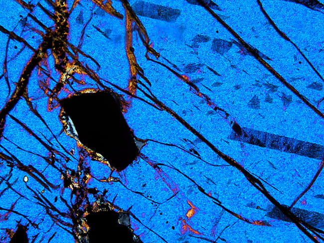
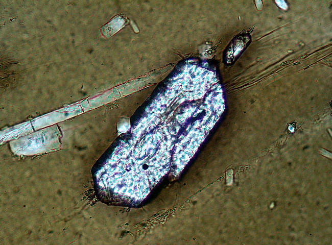
Zircon in a metaluminous granite. This is a rather blocky zircon, fully enclosed within biotite. Small apatite crystals surround it. Zircon has relief considerably higher than garnet, pyroxenes, and titanite. The uranium and thorium content of zircon causes development of pleochroic radiation halos around it. Zircon birefringence is typically in the 3rd order. This example has birefringence zoning that probably results from igneous compositional zoning. High uranium layers accumulate more radiation damage, and so become less birefringent. Plane- and cross-polarized light views. Field width is 0.6 mm. DIG-D |
|
 |
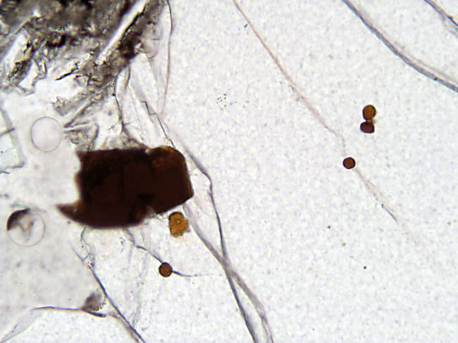
Zircon compared to monazite 1 Left: Zircon oriented N-S. Zircon is usually colorless in thin section. Right: Monazite oriented N-S. Monazite can be very pale yellow-green in thin section. This grain has developed a light-brown radiation halo (not pleochroic) in the adjacent garnet. Both are in plane-polarized light. Field widths are 0.3 mm. |
 |
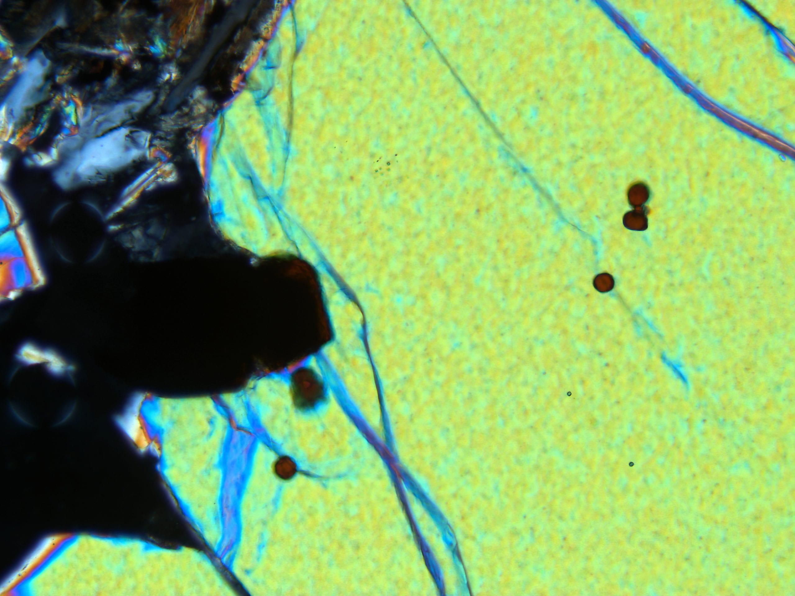
Zircon compared to monazite 2 Left: In the same orientation zircon is extinct, and so it has parallel extinction. Right: In the same orientation Monazite is not extinct, and so it must have inclined extinction, characteristic of its monoclinic crystal structure. Xenotime is tetragonal, like zircon, so it can’t be distinguished so easily. Both are in cross-polarized light. Field widths are 0.3 mm. |
|
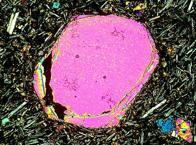
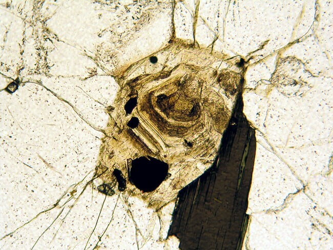
Allanite in a metaluminous granite. This allanite has concentric zoning, probably from changing mineral composition during successive stages of growth. Allanite contains substantial amounts of Th and U, and sustains considerable radiation damage over time. Swelling of the crystal as damage accumulates, and absorption of water, causes radial cracks that extend out into the surrounding minerals. This allanite grain mostly has low birefringence, a result of radiation damage that has essentially turned the crystal lattice into glass. Plane- and cross-polarized light views. Field width is 1.2 mm. 4.7.84G |


Secondary minerals
|
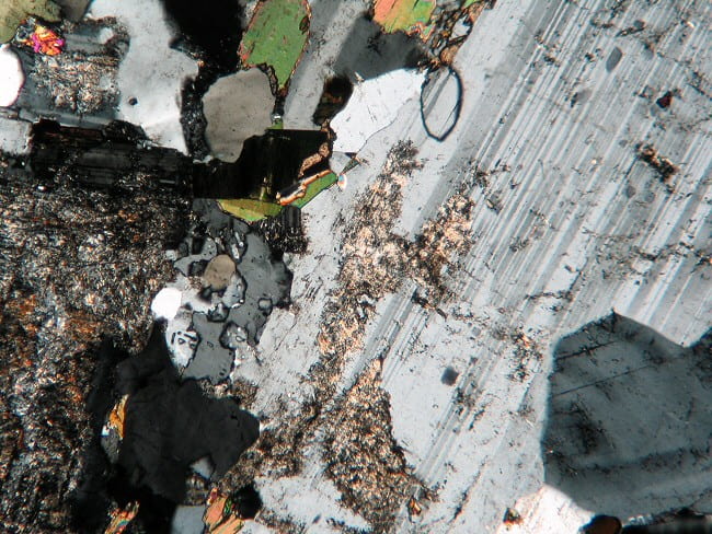
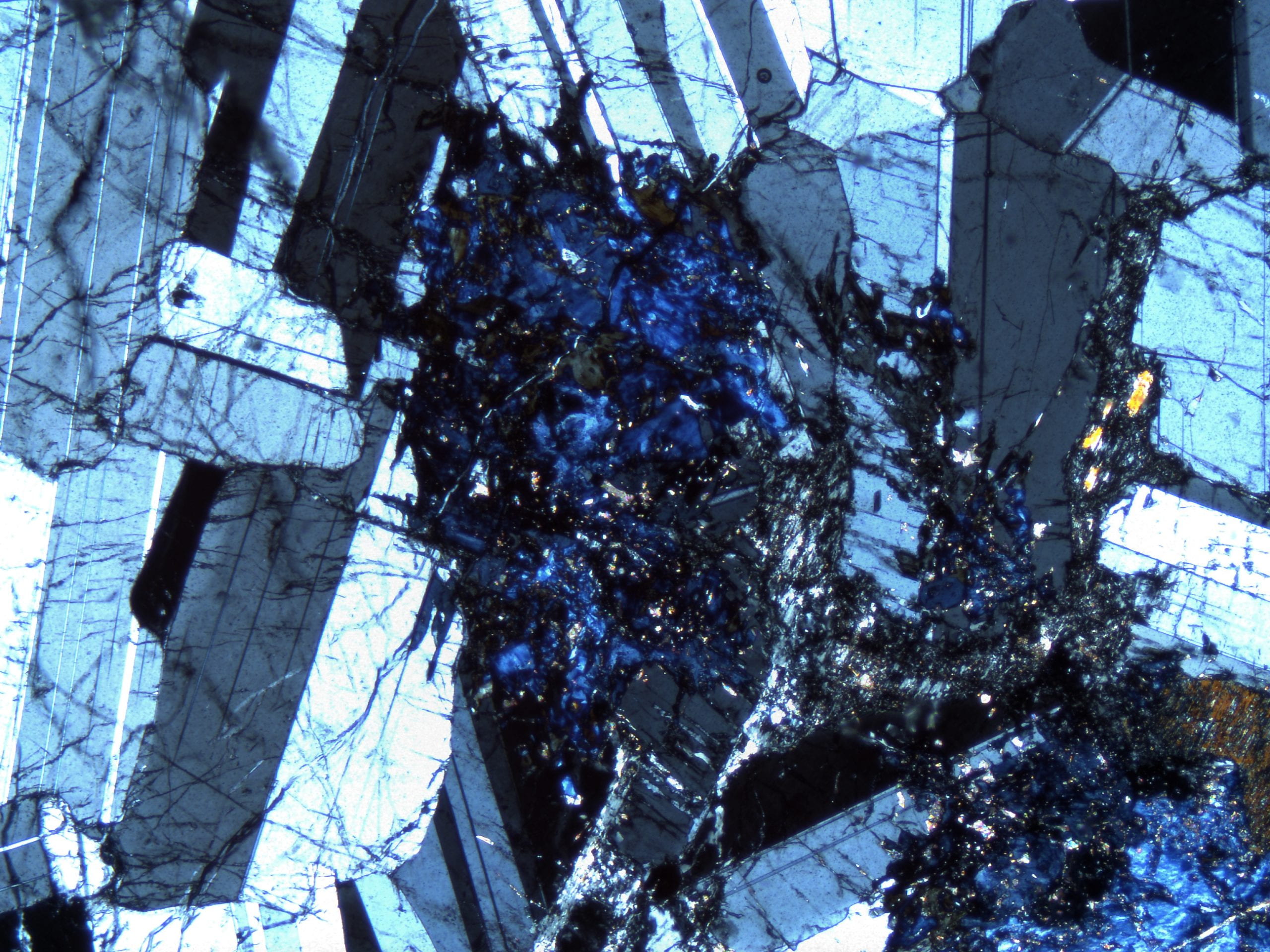
Chlorite replacing biotite in a muscovite granite. Small residual brownish patches of biotite still occur. Dark bits of titanite are abundant in the chlorite. Biotite can hold several weight percent TiO2 in solid solution, but secondary chlorite can accommodate ≪1%, so the titanium precipitates as secondary titanite. The low first order, anomalous Berlin blue interference color indicates that this is an Fe-rich chlorite. Residual biotite patches have higher birefringence. Plane- and cross-polarized light views. Field width is 3 mm. NEIGC84-A5-6 |
|
|
Chlorite replacing biotite in a metaluminous granite. Lenses of high-index colorless material (titanite?) occur in the chlorite, and a small amount of brown biotite survives in the lower part of the grain. The chlorite mostly has a low first order, anomalous brown interference color, indicative of Mg-rich chlorite. The regions with purple birefringence are more Fe-rich. The adjacent biotite has third order birefringence. Plane- and cross-polarized light views. Field width is 1.2 mm. DIG-D |
|
|
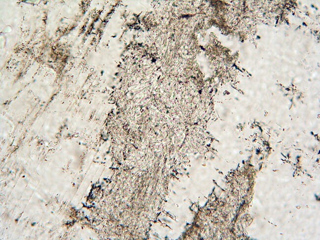
Sericite replacing plagioclase in a metaluminous granite. Sericite is grungy-looking fine-grained stuff that commonly replaces feldspars. Close examination of sericite reveals that at least some of it is fine-grained white mica. Because it is generally made up of very small crystals, its birefringence is irregular and generally low. Some of the larger crystals can be seen here to have first order birefringence. Albite twinning in the plagioclase is clearly visible. Plane- and cross-polarized light views. Field width is 3 mm. UN30A |
|
|
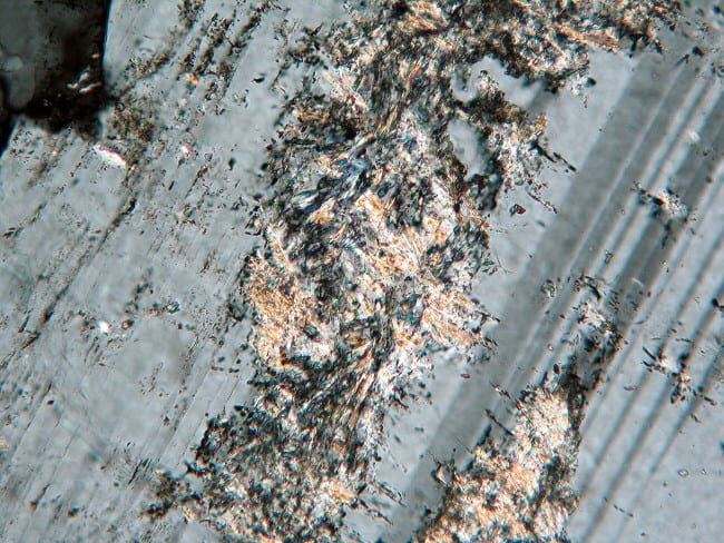
Sericite replacing plagioclase in a metaluminous granite. At higher magnification, some of the grungy-looking material resolves into little, colorless, platy crystals. The larger crystals have up to middle first order birefringence, with a positive sign of elongation, and so are probably small white micas that grew during subsolidus hydrothermal alteration. Plane- and cross-polarized light views. Field width is 1.2 mm. UN30A |
|
|
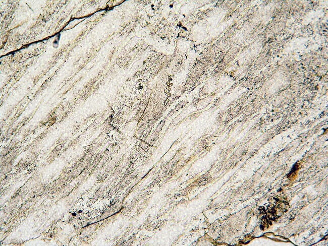
Serpentine in an altered harzbergite xenolith in a kimberlite dike from Pennsylvania. Much of this sample is made of calcite (colorless) and stained talc (darker browns). Serpentine is the yellowish material in thin veins. Calcite has very high birefringence, and the talc has irregular second and third order interference colors that are modified by the brown staining. The serpentine is most easily seen in the veins, where the fibers are perpendicular to the vein walls. Plane- and cross-polarized light views. Field width is 3 mm. Fayette Co. Penn. 33 |
|
|
Calcite in an alkaline granite. This shows two images with the same patch of calcite in two different orientations. In the first view, the calcite grain to the upper left is oriented so that a N-S line bisects the obtuse angle between its cleavages. This means the c axis of the calcite is approximately N-S, so the N-S polarized light is parallel to the low calcite refractive index and the grain has rather low relief. In the second image, the same calcite grain has been rotated so that a N-S line bisects the acute angle between the cleavages, so the c axis is approximately E-W. Thus, the N-S polarized light interacts with the high calcite refractive index, so it has high relief. Calcite and dolomite are the only two common minerals that can noticeably change from low to high relief on rotation in thin section. Both in plane-polarized light. Field width is 1.2 mm. NEIGC86-B2-7 |
|
|
Calcite in an alkaline granite. Calcite has very high birefringence, so interference order is difficult to judge from the high-order pastel colors. It is more reliable to look at a thin edge and count the number of magenta bands. The thin edge to the left of center of the first image has ~8th order birefringence. The second image shows this same pair of grains, with one rotated to extinction. Calcite is quite soft and undergoes substantial surface deformation during normal thin section grinding. The distorted crystal surface, combined with its high birefringence, results in incomplete, speckled extinction. Both in cross-polarized light. Field width is 0.6 mm. NEIGC86-B2-7 |
|
|
Zoisite developed in pagioclase in an anorthosite. Zoisite is essentially an orthorhombic, very low iron epidote. As an alteration product it commonly forms as masses or veins of irregular, ragged crystals in Ca-rich plagioclase. Zoisite has low first-order birefringence that is characteristically anamalous Berlin-blue and anomalous brown, caused by its very high dispersion of the 2V. Plane- and cross-polarized light views. Field width is 3.0 mm. STWR-8 |