
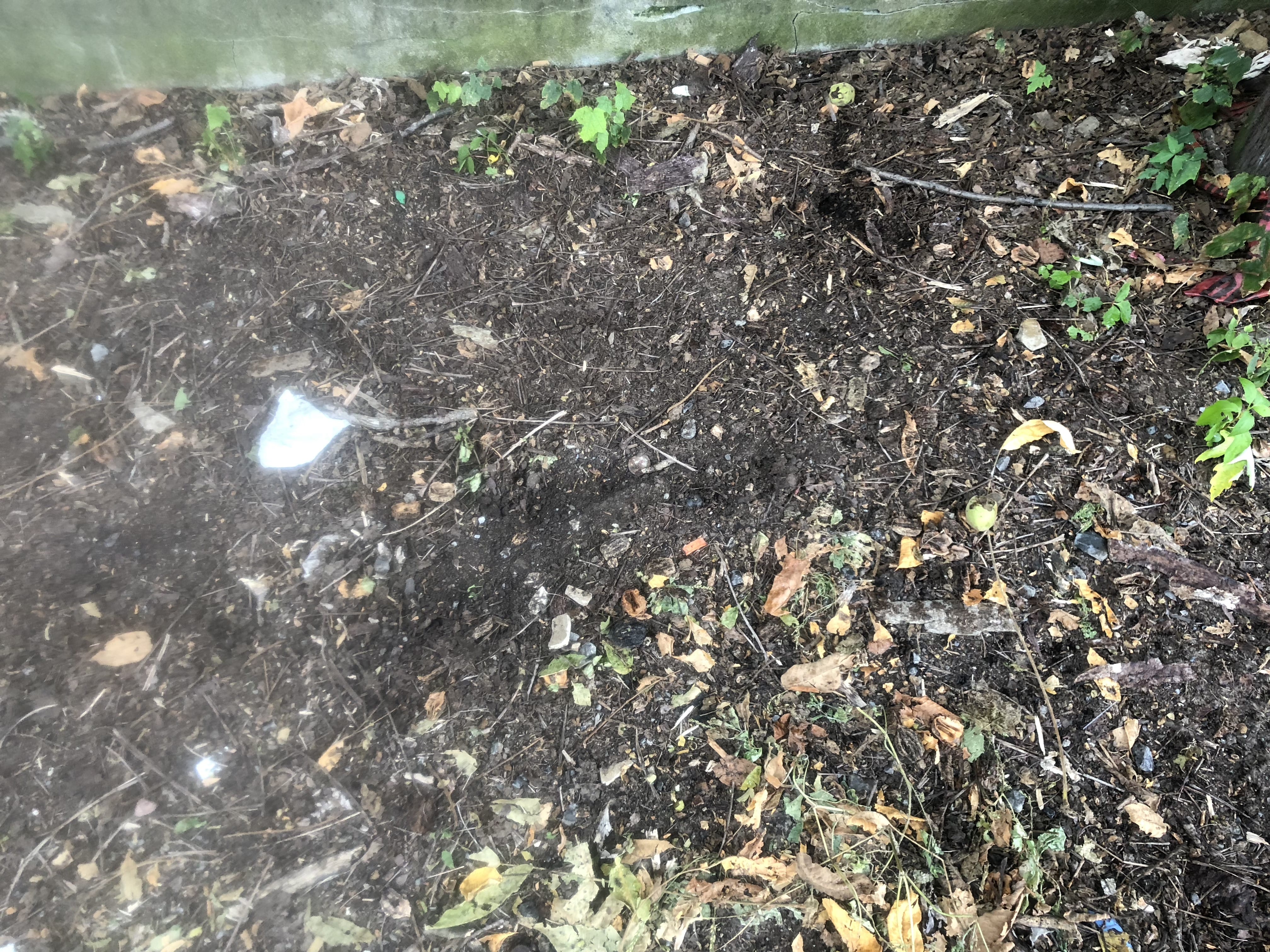
This is a picture of where I got my soil sample! There were lots of decomposing elements to this soil including rusty beer cans, paper, organic matter, etc. The soil was most likely in the A Horizon.

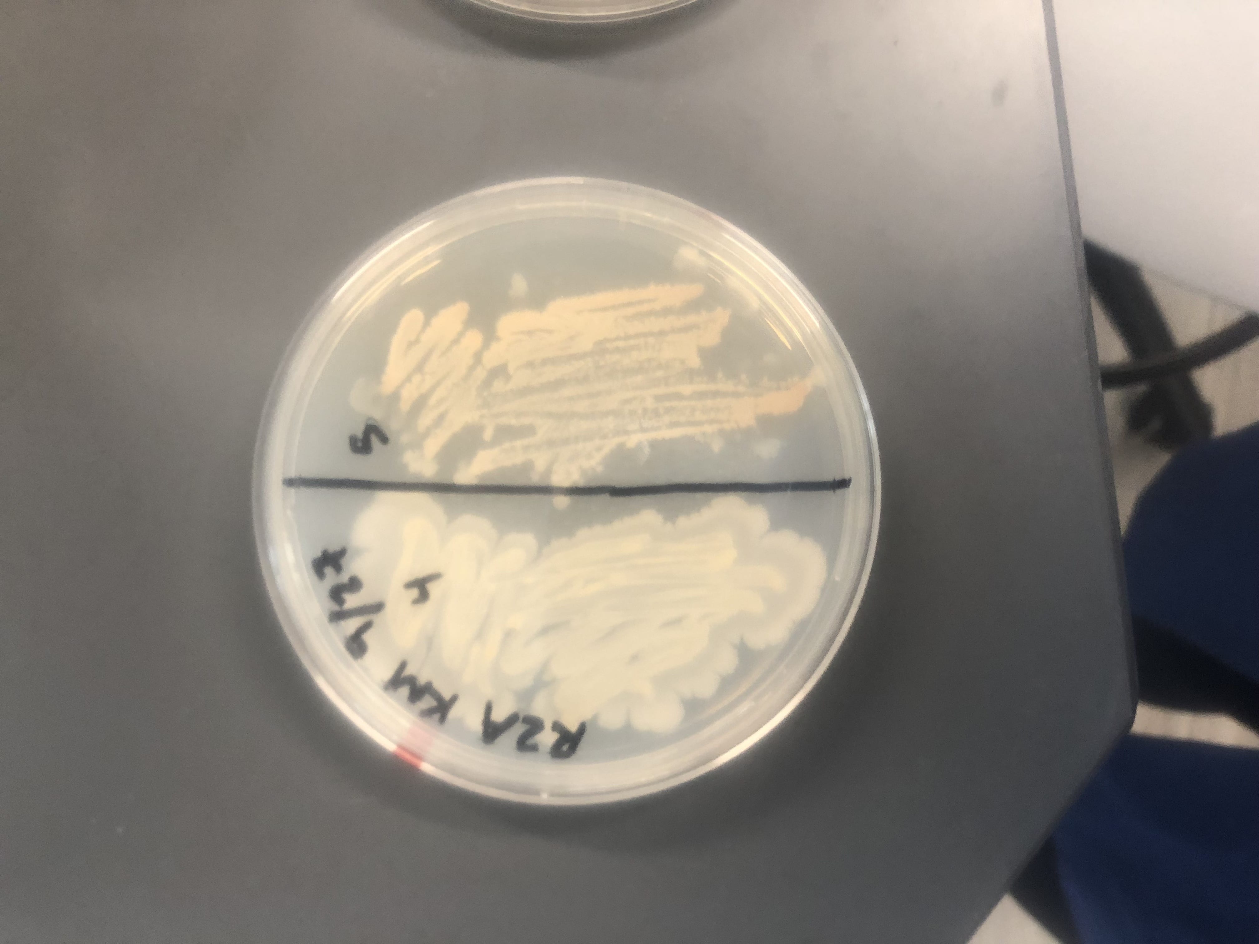
These are two of my streak plates with the four producers that I found in lab. (slightly blurry due to the plastic bag).
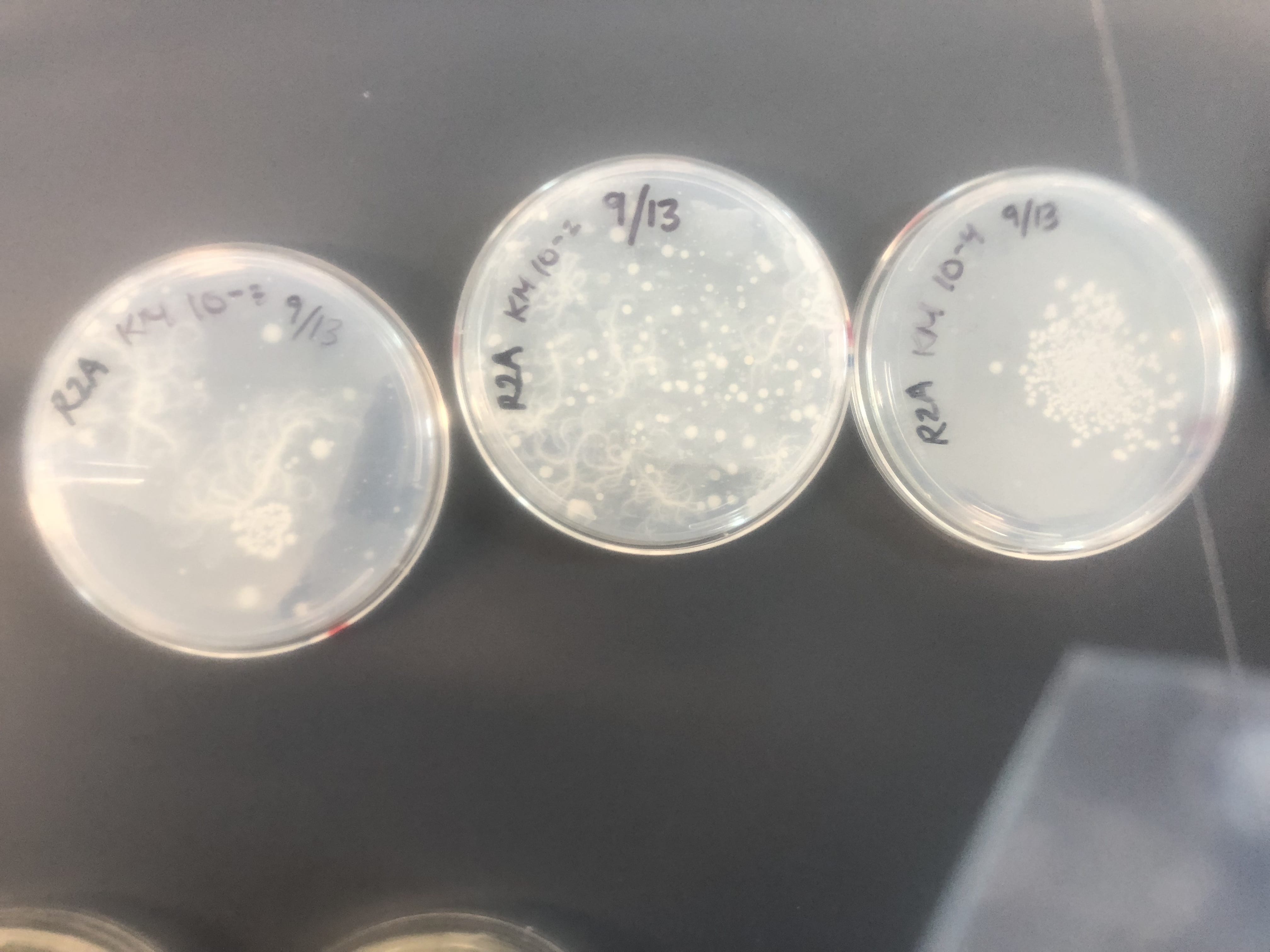
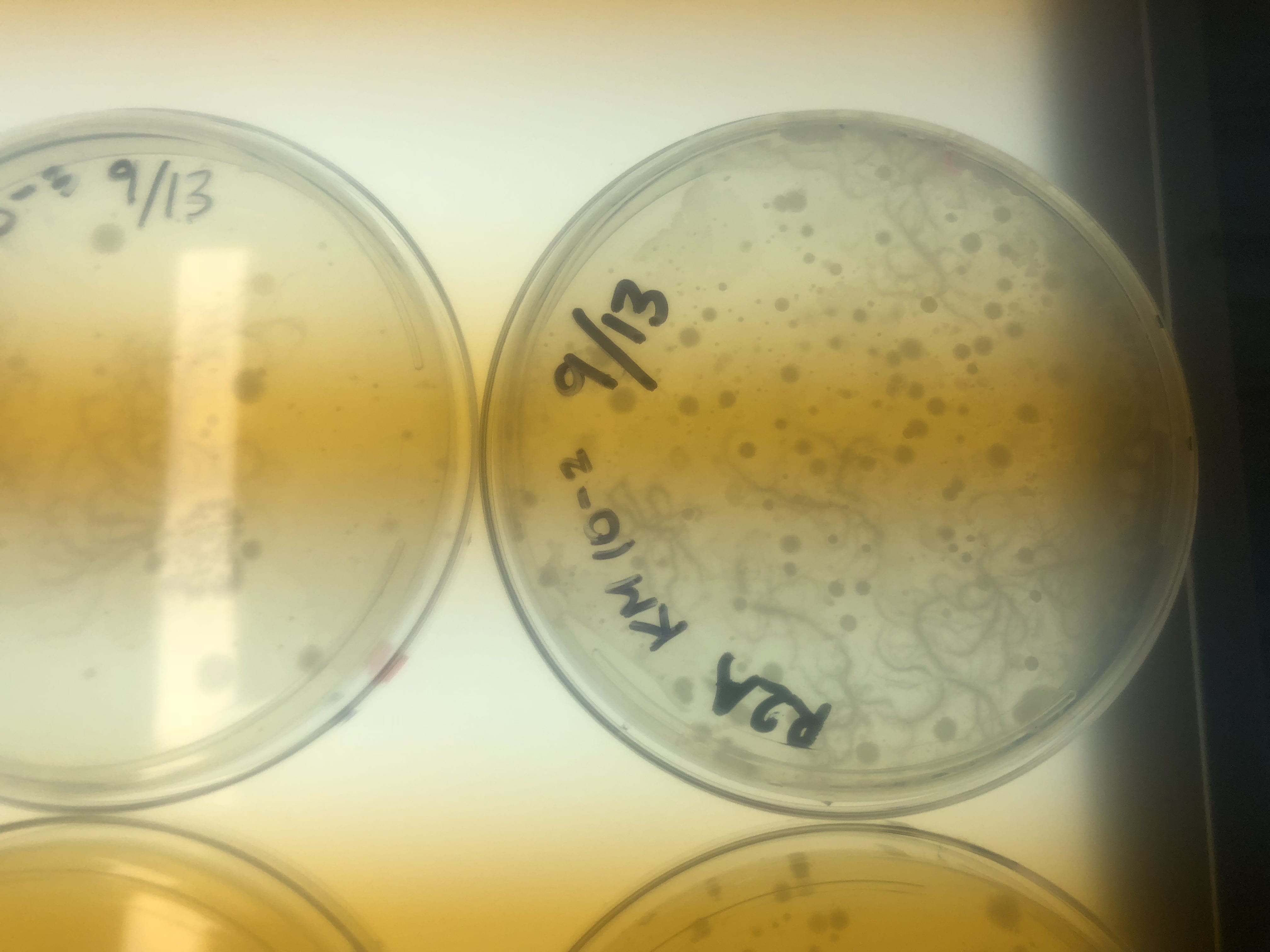
These are my R2A plates, where I found most of my producers. You can see the mycoides growing on the 10^-2 and 10^-3 dilution plates. The second photo is a close up of the mycoides on a light box at a dilution factor of 10^-2.

A close up of my R2A plate with a 10^-4 dilution. This photo was taken on the light box.
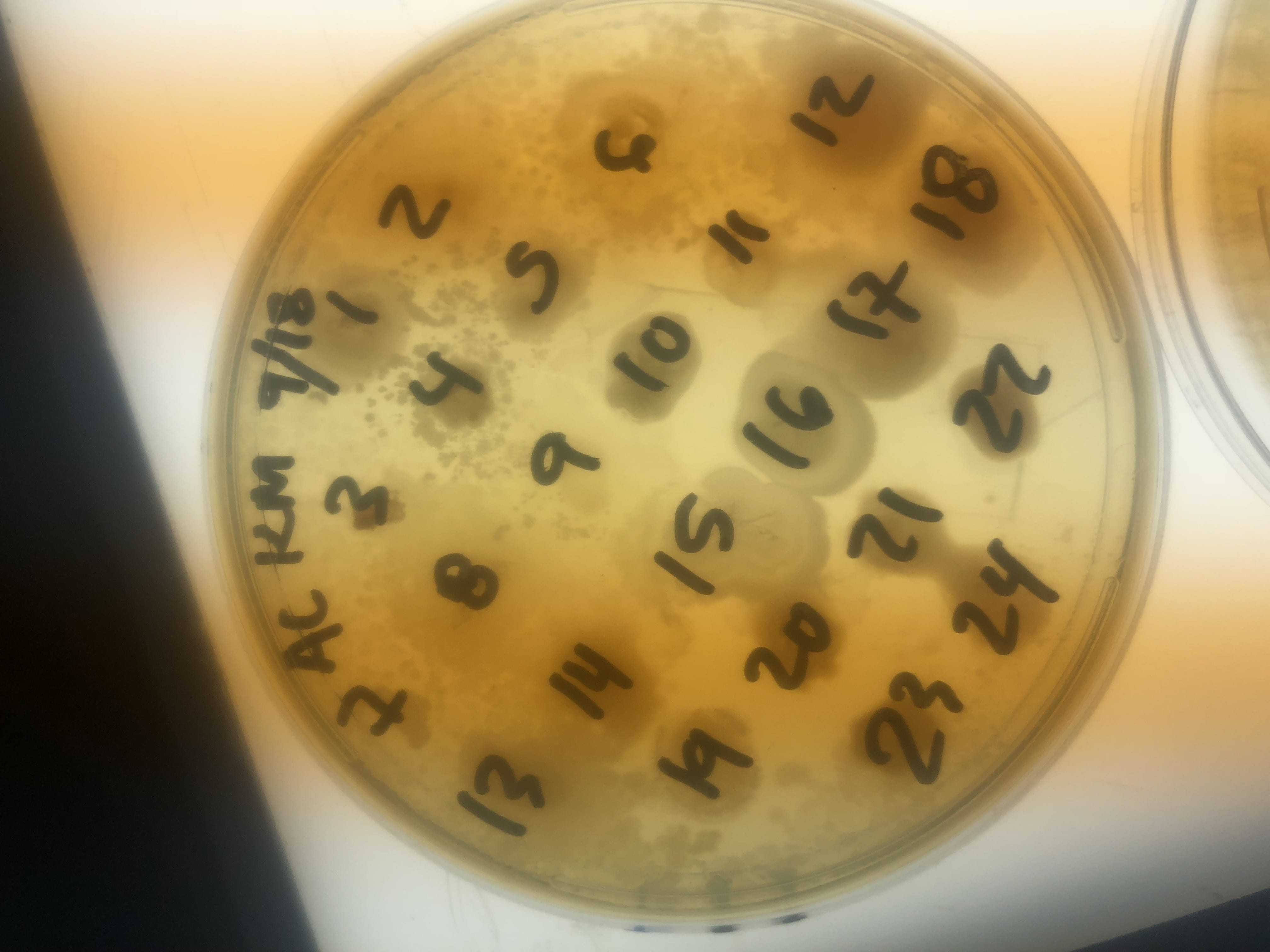
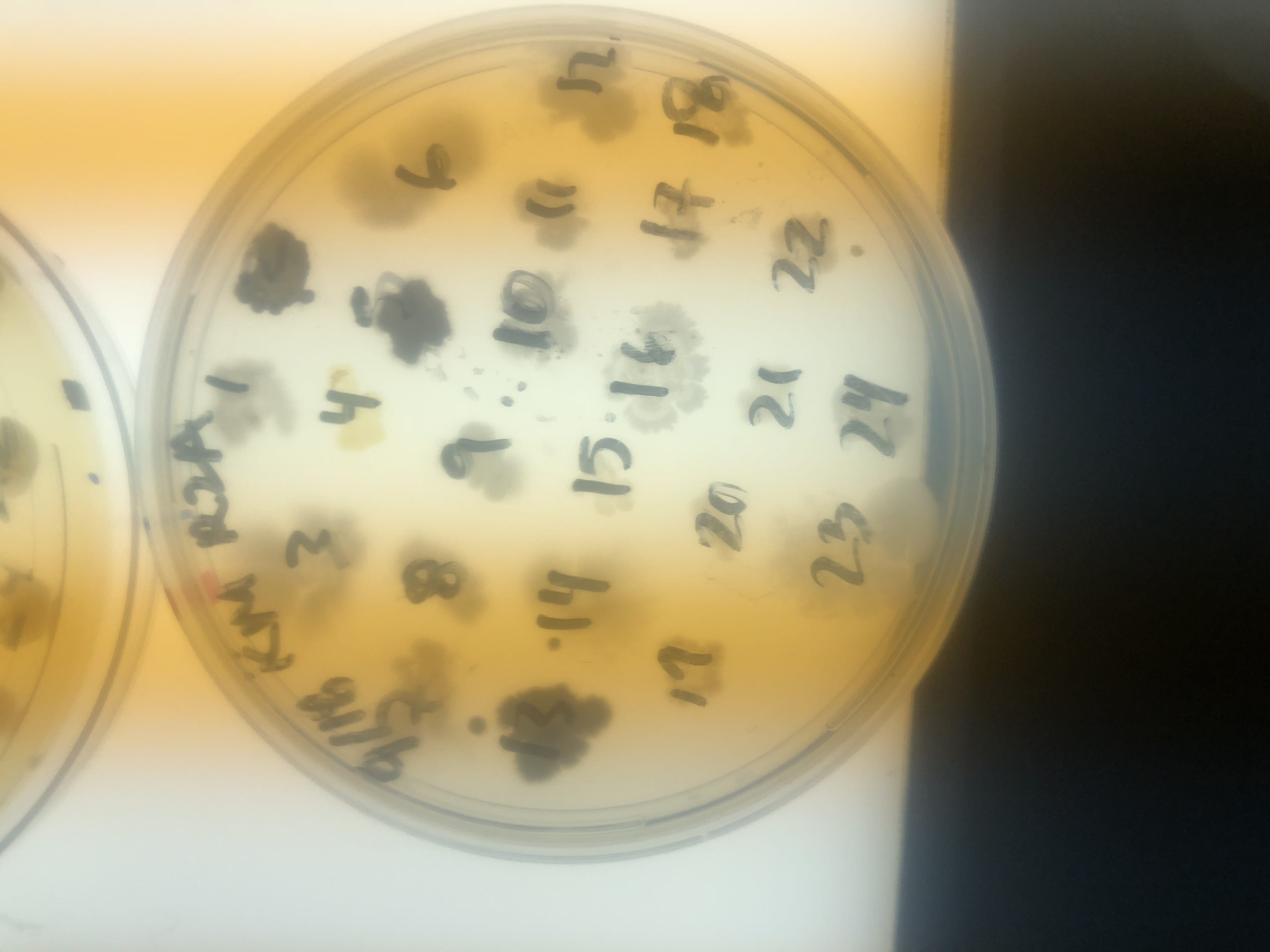
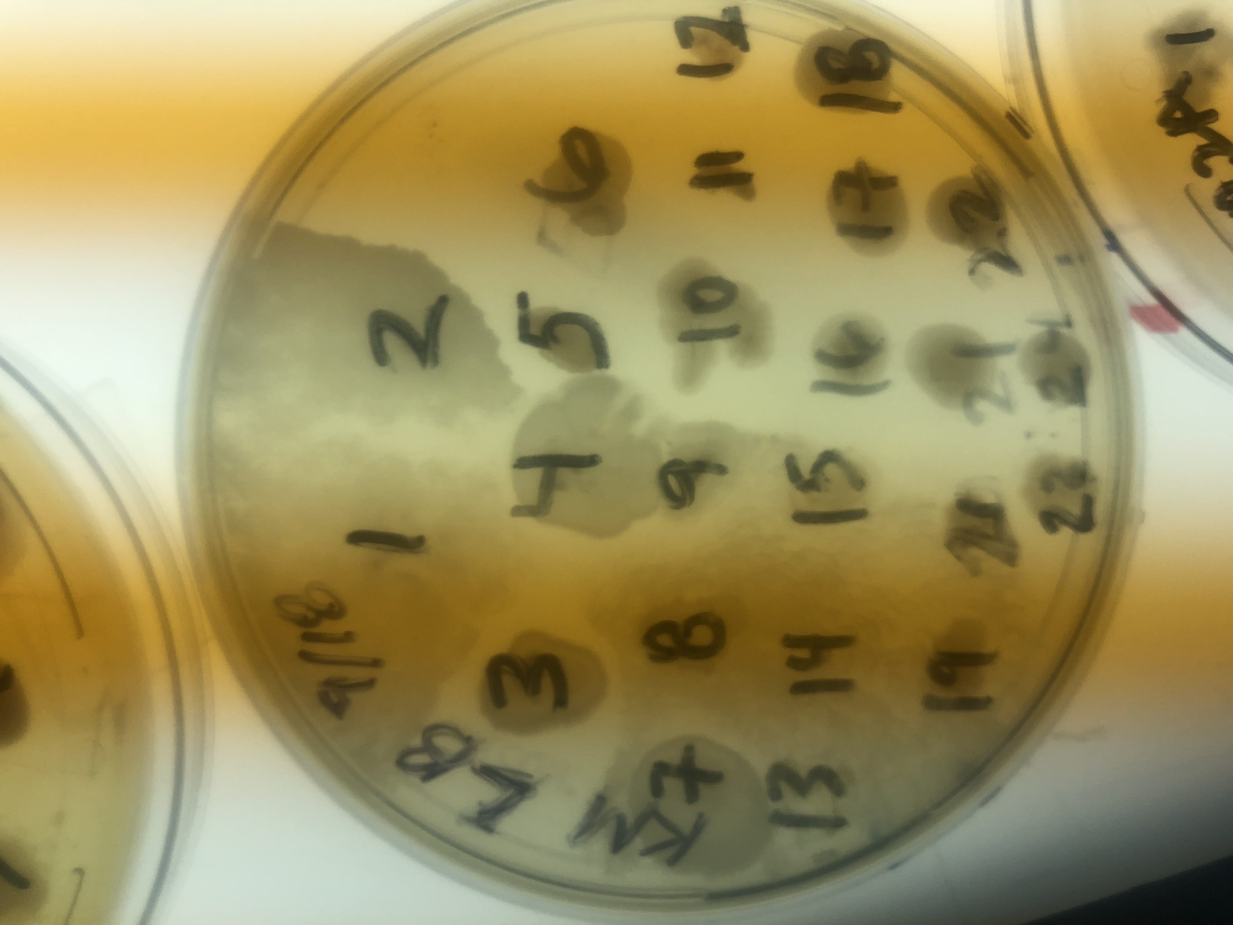
These are three examples of patch plates. These were the first set of patch plates that we did. These photos were all taken on the light box. In both the LB and AC media plates, you can see an overgrowth of mycoides. The mycoides overtook the patches in this section and rendered them unusable. I found minimum mycoides growth in the R2A plates throughout my experiment.

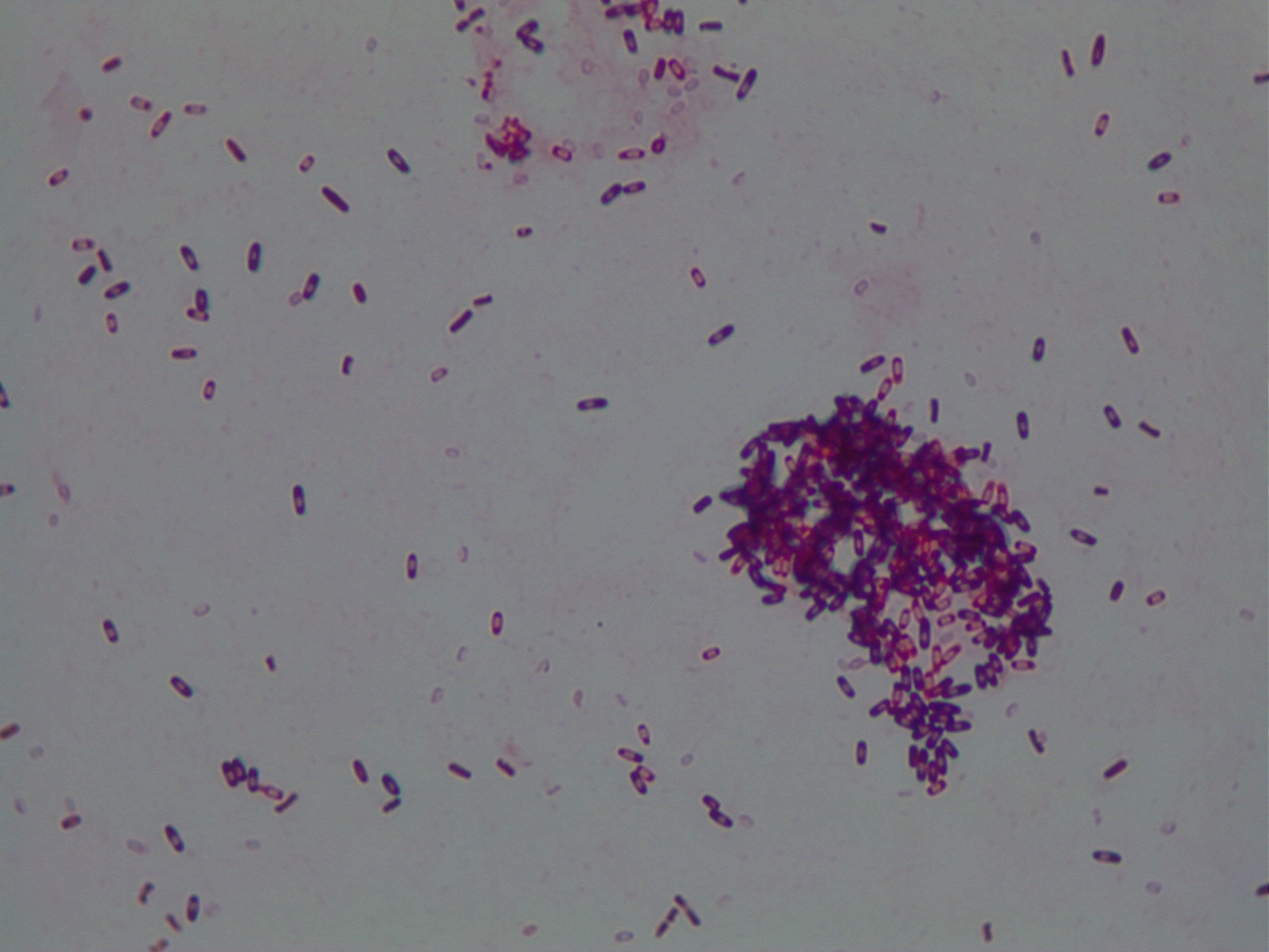
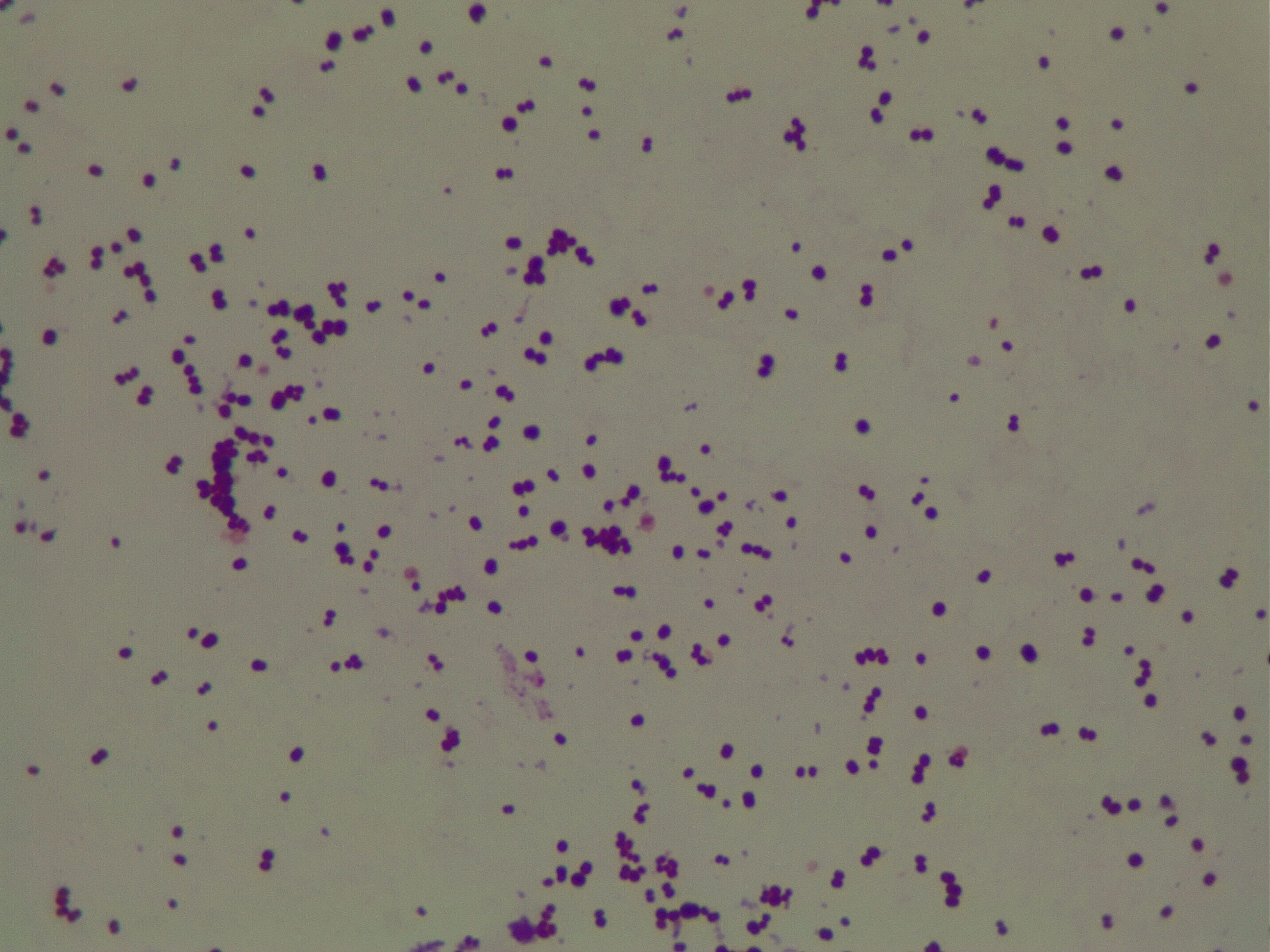
These are two pictures of my Gm+ bacteria. I know it is Gm+ due to the purple color. My bacteria are most likely bacillus because they are rods and spore forming. I know that they are spore formers due to the central pink spore between the purple ends of the rods. These photos were taken at 100x.
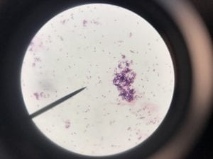
This is another photo of my bacteria. You can see a large aggregation of colonies on the right side of the slide. This photo was taken at 1000x.

These are pictures at 1ooox of my Gram positive control (S. epi).
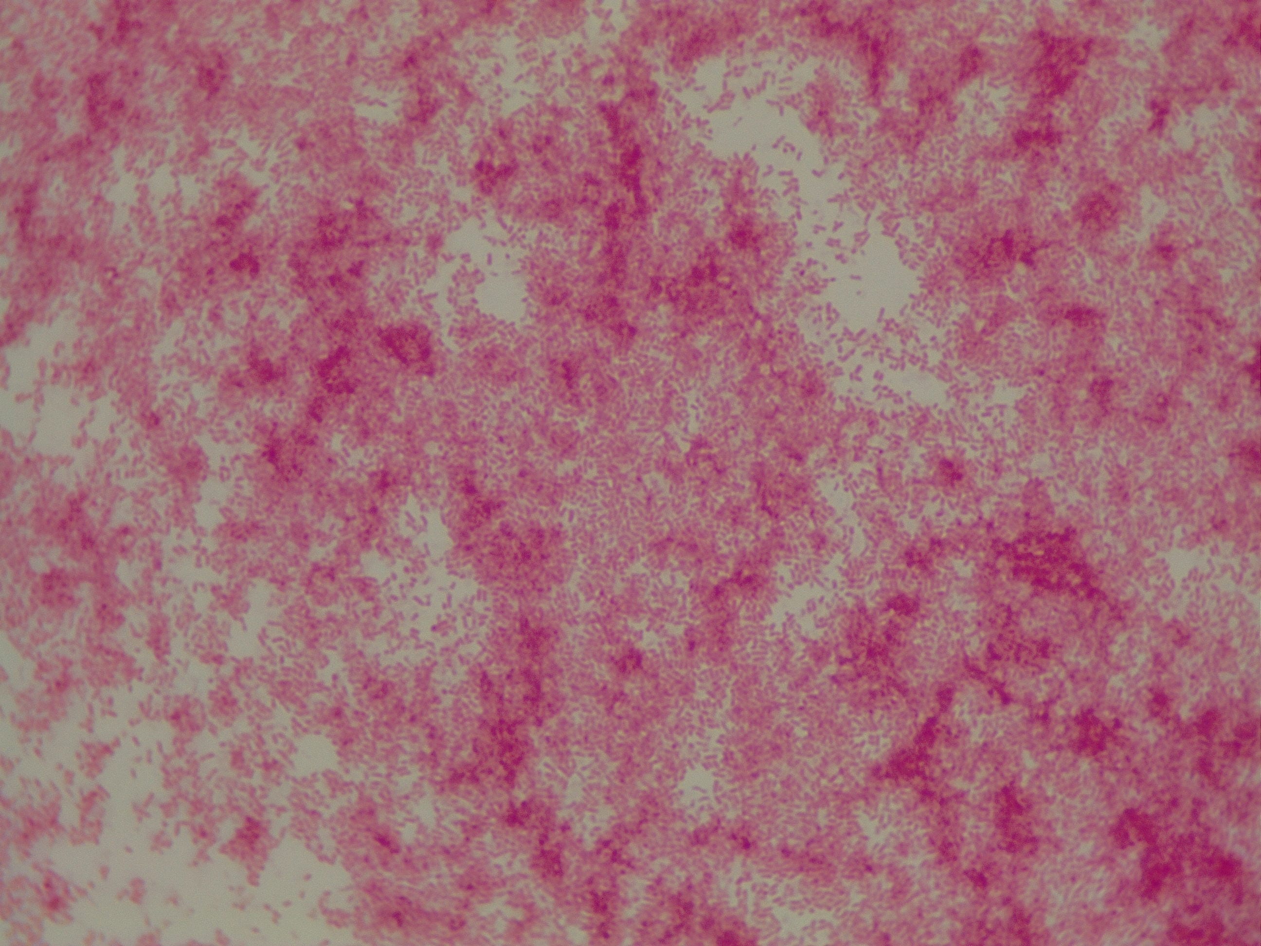
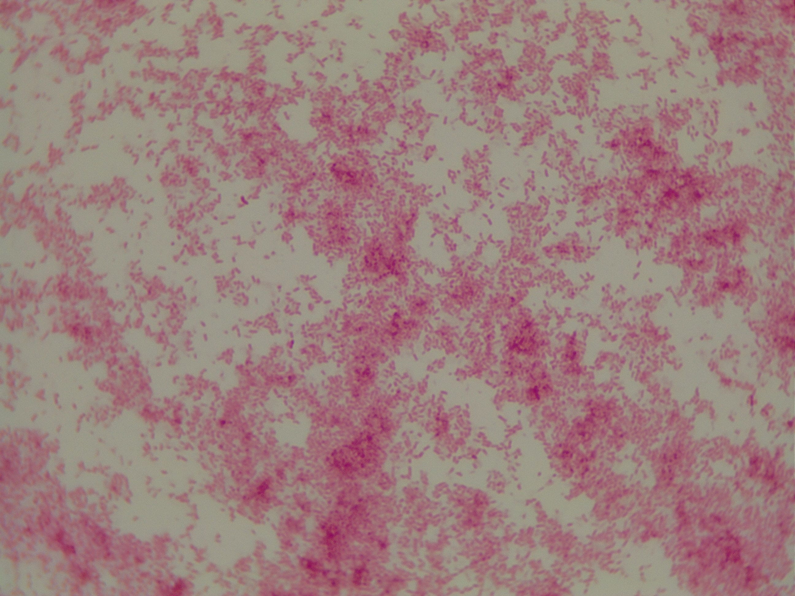
These are pictures at 1000x of my Gram negative control (P. putida).