And the name of my microbe is …Tommy

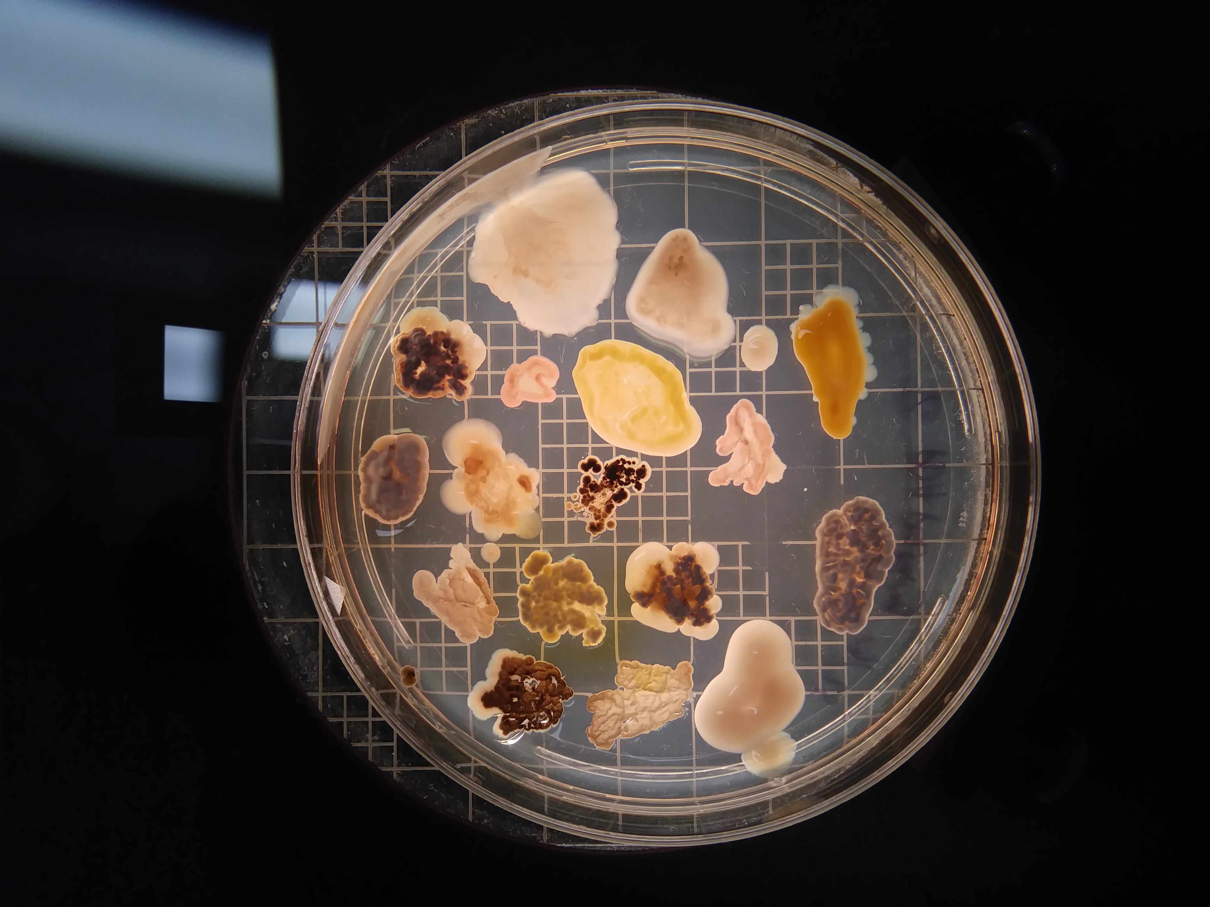
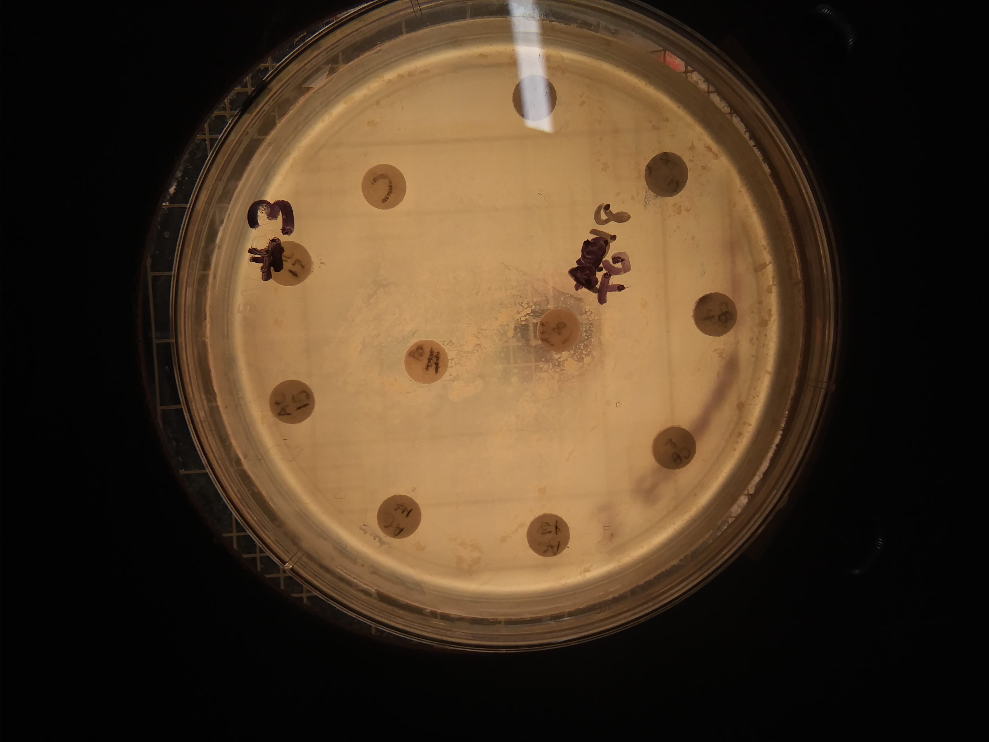
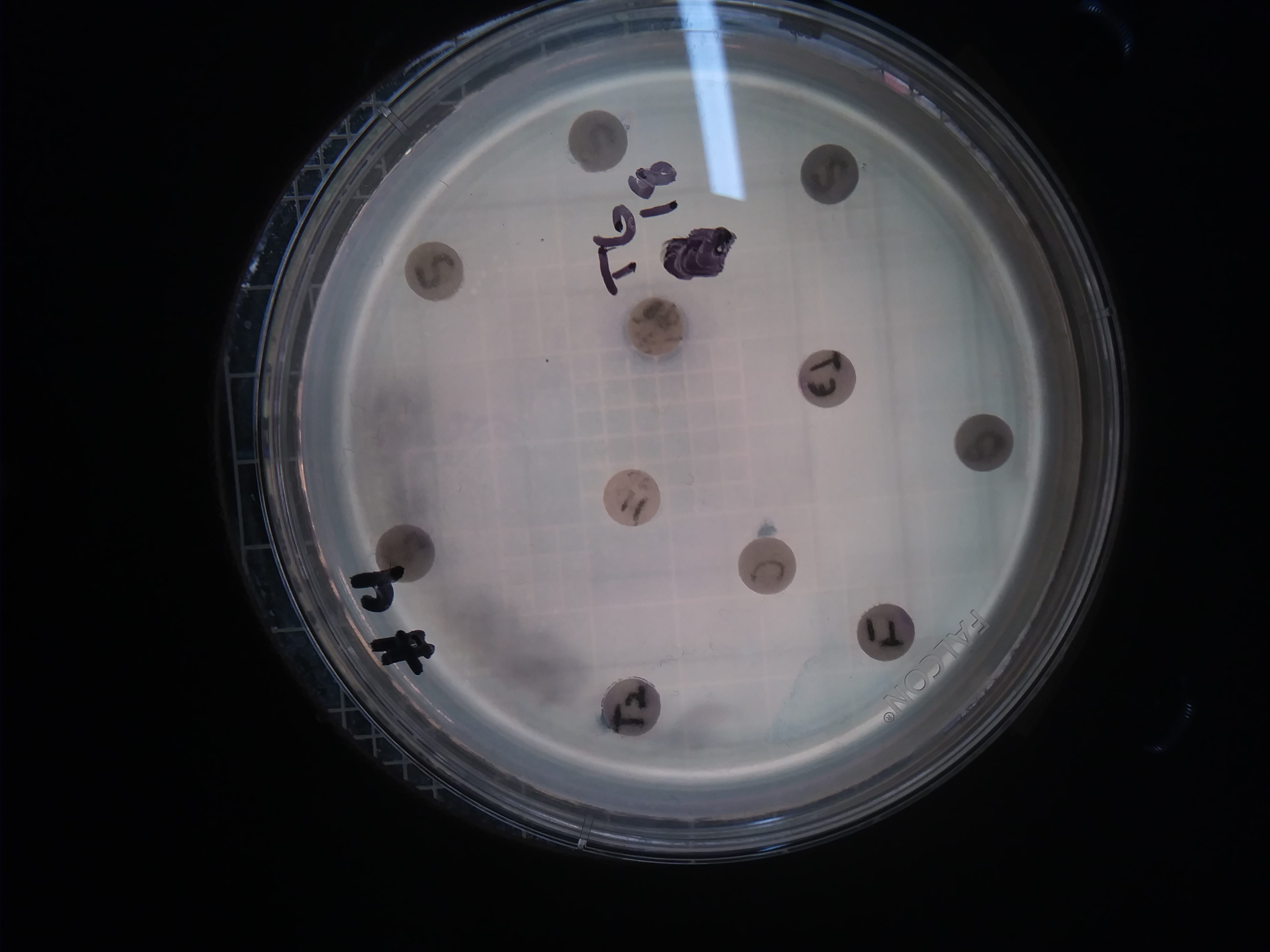
PCR resulted in successful amplification of two of my three isolates of interest (#5 and #11). Although #18 inhibited both Gram positive and Gram negative tester strains, BLAST sequence analysis could not be done to determine the genus, because no DNA was amplified to sequence! Additionally, neither Gram staining nor extraction were performed for isolate #5. Thus, here I identify and discuss the genus of isolate number 11 for which Gram stains and BLAST were both successfully performed.

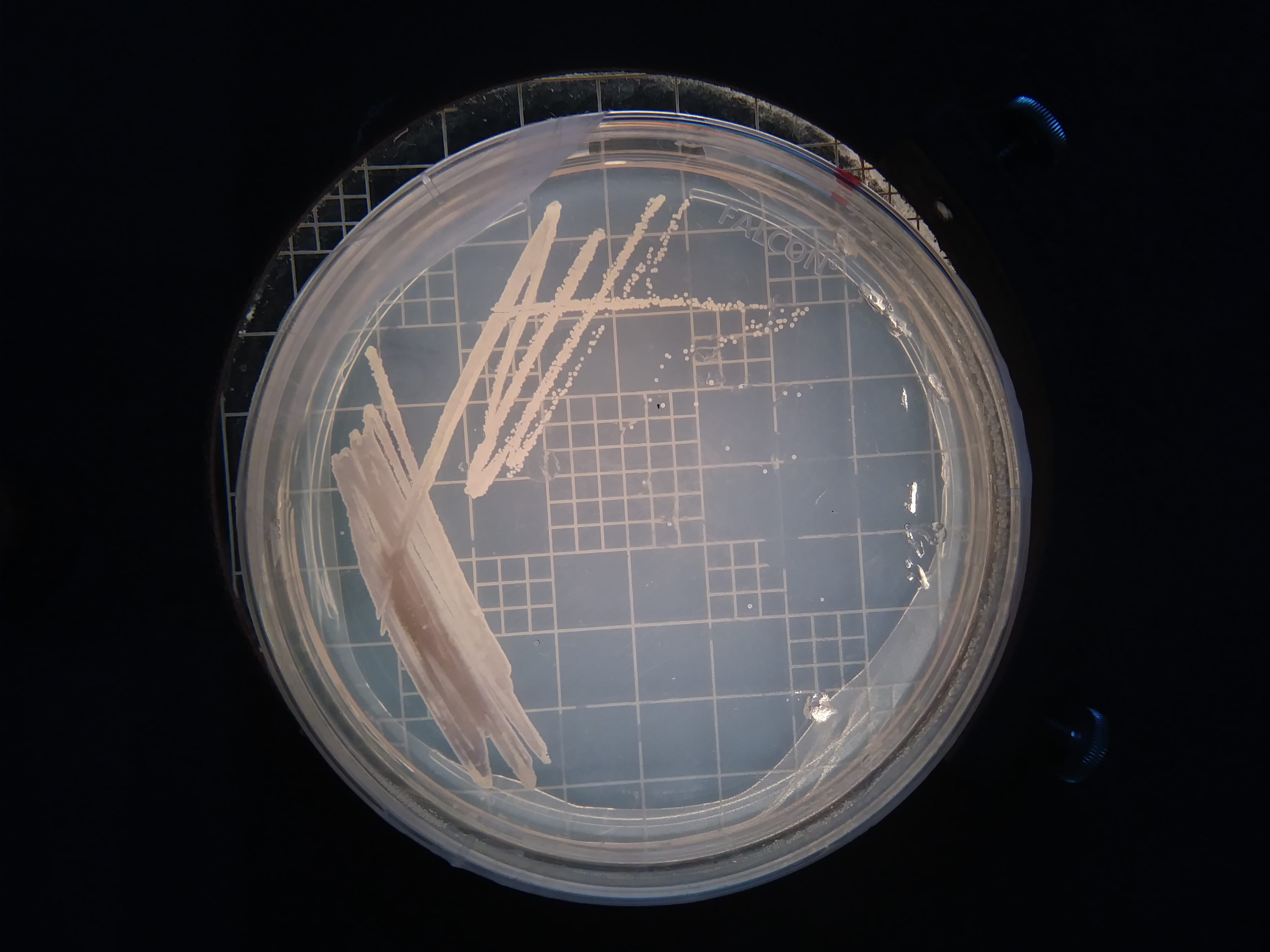
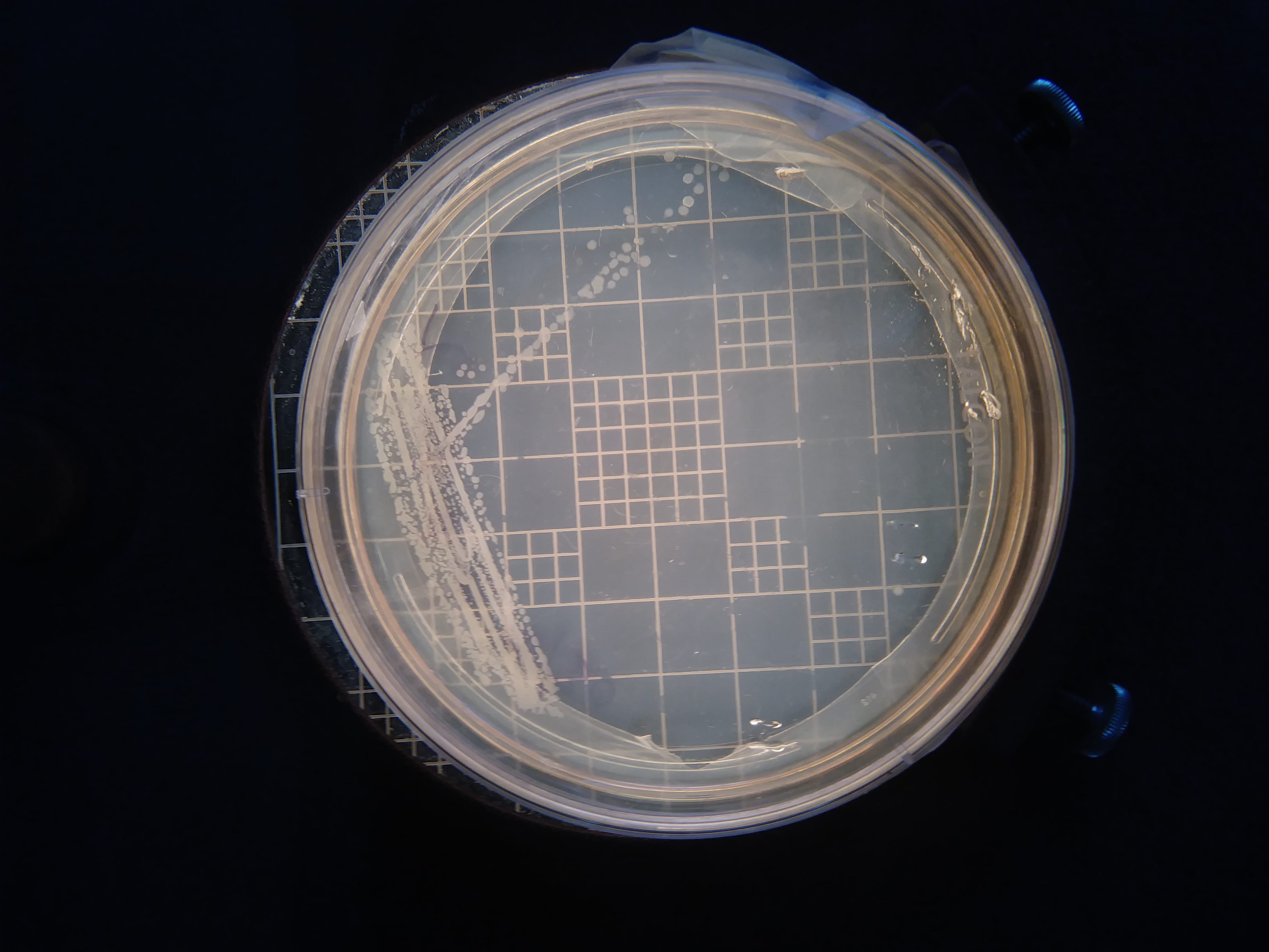
Streak plate of isolate 11.

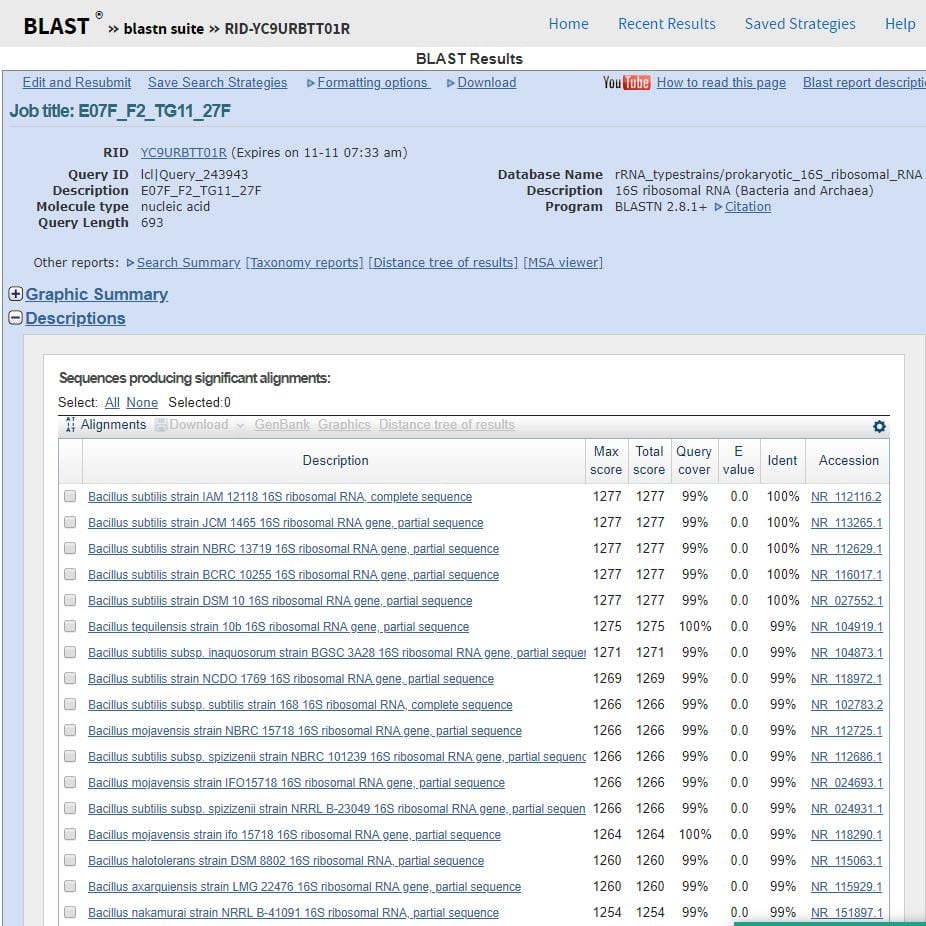
What were the results of your 16S analysis? 
BLAST retrieved a list of organisms (genus and species) that contain a high level of sequence similarity in the DNA region coding for the 16S rRNA subunit (693 base pairs analyzed). The results are seen below… and my microbe is…Bacillus! The genus Bacillus dominated the sequence similarity results in both BLAST and the Ribosomal Database Project (RDP) (results seen below). Both proposed a list of species and strains within the Bacillus genus that demonstrated similarity in 16S sequence. Bacillus subtilis was the most common species to appear in these lists, in a variety of strains. This is interesting because Bacillus subtilis was one of our safe-analog ESKAPE tester strains! A few such proposed strains were Bacillus subtilis NBRC 13719, Bacillus subtilis DMS 10, and Bacillus subtilis IAM 12118. However, Bacillus strain identification is limited to the species level in 16S rRNA analysis (1).

Another species that was listed as similar was Bacillus licheniformis, which has strains known to produce antibiotics such as bacitracin and bacteriocin (1). Since isolate 11 inhibited tester strains, it is possible that it was Bacillus licheniformis. Nevertheless, 16S results definitely point to Bacillus.

BLAST results listing organisms with highly similar sequences of DNA coding for 16S rRNA. Genus and species of similar organisms are included.

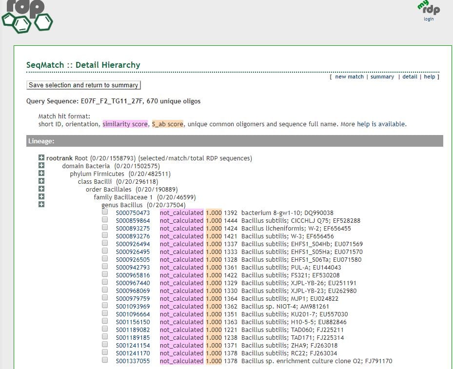
RDP results, affirming the genus as Bacillus in accordance with BLAST. Bacillus subtilis appears often in the list of similar strains, as it did in BLAST. This suggests isolate 11 may be a strain of Bacillus subtilis.

Does your gram stain agree? 
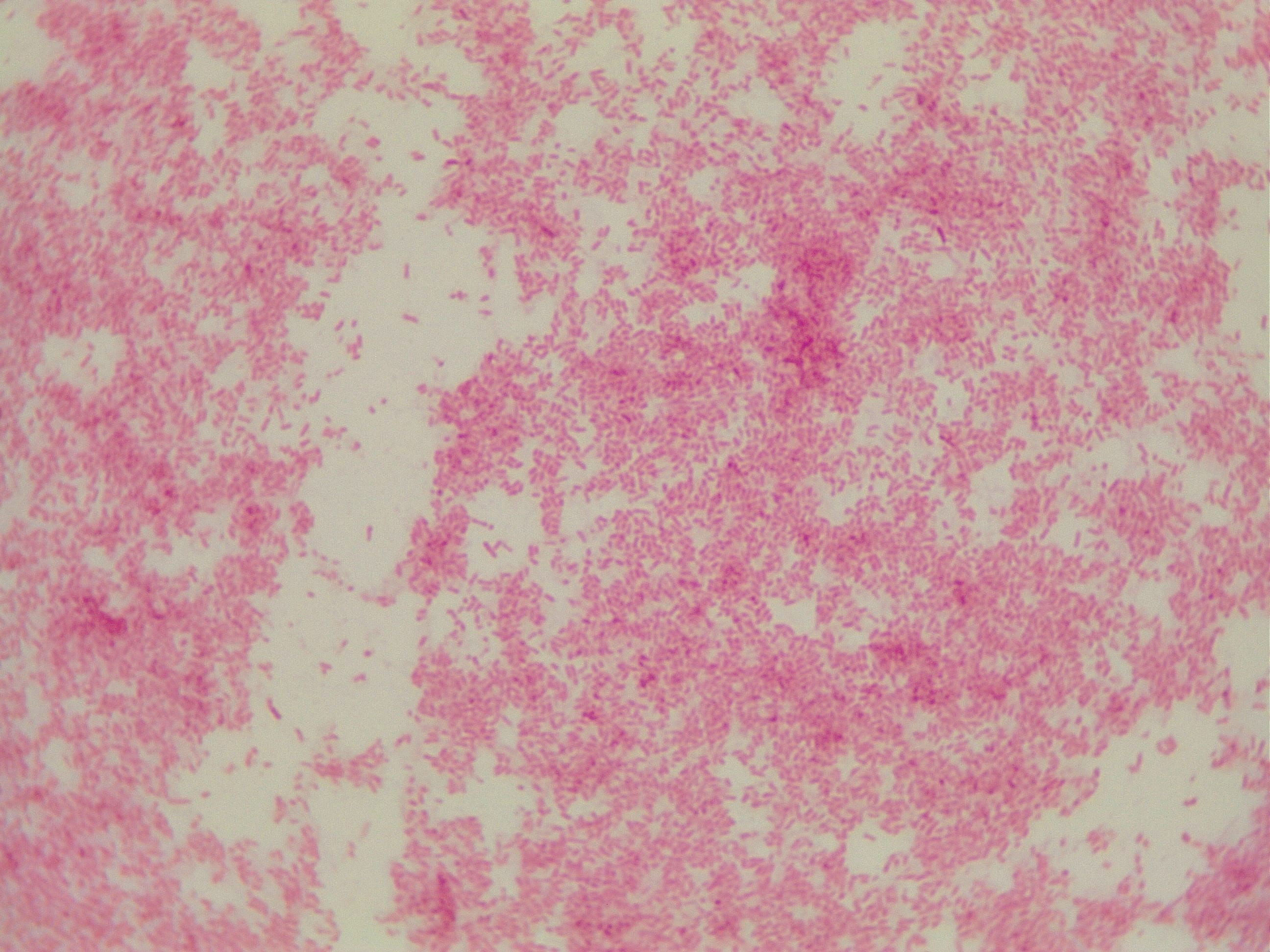
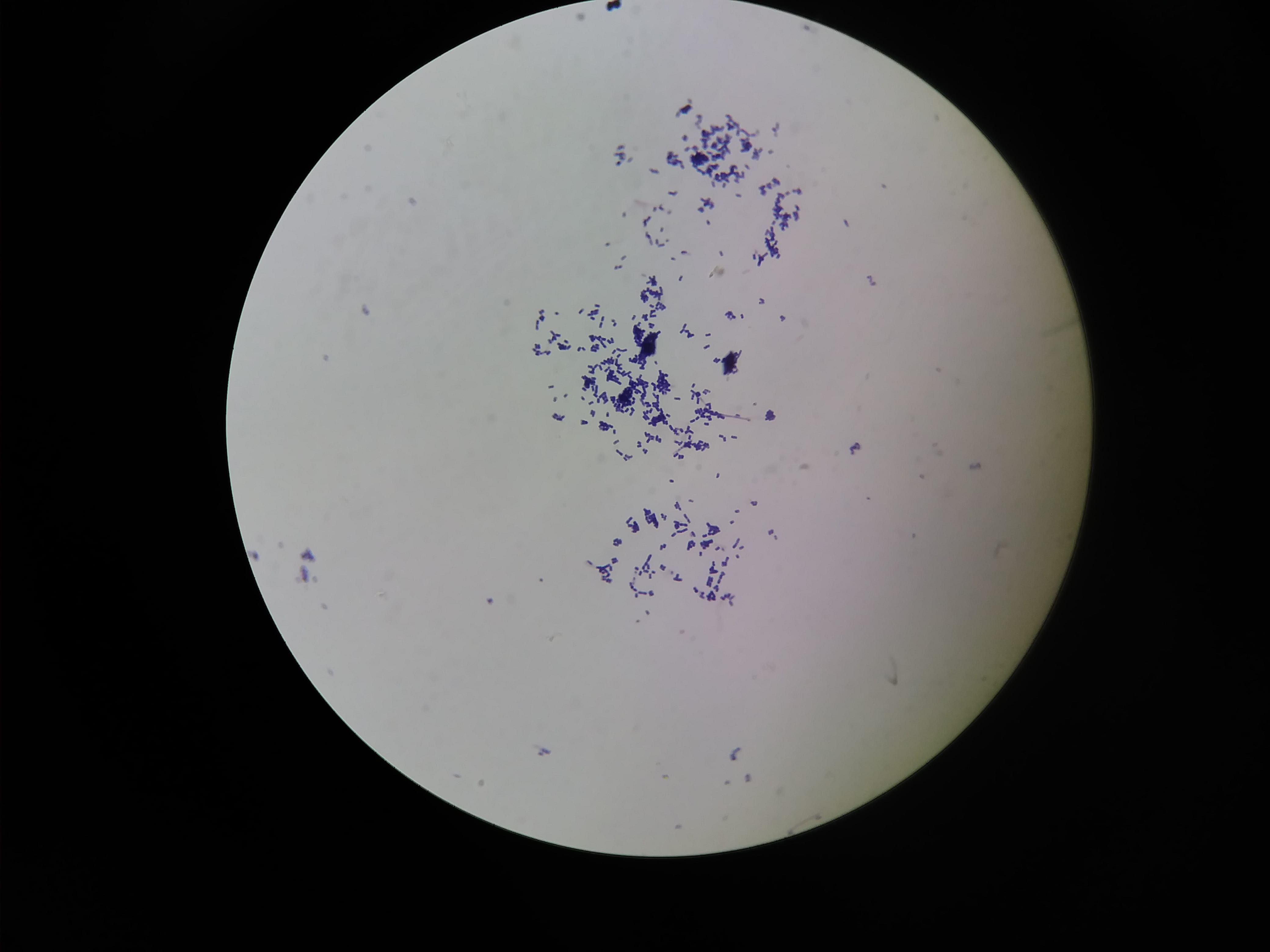
YES! My Gram stain showed that isolate 11 was Gram positive rods and upon close examination it appeared that they formed spores. We also know that this isolate must have been aerobic since it grew in our aerobic conditions.

The genus of Bacillus contains Gram-positive, spore-forming, rod-shaped, aerobic bacteria (1). All of this aligns with what we know to be true about my isolate 11! Thus, I feel comfortable claiming that isolate 11 is some species of Bacillus.

Isolate number 11. Gram positive rods. Looks like they might be spore formers.

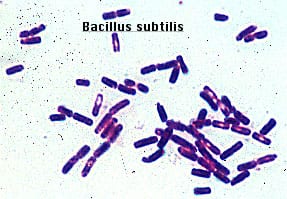
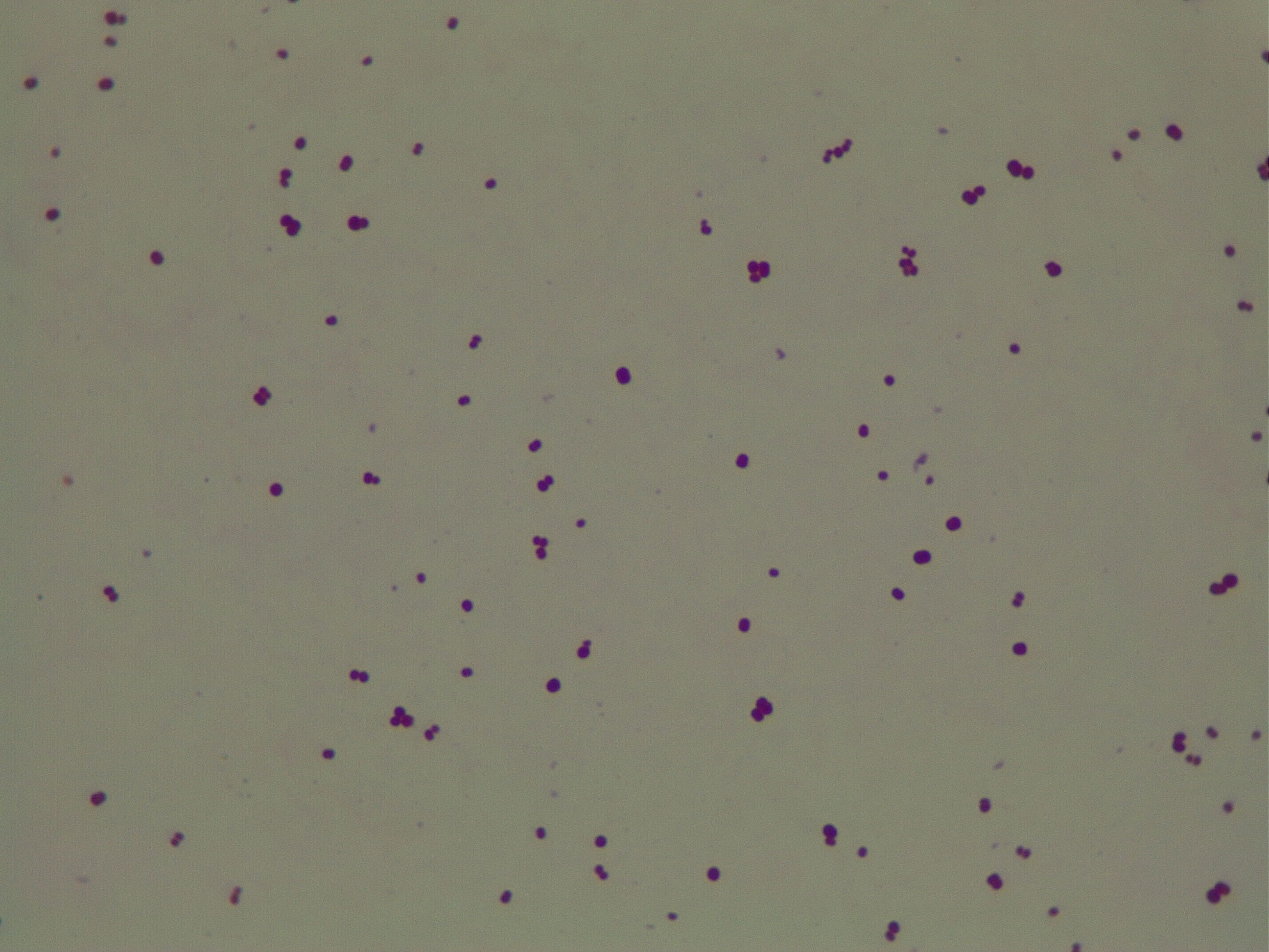
Gram positive rods, just like isolate 11. Form endospores which resemble the spore-looking characteristic of isolate 11. https://www.medschool.lsuhsc.edu/Microbiology/DMIP/dmex17.htm

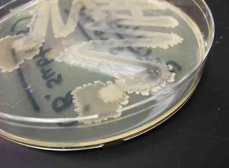
This streak plate of Bacillus subtilis found online resembles closely the morphology of my isolate 11 streaks in particular the color and unique pattern of growth on the edges of colonies. https://www.scienceprofonline.com/science-image-libr/sci-image-libr-bacterial-colonies.html

a) General cellular and morphological characteristics of the genus (taxonomic classification, nutrition, cell shape, habitat). 

Taxonomic classification. Domain: Bacteria; Phylum: Firmicutes; Class: Bacilli; Order: Bacillales; Family: Bacillaceae 1; Genus: Bacillus

These are Gram-positive, spore-forming, rod-shaped, aerobic bacteria (1). They are widely distributed in the soil as well as the aquatic environment (2). Their ability to inhabit diverse habitats results in part due to their spore forming ability (2). Some Bacillus species inhabit extreme environments such as high temperature or extreme pH since spores can survive dormant in extreme environmental conditions (3). Bacillus can also survive in low-nutrient environments, including a lack of elements such as phosphorus, nitrogen or oxygen, provided that the organism has a sufficient supply of carbon sources (1). Bacillus represent heterogeneous species that also form bioactive polymers useful in industry including medicine, biodefense, biofuels, and bio-pesticides. For example, Bacillus thuringiensis is used for biological control of insects to protect crops (1). Spores of Bacillus subtilis have also been used as probiotics in humans (1).

b) Information regarding antibiotic production in this genus. 

The number of antibiotics produced by the genus Bacillus is in the hundreds (4). Many of these antibiotics are peptide antibiotics, which tend to be smaller than proteins but range in molecular weights from 270 (bacilysin) to 4500 (licheniformin) (4). 66 different peptide antibiotics are elaborated by strains of Bacillus subtilis (4) and 23 by Bacillus brevis (4). Most of these peptide antibiotics are made exclusively from amino acids, but some contain additional substituents (4). As mentioned above, the species Bacillus licheniformis is known to contain strains that produce antibiotics including bacitracin and bacteriocin (1). Additionally, polymyxin and gramicidin S have been isolated from species of Bacillus. Antibiotics of Bacillus have been seen to be more effective against Gram positive bacteria (4), which is consistent with my observations of isolate 11.

References

(1) Porwal S., Lal S., Cheema S., Kalia V.C. (2009). Phylogeny in Aid of the Present and Novel Microbial Lineages: Diversity in Bacillus. Plos One.

(2) Parvathi A., Krishna K., Jose J., Joseph N., Nair S. (2009). Biochemical and molecular characterization of Bacillus pumilus from coastal environment in Cochin, India. Brazilian Journal of Microbiology. 40(2):269-275.

(3) Nicholson W.L., Munakata N., Horneck G., Melosh H.J., Setlow P. (2000). Resistance of Bacillus endospores to extreme terrestreial and extraterrestrial environments. Microbiology and Molecular Biology Reviews. 64(3):548-572.

(4) Katz E. and Demain A.L. (1977). The peptide antibiotics of Bacillus: Chemistry, Biogenesis, and Possible Functions. Bacteriological Reviews. 41(2):449-474.

Meet my microbes! – Tommy

Here is a photo of my PDA patch plate. PDA plates gave me a lot of mold over the course of the term, but also displayed some really interesting and unique microbe morphologies. Unfortunately, none of these inhibited the ESKAPE tester strains – neither Gram positive nor Gram negative. But they look awesome!


Gram negative control.


Gram positive control. Cocci.


Streak plate of isolate 18. This is the isolate whose extraction strongly inhibited a Gram positive tester strain and a Gram negative tester strain. It was my best producer!


Unknown 18. Gram positive. Appear to be diplococci that form some longer chains as well and like to aggregate. This is what my best antibiotic producer looks like up close!


Streak plate of isolate 11


Isolate number 11. Gram positive rods. Looks like they might be spore formers.

Extract news! – Tommy

On October 25th, we performed an organic extraction of our isolates. I had two isolates that I attempted extractions on. After letting the extraction vials dry, a residue was present which was weighed and subsequently resuspended in methanol. Not all of the residue dissolved, but 20 microliters of this resuspended methanol/extract solution was put on little circles to put on tester strains to see if they inhibited tester strains. If the tester strains were inhibited, it would mean we were successful in extracting an antibiotic from the bacteria!

We tested our extractions on two Gram positive and two Gram negative tester strains. Both of my isolates had inhibited at least one Gram positive and one Gram negative tester strain. To test extractions, I chose testers 1, 3, 4, and 5 (Staphylococcus epidermidis, Erwinia carotovura, Enterococcus raffinosus, and Actinetobacter baylyi, respectively). Testers 1 (Gram positive) and 5 (Gram negative) were previously inhibited by isolate number 11 and testers 3 (Gram negative) and 4 (Gram positive) were previously inhibited by isolate number 18.

Below are pictures of the results of testing the extracts on testers 3 and 4 (Erwinia carotovura and Enterococcus raffinosus). Extract from isolate number 18 inhibited both testers as seen by the clearing of growth of the tester bacteria around the 18 extract circle. This is interesting because I chose these two testers based on inhibition that I saw from isolate number 11, not isolate 18, but extract 18 showed inhibition while isolate 11 did not. However, extract 18 also inhibited testers 1 and 5 (Staphylococcus epidermidis and Actinetobacter baylyi), albeit not as robustly. Extract 11 did not show inhibition of any tester strains. The fact that extract 18 inhibited tester strains indicates that an antibiotic was successfully extracted!

Tester 3 – Erwinia carotovura – with organic extractions. The extraction from my isolate 18 shows a clearing of growth around it, indicating inhibition of the tester.


Tester 4 – Enterococcus raffinosus – with organic extractions. The extraction from my isolate 18 shows a small inhibition of tester growth around it.

Meet the ESKAPE Pathogens – Tommy

Assigned ESKAPE Pathogen

Enterococcus faecium

Why is this ESKAPE Pathogen of interest (in brief)

Enterococcus faecium can exist commensally within human or animal gastrointestinal tracts but can also be pathogenic in that it can cause neonatal meningitis and endocarditis (1). There have been strains of Enterococcus faecium identified as resistant to vancomycin – coined Vancomycin-Resistant E. faecium (VRE). Enterococcus faecium has also show tolerance to handwash alcohols in hospitals (2). Since alcohol-based disinfectants are used to control hospital infections worldwide, it is frightening that the multidrug-resistant Enterococcus faecium displays tolerance to commonly used hand rub alcohol solutions. VRE can survive on inanimate surfaces for weeks – including medical equipment – which explains how most VRE infections are acquired nosocomially (3).

General Cellular and Morphological Characteristics of the Organism (taxonomic classification, nutrition, cell shape, habitat)

Enterococcus faecium belongs to Domain: Bacteria, Class: Bacilli, Order: Lactobacillales, Family: Enterococcaceae, Genus: Enterococcus and Species: faecium. This ESKAPE pathogen are Gram positive cocci (spherical in shape) that form short to medium length chains. They can also exist in pairs or single cells. E. faecium are also Facultative anaerobes that are ovoid in shape (1). They do not have cytochrome enzymes and are catalase negative (1). Enterococci are found in the feces of most healthy adults – where there are more faecalis than faecium although both are present. Lower percentages were found in oral cavities in healthy students (1). These bacilli tend to live in the gastrointestinal tract of humans and animals and live in feces and sewage. They are able to withstand harsh environmental conditions including high temperatures, periods of drying, and some antiseptics (1).

Clinical Importance and Prevalence

E. faecium’s high level of inherent and acquired resistance (especially VRE’s) along with the pathogen’s ability to survive on surfaces in hospitals makes it an important bacterium to study clinically. VRE constitute about 43% of all Enterococci isolates (4) making it a severe threat across the world, especially in hospitals that rely upon vancomycin or related antibiotics to combat infection. The bacterium has shown tolerance to hand-rub alcohols, making it a greater threat in hospitals (2). Additionally, this pathogen affects largely older adults with comorbidity, leading to heightened mortality rates (3). Furthermore, Enterococci harbor transferable genetic elements, meaning that resistant genes can be passed to both Gram positive and negative bacteria by conjugation systems with plasmids and transposons (4). This potential of E. faecium to pass on its multidrug resistance to other bacteria in a horizontal fashion is particularly frightening.

Infection (How does the infection occur and where is it localized?), Pathology (What disease is caused? What are the symptoms?)

E. faecium is known to cause urinary tract infections, intraabdominal, pelvic and wound infections, superinfections, and bacteremias (often with other organisms) (5). Lower urinary tract infections including cystitis, prostatitis, and epididymitis are seen in older men (5). Enterococci can also cause infection in the abdominal lining in conjunction with liver cirrhosis or in patients with chronic peritoneal dialysis (5). This ESKAPE pathogen is the third most common organism seen in nosocomial infections (3). Another notable infection of E. faecium is endocarditis. Bacteremia and endocarditis are common infections of Enterococci. The mortality associated with these infections is likely partly due to the demographic of patients who present: older adults with multiple underlying diseases such as diabetes. Synergistic, bactericidal attack is required for treatment of endocarditis (5). Multidrug resistant Enterococci are arising, including Vancomycin Resistant Enterococcus (VRE). Enterococcal surface proteins are virulence factors that contribute to infection in humans (5).

Ineffective Antibiotics (Antibiotics to which the organism has acquired resistance)

E. faecium has shown greater antibiotic-resistance than E. faecalis (3). More than half of its pathogenic isolates express resistance to vancomycin, ampicillin, and high levels of aminoglycosides (3). Additionally, the pathogen has shown resistance to some handwash alcohols (2). Enterococci also exhibit inherent and acquired resistance to cephalosporins, clindamycin, tetracycline, and penicillins. A mutation in the domain V of the 23 S rRNA of E. faecium appears responsible for linezolid resistance (5). Additionally, resistance to quinupristin-dalfopristin may be due to enzyme modification, and drug efflux (5).

Effective Antibiotics (Antibiotics known to inhibit the organism)

VRE can be successfully treated with sultamicillin (5). For most Enterococcal infections, single-drug therapies with penicillin, ampicillin, or vancomycin is adequate although resistance against these antibiotics has been observed (3). Currently, linezolid, daptomycin, tigecycline and the streptogramins (quinupristin/dalfopristin) have shown activity against VRE’s (3).

Corresponding Safe Relative

Enterococcus faecium’s safe relative is Enterococcus raffinosus (6). This non-faecialis and non-faecium enterococcus has rarely been connected to human infections (6). One of the first and only reported cases of E. raffinosus infection was endocarditis in an 85 year old man described in 2009 by Antonio Mastroianni in Le Infezioni in Medicina (6).

References

(1) Murray BE. (1990). The life and times of the Enterococcus. Clinical Microbiology Review. 3(1):46-65.

(2) Pidot SJ., Gao W., Buultjens A.H., Monk IR., Guerillot R., Carter GP., Lee JY., Lam, M., Grayson L., Ballard SA., Mahony AA., Grabsch EA., Kotsanas D., Korman TM., Coombs GW., Robinson JO., Silva A., Seemann T., Howden BP., Johnson PD., Stinear TP. (2018). Increasing tolerance of hospital Enterococcus faecium to handwash alcohols. Science Translational Medicine. 10(452).

(3) Dobbs TE., Patel M., Waites KB., Moser SA., Stamm AM., Hoesley CJ. (2006). Nosocomial Spread of Enterococcus faecium Resistant to Vancomycin and Linezolid in a Tertiary Care Medical Center. Journal of Clinical Microbiology.

(4) Olawale KO., Fadiora SO., Taiwo SS. (2011). Prevalence of Hospital-Acquired Enterococci Infections in Two Primary-Care Hospitals in Osogbo, Southwestern Nigeria. African Journal of Infectious Diseases. 5(2):40-46.

(5) Higuita NA. and Huycke MM. (2014). Enterococcal Disease, Epidemiology, and Implications for Treatment. Print.

(6) Mastroianni A. (2009). Enterococcus raffinosus endocarditis. First case and literature review. Le Infezioni in Medicina. 1:14-20.

Fun with Soil – Tommy

Where did you obtain your soil sample?

My soil sample was collected under a tree on the North side of the Rugby field at Union. I dug down underneath a layer of mulch to reach the soil, which was held together by lots of roots. It was a dark, damp soil and contained wood chips from the relatively fresh mulch above.


Why did you choose this location?

Before inauguration, I saw groundskeepers putting fresh mulch around the trees and bushes around campus. I figured these frequently cared for soils might contain diverse microbe populations because of the recent layers of material deposited upon them. Part of the reason I was attracted to this soil was because its dark color reminded me of topsoil which I think of as being fertile with rich microbial life.


Do you expect a lot of isolates? Why or why not?

Hopefully some. It is encouraging that Penicillin-resembling molds grew on my PDA plates because if this antibiotic producing fungus lives in the soil, then possibly microbes that produce antibiotics will be selected for. The dark colour of the soil leaves me hoping for diverse microbial life some of which could be antibiotic producing. However, the fact that it was damp implies that some anaerobic microbes most likely were present, and I don’t expect these will grow on the plates much.  


Have you initial observations supported this?

Yes, lots of diverse colonies grew even though plates were mostly filled with one or two predominant types of colonies. PDA plates grew mold, R2A plates grew swarmers and mycoides, and LB plates grew big white colonies, but all plates had some unique colonies and even ones with rings of no growth around them indicating inhibition of other colonies. One small white colony with a distinctive texture on an R2A plate showed clear inhibition of a larger colony, but didn’t seem to inhibit another smaller colony nearby. I patched any colony that seemed to show inhibition and also any colony that looked unique, so hopefully some of these patched colonies turn out to be antibiotic producers. I calculated 170 cfu’s per gram from the 10^-3 dilution sample.


What media did you choose? How did you sample differ on the different media?

I chose PDA, R2A and LB because I wanted to use less nutritious media in addition to LB. In PDA and R2A, my dilutions were 10^0, 10^-1, and 10^-2, while in LB my dilutions were 10^-1, 10^-2, and 10^-3 because I didn’t want the LB plates to be too crowded nor the less nutritious plates to lack growth. Plates with higher dilutions showed less growth, but seemed more diverse – for example the 10^-3 dilution for the LB was the most diverse plate I had. The distinctions between dilutions blurred with time – by 6 days of growth all plates had a lot going on. PDA plates grew SERIOUS mold which resembled Penicillin producing molds. I even had to seal these plates up so I couldn’t patch much from these. This mold probably grew because of the mulch content in the soil. The R2A plates were overrun by small pinpoint colonies and swarmers, but this was less apparent in the most dilute sample. These also showed mycoides. LB plates were the most diverse and less dominated by only one or two types of colonies (although a large white type of bacteria enjoyed growth). 170 cfu/g in largest dilution. Most varied pigments showed up in LB – orange, brown, white, yellow. LB had the most colonies yielding inhibition.

What dilutions? (see above)

Will you need to redo any?

All plates had too many colonies to count, except LB at 10^-3 dilution which was also the most diverse plate. Thus, I plated two of each media at this dilution in hopes that I will be able to get CFU counts and more inhibiting colonies. At two days growth, R2A was more diverse than LB or PDA – interesting! Also, each plate (regardless media) showed around 40 colonies at 2 days growth.