And the name of my microbe is… Kelsey

What were the results of your 16S analysis?

The 16S analysis results showed the genus of my species is Bacillus. The closest relative of my bacteria, determined by BLAST, is Bacillus subtilis. This relative had a percent identity of 99% and the most common sequence alignment. The phylogenetic tree produced by BLAST explains that other relatives with high percent identity values are close together in the tree. For example, Bacillus glycinifermentans, Bacillus haynesii, and Bacillus sonorensis are all of 99% identity values, but had less similar “hits” or or sequence comparisons than B. subtilis.

Figure 1. Phylogenetic tree, produced by BLAST, showing the relationship between bacteria that produce 99% identity matches. Although they produce similar % identities to B. subtilis, they have less overall sequence similarities to my unknown sample.

Does your gram stain agree?

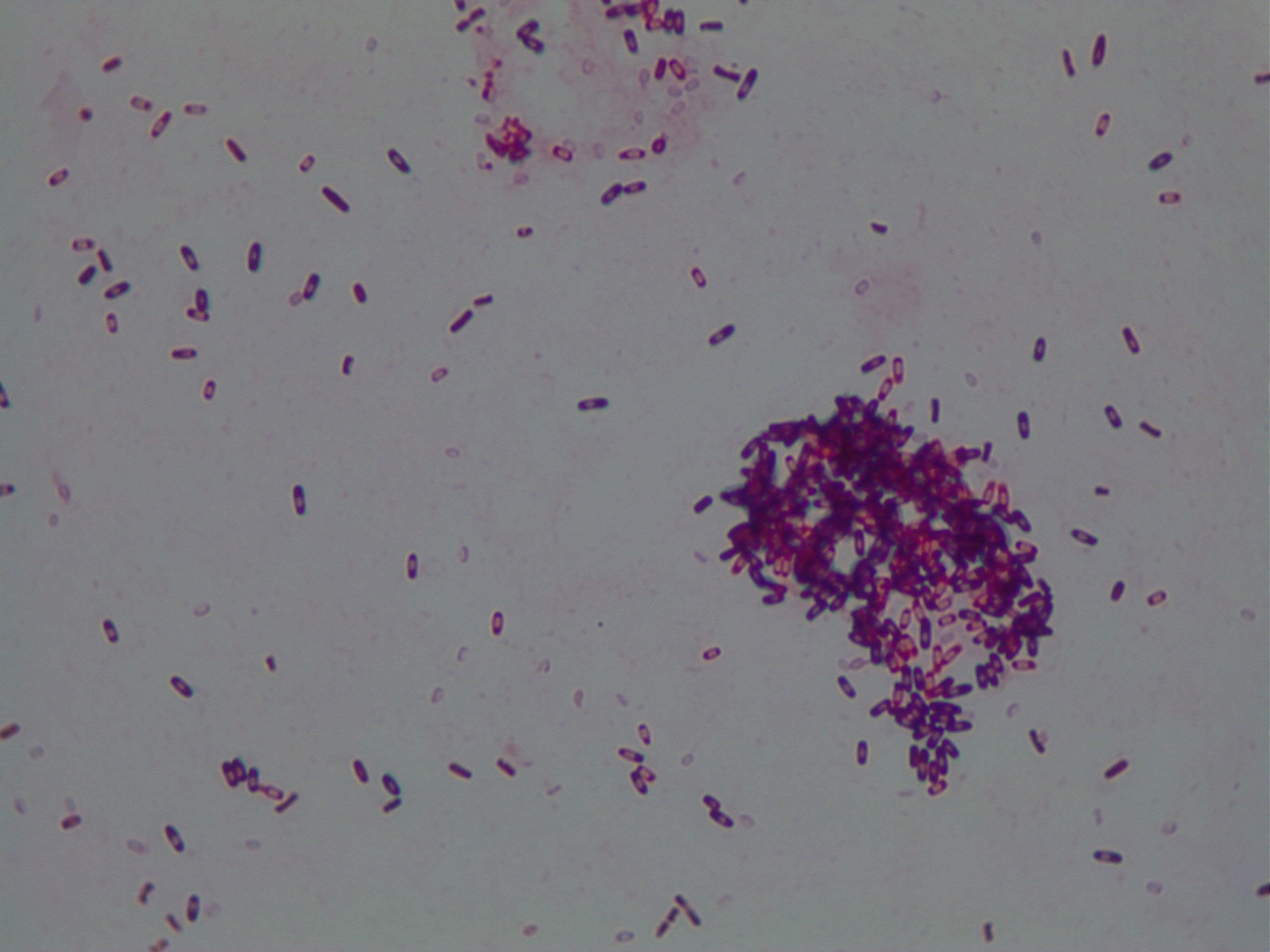
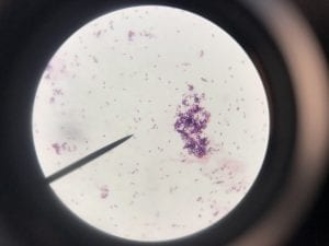
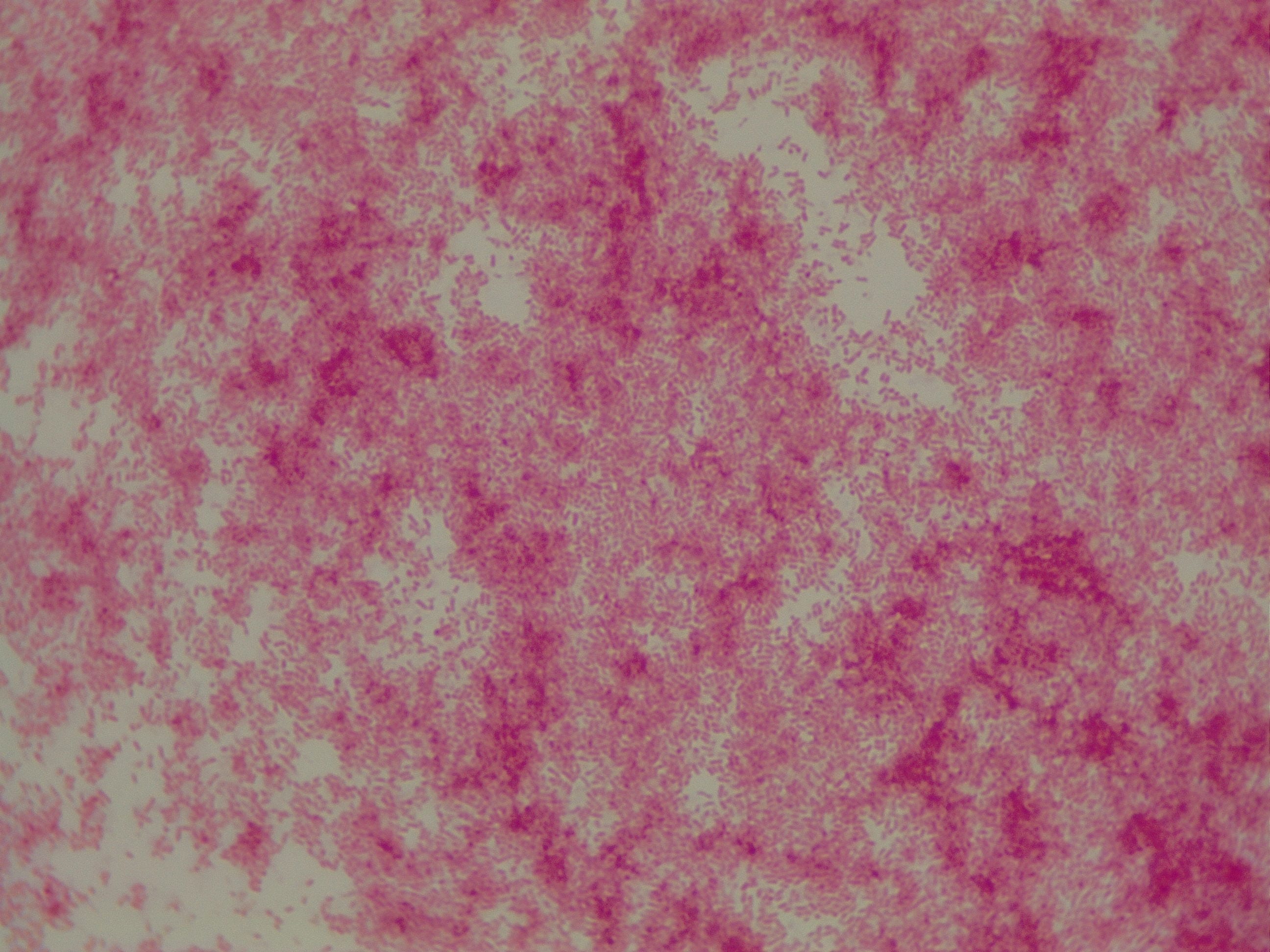
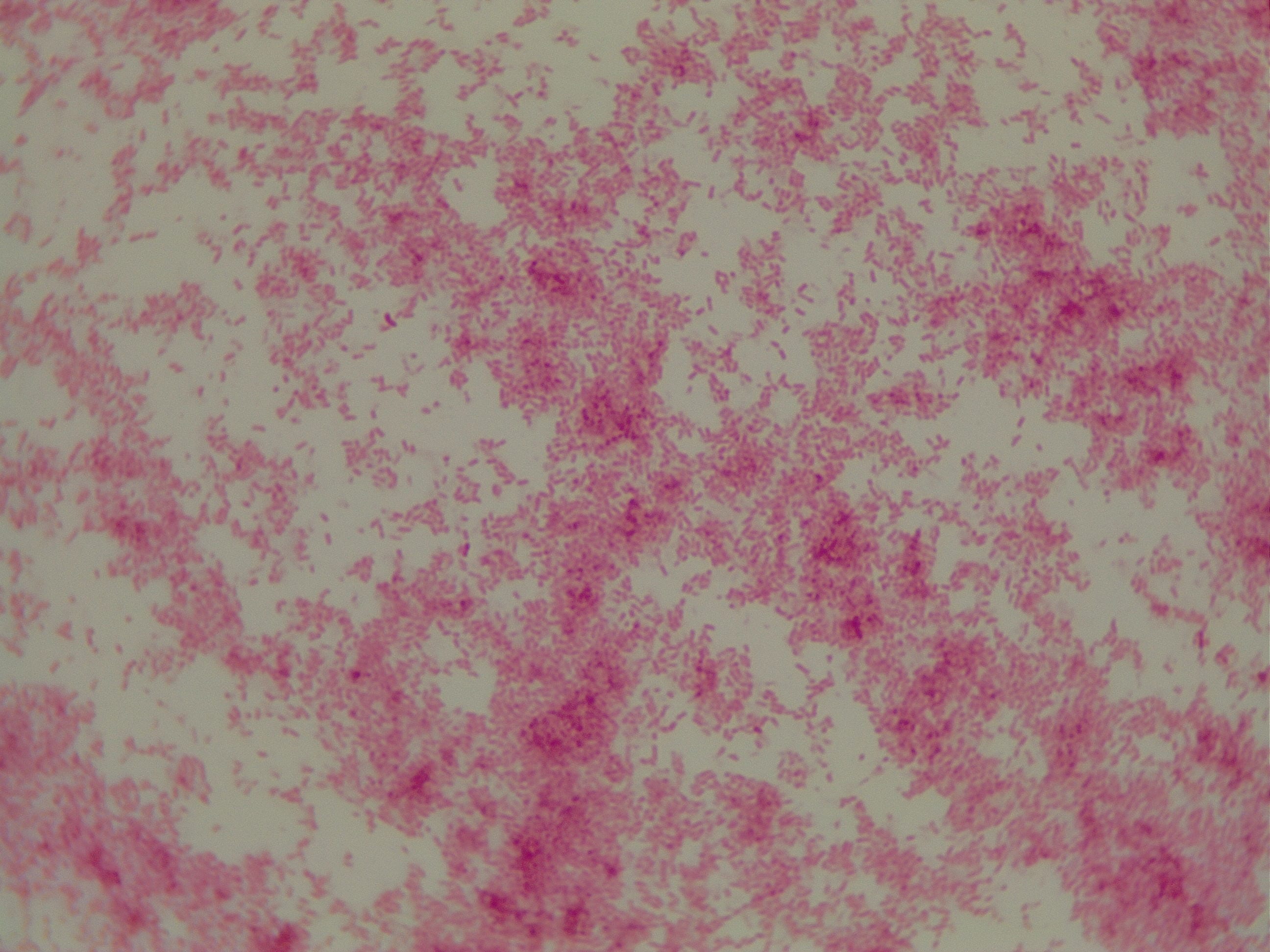
My gram stain agrees! Bacillus is Gram positive, rod-shaped, spore forming bacterium. My Gram stain shows all of these features. The image below shows the Gram stain of my bacteria. The rods are purple in color with a pink center. The purple color signifies a Gram positive bacteria. The pink center shows that the bacteria is spore-forming.

Pick one of your isolates and find out more about the genus (it is unlikely you will be able to determine the species).

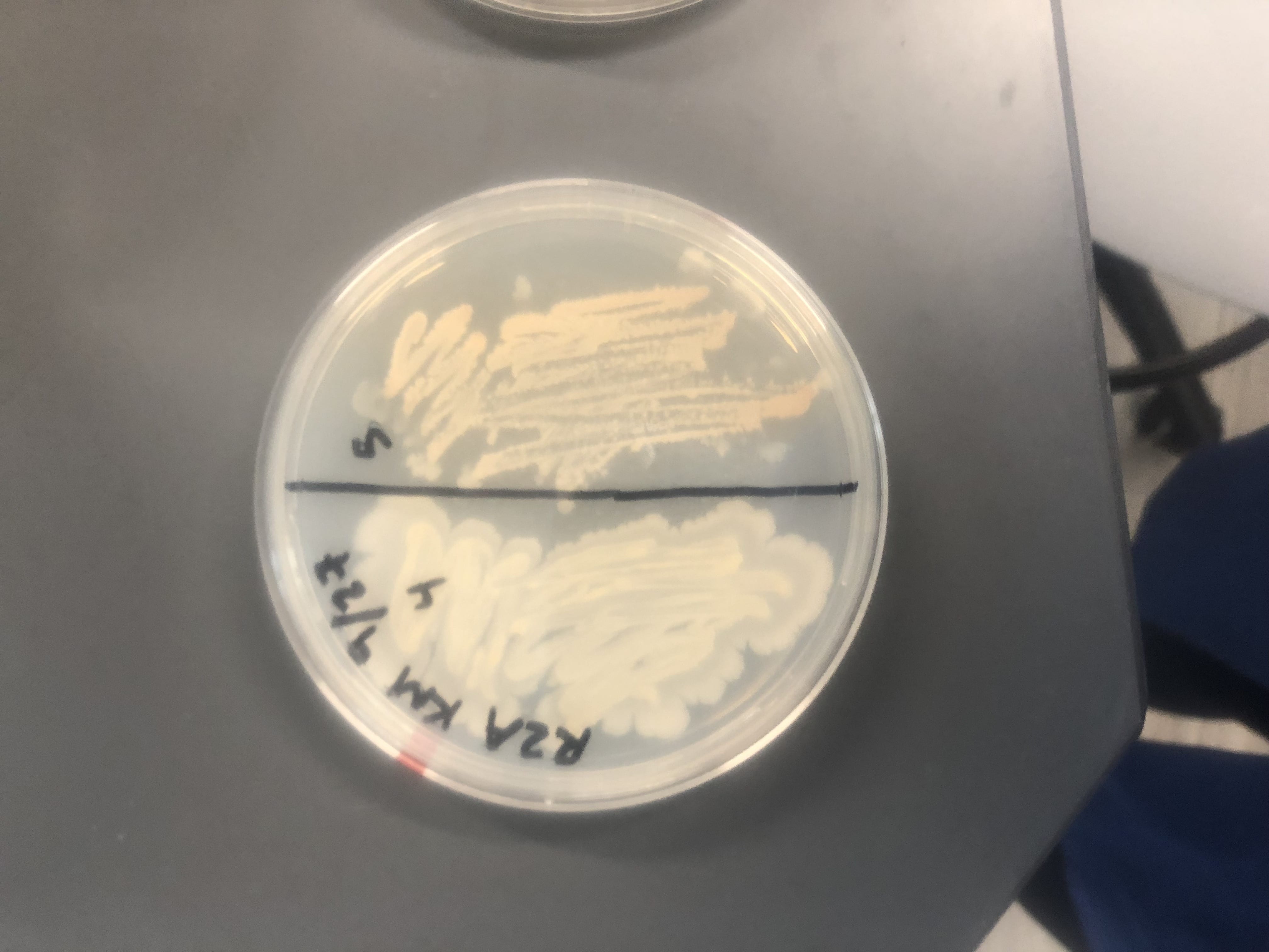
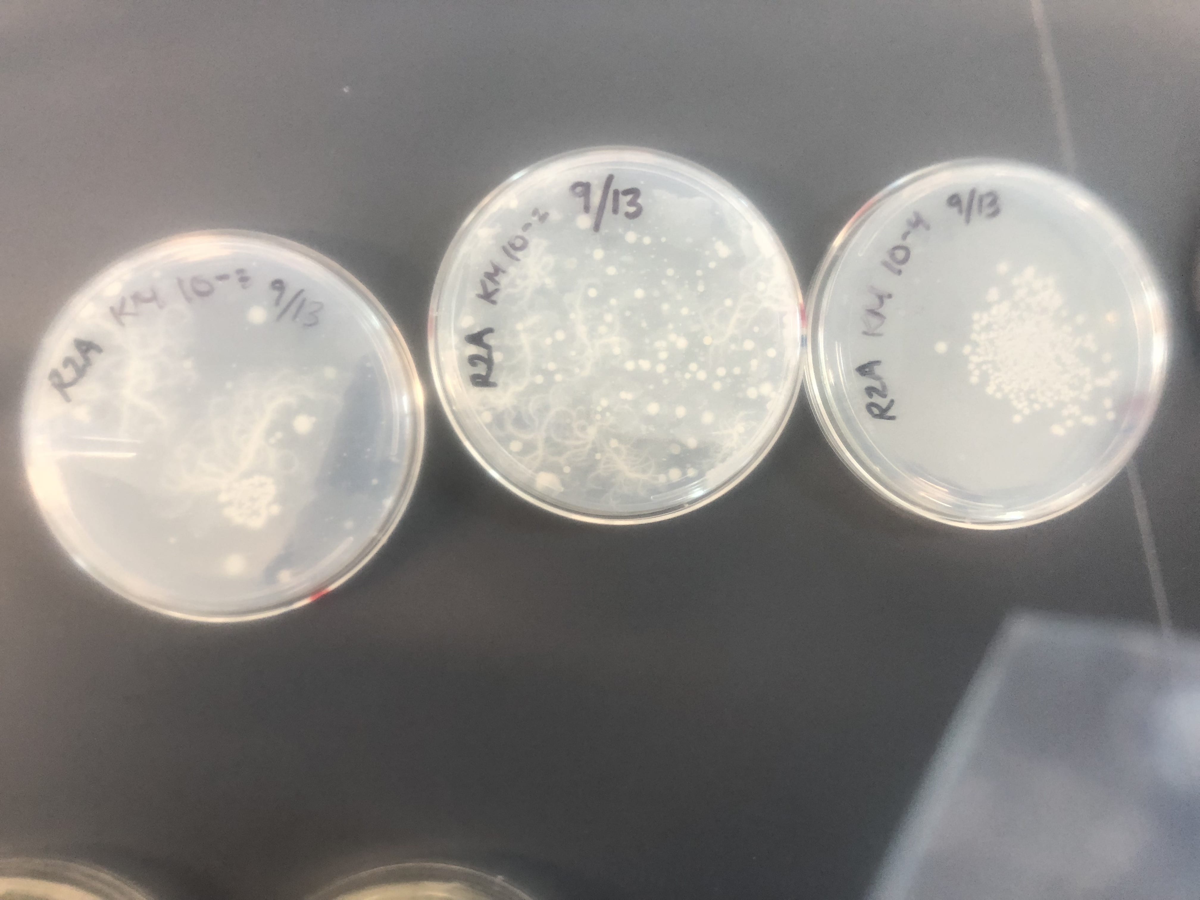
I only have one isolate, so that is the one that I have picked (KM5). The genus is Bacillus. 

a) General cellular and morphological characteristics of the genus (taxonomic classification, nutrition, cell shape, habitat).

There are over 200 known species of Bacillus, but only two are known to impact humans. Bacillus can be classified as follows: Kingdom- Bacteria, Phylum- Firmicutes, Class- Bacilli, Order- Bacillales, Family- Bacillaceae. Bacillus are rod-shaped, Gram positive, endospore-forming bacteria. The bacteria that has been isolated in lab, as well as most of the Bacillus genus is aerobic. There are some species that act as facultative anaerobes. Due to most species being aerobic, their nutritional well-being depends on having oxygen. To achieve optimal growth in a lab setting, Bacillus prefers a rich media. With that being said, the genus would grow best in its natural environmental conditions and it is hard to replicate those. Their optimal temperature range for growth and life is between 25-35 degrees Celsius. Finally, the genus are known to inhabit soil. However, they can also be found in aquatic environments. Some species, such as B. subtilis can be found naturally in the human gut.

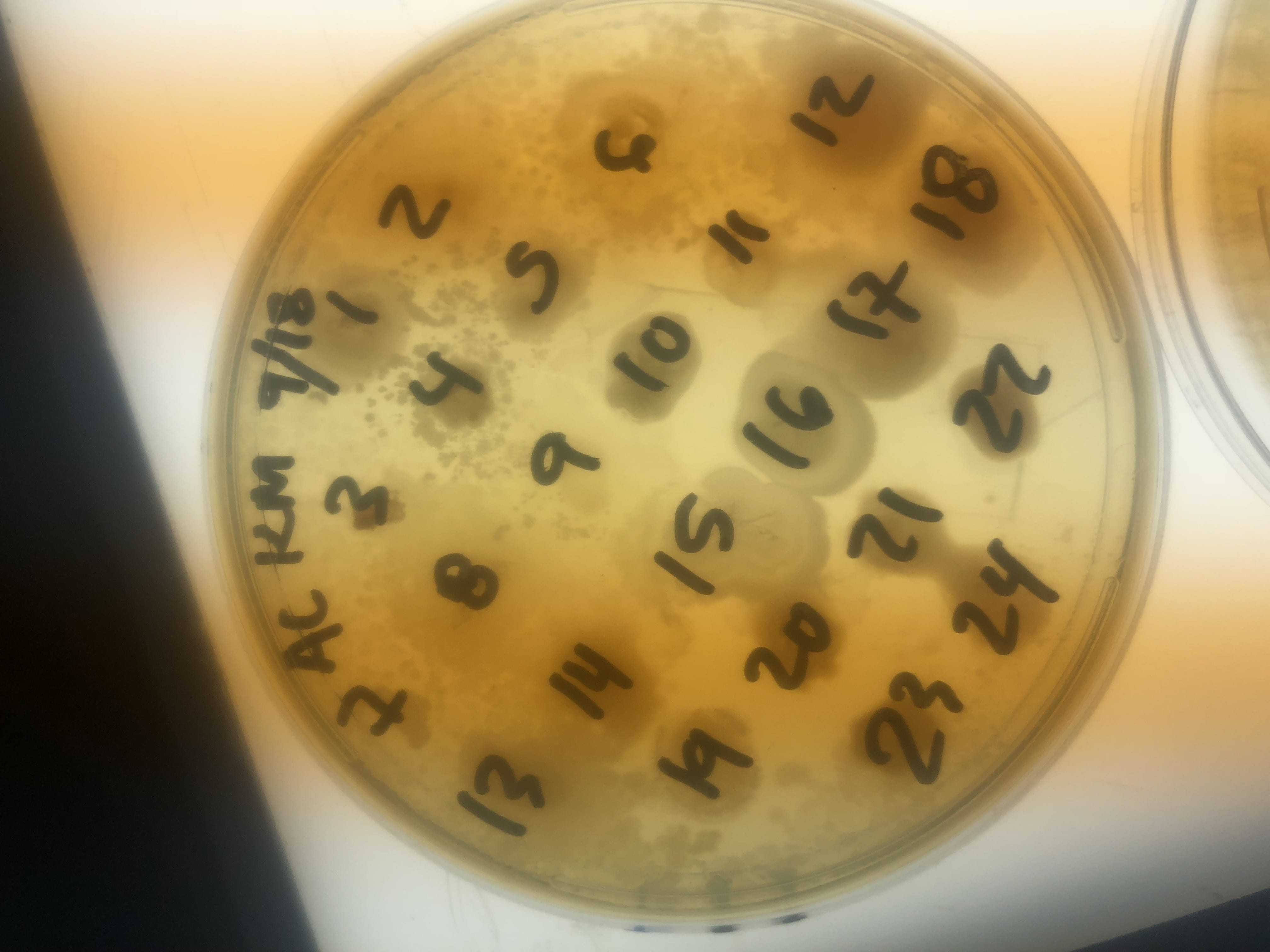
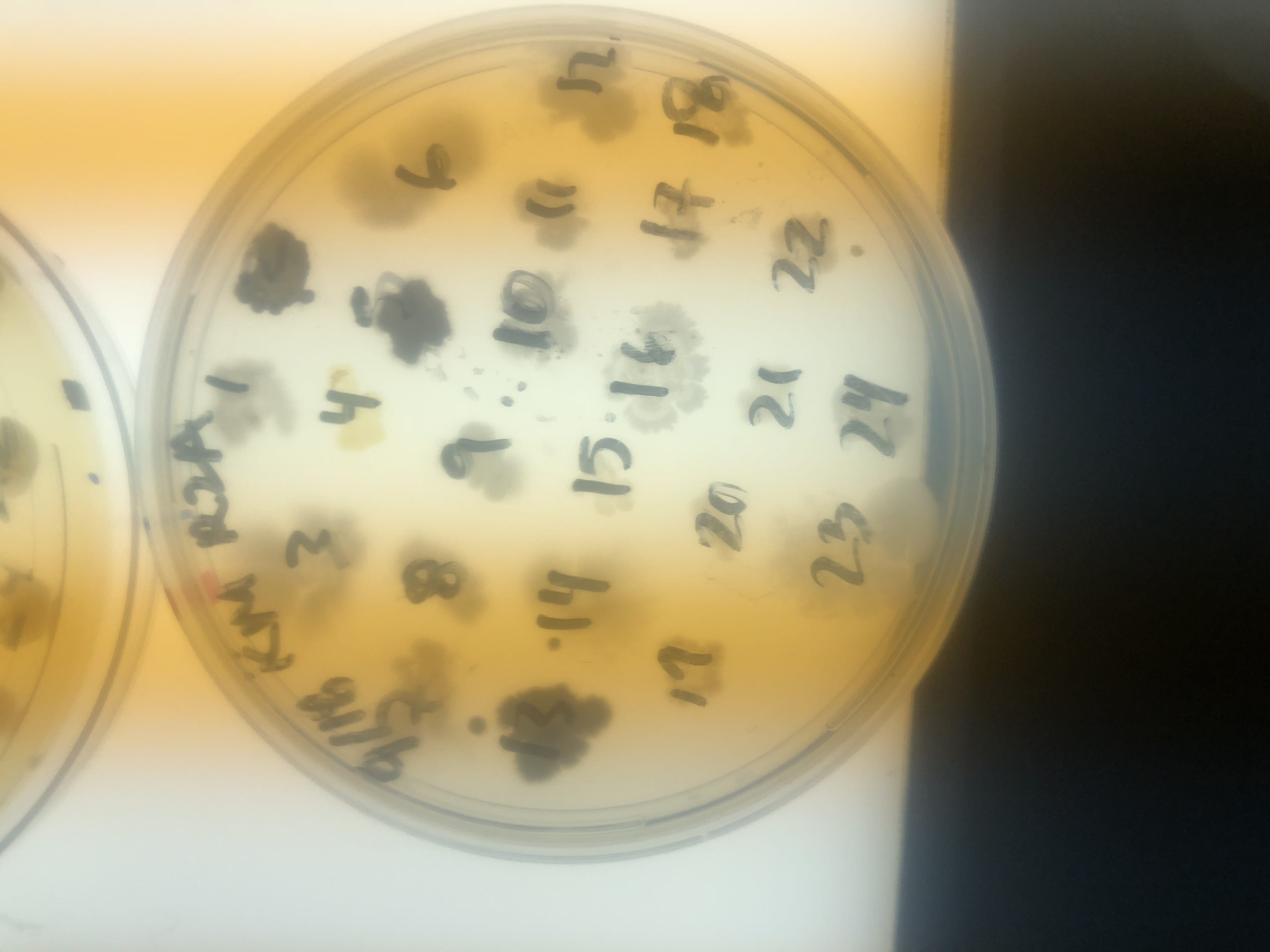
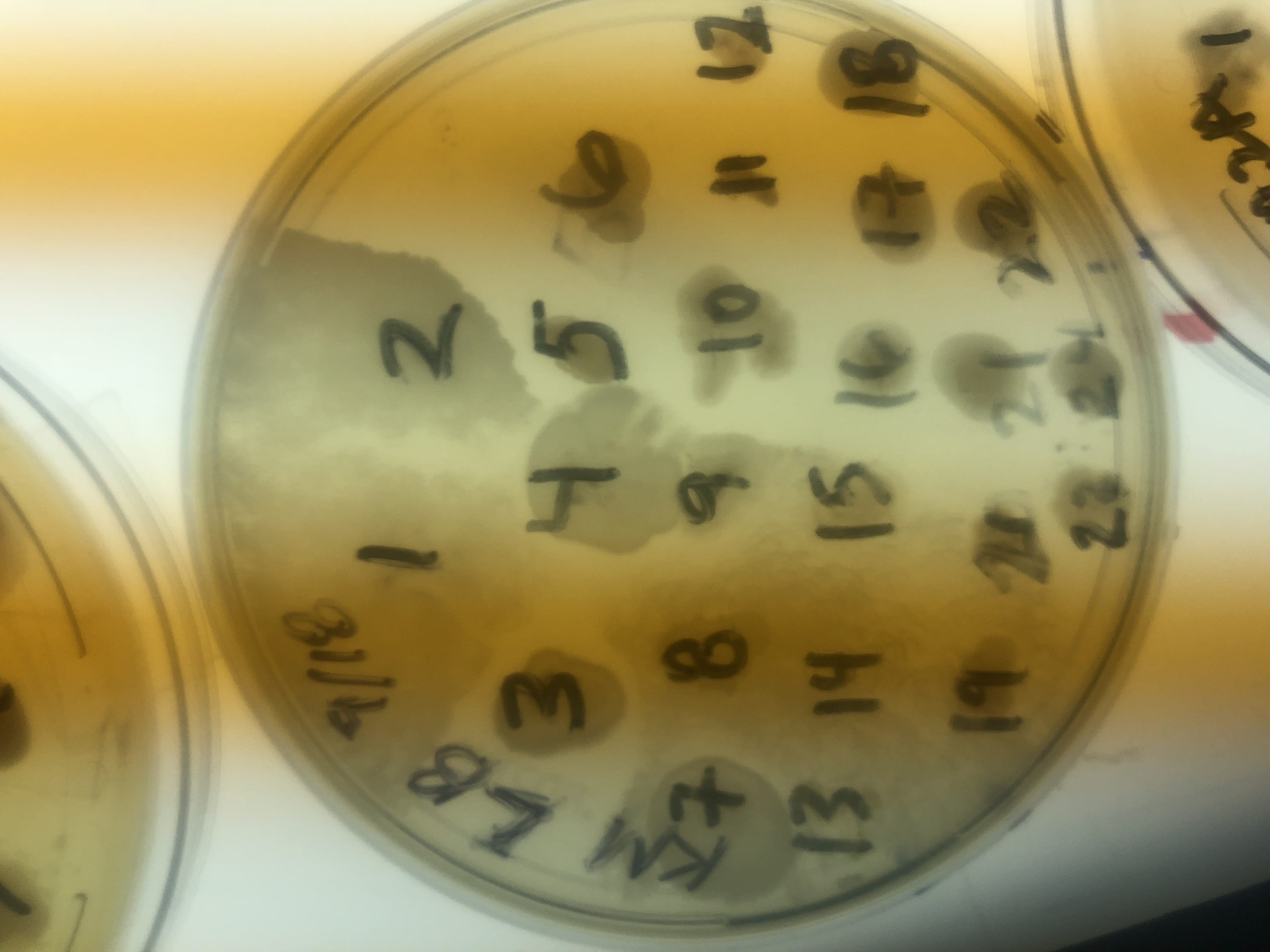
b) Information regarding antibiotic production in this genus.

The Bacillus species produces antibiotics. Antibiotics act as the bacteria’s “fight” mechanism. Some examples of common antibiotics that the Bacillus genus excretes are polymyxin, bacitracin, and gramicidin. The antibiotics that the Bacillus genus produces are effective against both Gram positive and Gram negative bacteria. From my experiments, I determined that my isolate was only effective against Gram positive bacteria. This is not unusual for a variety of species of Bacillus. The genus can also have other antimicrobial uses, such as being an anti-fungal.

Sources:

https://www.sciencedirect.com/science/article/pii/S0944501305000704

https://www.ncbi.nlm.nih.gov/books/NBK7699/

https://www.flandershealth.us/microbiology/i-characteristics-of-bacillus-subtilis.html

http://www.heathermaughan.ca/resources/Maughan%26vanderAuwera11.pdf

https://microbewiki.kenyon.edu/index.php/Bacillus_subtilis

https://www.ncbi.nlm.nih.gov/blast/treeview/treeView.cgi?request=page&blastRID=Y38Z9EVS014&queryID=lcl|Query_28503&entrezLim=&ex=&exl=&exh=&ns=100&screenWidth=1280&screenHeight=800

Extract News- Kelsey

I tested two extracts. One extract was an isolate of mine on 10% TSA and the other extract was the same isolate on R2A. I tested the extracts on two Gram negative strains, E. carotova and E. coli. I also tested the extracts on two Gram positive strains, E. raffinosis and B. subtilis. When testing the extracts, I found no resistance observed to any of the strains. When the two isolates were tested, they had observed resistance against E. raffinosis and B. subtilis. If I were to further test the extracts, I could try different media or the same media again. I could also leave the plates out for longer to see if any antibiotic activity would show over time. However, at this point, I conclude that the isolates did not secrete any antibiotic activity into the media on which they were plated.

Meet My Microbes- Kelsey


This is a picture of where I got my soil sample! There were lots of decomposing elements to this soil including rusty beer cans, paper, organic matter, etc. The soil was most likely in the A Horizon.


These are two of my streak plates with the four producers that I found in lab. (slightly blurry due to the plastic bag).


These are my R2A plates, where I found most of my producers. You can see the mycoides growing on the 10^-2 and 10^-3 dilution plates. The second photo is a close up of the mycoides on a light box at a dilution factor of 10^-2.


A close up of my R2A plate with a 10^-4 dilution. This photo was taken on the light box.


These are three examples of patch plates. These were the first set of patch plates that we did. These photos were all taken on the light box. In both the LB and AC media plates, you can see an overgrowth of mycoides. The mycoides overtook the patches in this section and rendered them unusable. I found minimum mycoides growth in the R2A plates throughout my experiment.


These are two pictures of my Gm+ bacteria. I know it is Gm+ due to the purple color. My bacteria are most likely bacillus because they are rods and spore forming. I know that they are spore formers due to the central pink spore between the purple ends of the rods. These photos were taken at 100x.


This is another photo of my bacteria. You can see a large aggregation of colonies on the right side of the slide. This photo was taken at 1000x.


These are pictures at 1ooox of my Gram positive control (S. epi).


These are pictures at 1000x of my Gram negative control (P. putida).

Eskape Pathogens- Kelsey and Nicole D

Assigned ESKAPE Pathogen

#6 Enterobacter Species


Why is this ESKAPE Pathogen of interest (in brief)

The Enterobacter species is of interest due to the high rates of hospital-acquired infection that it causes. A study from the National Nosocomial Infections Surveillance System showed that the Enterobacter species is the third most common cause of pneumonia in ICUs. It is of increasing interest because the species has extremely high rates of antibiotic resistance. This is of concern due to the prevalence of infection and the mortality rates associated with the infections it causes. Crude mortality rates range from 15-87%. This is an extremely wide range, making the infections caused by the Enterobacter species highly unpredictable.


https://www.britannica.com/science/Enterobacter

https://emedicine.medscape.com/article/216845-overview

https://www.hopkinsguides.com/hopkins/view/Johns_Hopkins_ABX_Guide/540201/all/Enterobacter_species


General Cellular and Morphological Characteristics of the Organism (taxonomic classification, nutrition, cell shape, habitat)

The Enterobacter species is a rod-shaped bacteria that is gram negative. The size of each colony is 0.6-1.0 micrometers by 1.2-3.0 micrometers. The species is not capable of forming spores. It is motile due to flagella. The taxonomic classification is Bacteria; Proteobacteria; Gammaproteobacteria; Enterobacteriales; Enterobacteriaceae. The species are facultative anaerobes; meaning the presence of oxygen is not required. They are capable of fermenting glucose and lactose. Through this fermentation, gases are produced. They are commonly found in intestinal tracts and in soil, water, and sewage. They are not harmful when found in the gut. Less commonly, they are found in urine, pus, and bodily fluids.


https://microbewiki.kenyon.edu/index.php/Enterobacter

https://www.britannica.com/science/Enterobacter


Clinical Importance and Prevalence

This Eskape pathogen is known to cause nosocomial infections. A nosocomial infection is caused due to a bacterium’s prevalence in certain locations. A nosocomial infection can also be called a hospital-acquired infection. The Enterobacter species is prevalent in ICUs and also commonly survives in equipment with water. This is because the species is able to live for a long time on surfaces. In wet environments, it is able to replicate rapidly. The most common source of spreading the bacteria is a lack of cleanliness, i.e. a lack of handwashing. This is a major source of infection in hospitals, with about 50% of infection in the ICU caused by the Enterobacter species. The species causes infections in the respiratory tract, urinary tract, intra abdominal cavity, intravascular devices, and can lead to sepsis.


https://www.healthline.com/health/hospital-acquired-nosocomial-infections


Infection (How does the infection occur and where is it localized?)

The Enterobacter species cause nosocomial infections in immunocompromised patients. The most common types of Enterobacter that cause human infection are E. cloacae, E. aerogenes, E. gergoviae, and E. agglomerans. Infections occur endogenously or exogenously. Endogenous sources of infection are the most common type of nosocomial infections caused by the Enterobacter species. The endogenous sources include the skin, respiratory tract, urinary tract, and gastrointestinal tract. The endogenous source tend to be the site of localization for the species.


https://emedicine.medscape.com/article/216845-overview


Pathology (What disease is caused? What are the symptoms?)

An infection caused by the Enterobacter species include a heart rate exceeding 90 bpm, a respiration rate greater than 20, and a fever above 100.4 °F or below 96.8°F. Other symptoms include hypotension, septic shock, cyanosis, and hypoxemia. Diseases and conditions that can be caused by this species are eye and skin infections, meningitis, bacterial blood infections, pneumonia, and urinary tract infections.


https://emedicine.medscape.com/article/216845-overview

http://www.antimicrobe.org/b97.asp

https://www.britannica.com/science/Enterobacter


Ineffective Antibiotics (Antibiotics to which the organism has acquired resistance)

Effective Antibiotics (Antibiotics known to inhibit the organism)

This species has developed resistance to a number of different antibiotics, especially in hospital settings. Infections and diseases caused by the Enterobacter species have been treated with an aminoglycoside, a fluoroquinolone, a cephalosporin, or imipenem. Over time, however, Enterobacter species has developed resistance to most beta lactam drugs and many other different drug types. Another source suggests that “third generation” cephalosporins, penicillins, and quinolones have become ineffective antibiotics as a result of increasing resistance. However, there are some antibiotics that are still effective. “Fourth generation” cephalosporins and carbapenems remain a viable option for treatment. Aminoglycosides are noted to be viable as well, but need to be combined with another type of antibiotic to allow for successful treatment. Lastly, quinolones are able to be used against many strains of the Enterobacter species, but similar to the trend of other drugs, emerging resistance is of concern. More specifically, Polymyxin B, Levofloxacin, Doripenem, Imipenem, Meropenem, Cefepime, Ciprofloxacin, Trimethoprim-sulfamethoxazole, Ertapenem, and Tigecycline are all examples of antibiotics that seem to remain effective and are largely avoiding resistance for now.


http://www.antimicrobe.org/b97.asp

https://www.britannica.com/science/Enterobacter

https://catalog.hardydiagnostics.com/cp_prod/Content/hugo/Enterobacter.htm


Corresponding Safe Relative

The corresponding safe relatives are the Enterobacter aerogenes and the Erwinia carotovora.


(Source: Lab handout)


Images:


Enterobacter cloacae

https://jamanetwork.com/journals/jamadermatology/fullarticle/413232


https://www.researchgate.net/figure/A-localized-infection-of-Enterobacter-cloacae-developed-on-the-patients-left-forehead_fig4_258503948


Sources:

https://microbewiki.kenyon.edu/index.php/Enterobacter

http://www.antimicrobe.org/b97.asp

https://www.britannica.com/science/Enterobacter

https://emedicine.medscape.com/article/216845-overview

https://www.hopkinsguides.com/hopkins/view/Johns_Hopkins_ABX_Guide/540201/all/Enterobacter_species

https://catalog.hardydiagnostics.com/cp_prod/Content/hugo/Enterobacter.htm


Davin-Regli, Anne, and Jean-Marie Pagès. “Enterobacter Aerogenes and Enterobacter Cloacae; Versatile Bacterial Pathogens Confronting Antibiotic Treatment.” Frontiers in Microbiology 6 (2015): 392. PMC


Davis, Elizabeth et al. “Antibiotic Discovery throughout the Small World Initiative: A Molecular Strategy to Identify Biosynthetic Gene Clusters Involved in Antagonistic Activity.” MicrobiologyOpen 6.3 (2017): e00435. PMC


Santajit, Sirijan, and Nitaya Indrawattana. “Mechanisms of Antimicrobial Resistance in ESKAPE Pathogens.” BioMed Research International 2016 (2016): 2475067. PMC


Fun With Soil- Kelsey

Where did you obtain your soil sample?
I collected my soil from my backyard here at school. I live at 22 Union Ave. If you walk into my back yard there is a parking lot that leads to two other houses– 20r and Carriage House. I collected the soil next to the cement wall that separates our houses from a house nearby. I have attached a picture below! I dug about 2.5 inches down to get a better soil, rather than a sandy, beer can infested selection. 

Why did you choose this location?

I chose this location because the two houses behind my own tend to have a lot of outdoor parties. I thought that I would get a lot of interesting bacteria due to the wastes and circumstances this environment is exposed to.

Do you expect a lot of isolates? Why or why not?

YES! I think that I will get tons of cool stuff from this dirt. I think that because this location is exposed to both human and animal waste that I will get a lot of interesting isolates. I also think that because there was some rusty old objects mixed in with the dirt, I am even more apt to get some interesting isolates.


Have you initial observations supported this?

Yes, I have had a lot of growth on my plates! The smallest CFU that I have is 2.16×10^6. I think this is a great number. I have had a lot of diversity with my plates as well. Lots of different shapes and colors of the colonies.

What media did you choose?
I chose three types of media– LB, R2A, and AC. I found that LB had the best growths, AC had a lot of overgrowth, and R2A provided the most diverse growths (but had mycoides at the lowest dilution).


What dilutions? 

I plated 3 different dilutions on 3 medias– totaling 9 plates. In terms of the books explanation the three dilutions were 10^-1, 10^-2, and 10^-3.

Will you need to redo any? 

I did not necessarily need to redo any, but I decided to plate 1 plate of R2A with a 10^-3 dilution, 1 plate of AC with a 10^-2 dilution, and 1 plate of LB on a 10^-3 dilution. I decided to do this in order to achieve even more diversity with my plates!

How did you sample differ on the different media? 

The sample differed across media in a few ways. AC with the sample caused a major overgrowth of one colony. The R2A and sample had mycoides growth across all dilutions except for 10^-3. The LB had the most variety of growth. I think that R2A provided the most diversity, with LB a close second. I would choose them to plate more if my new plates are unsuccessful.