Meet My Microbes! Troy

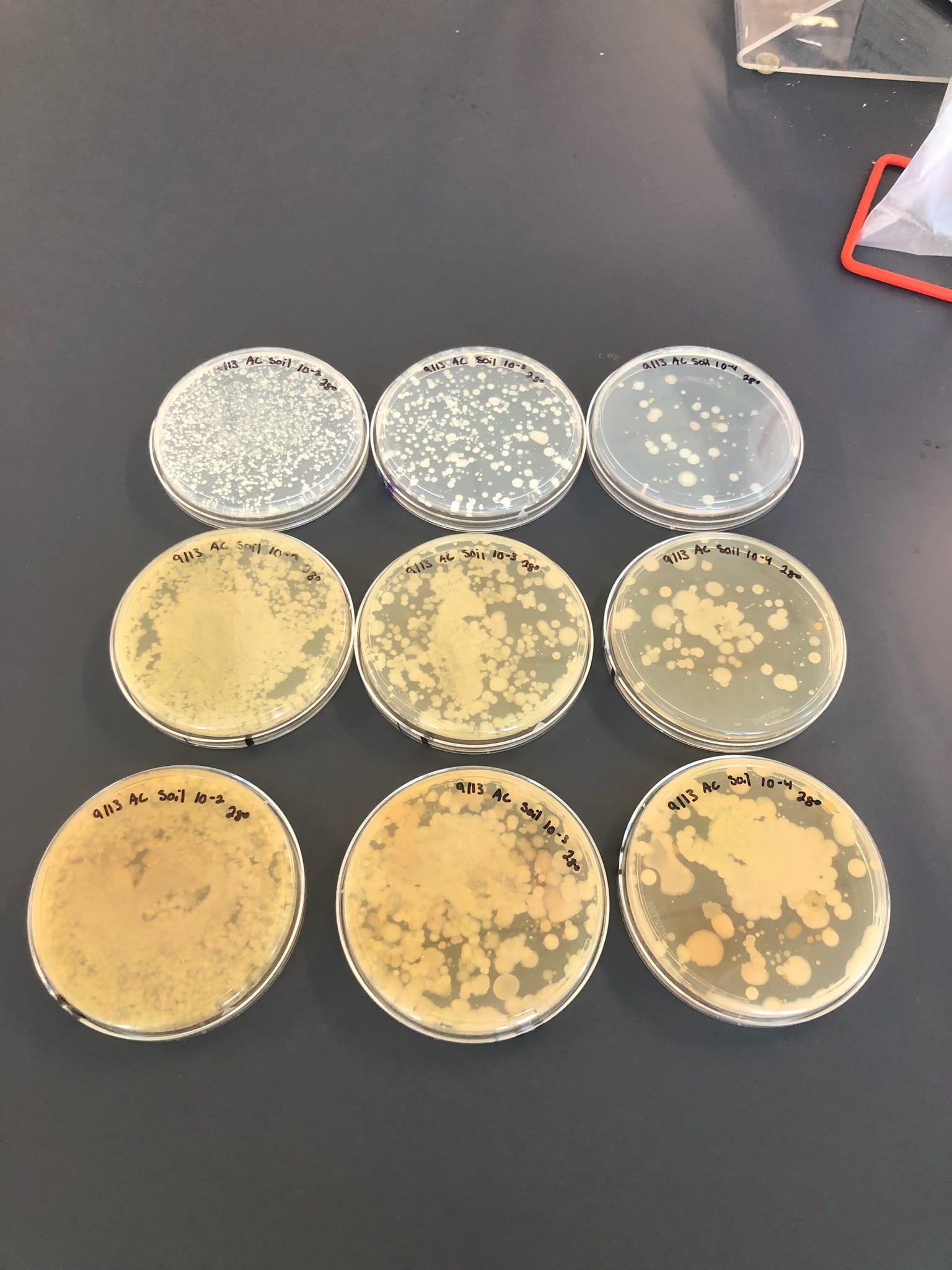
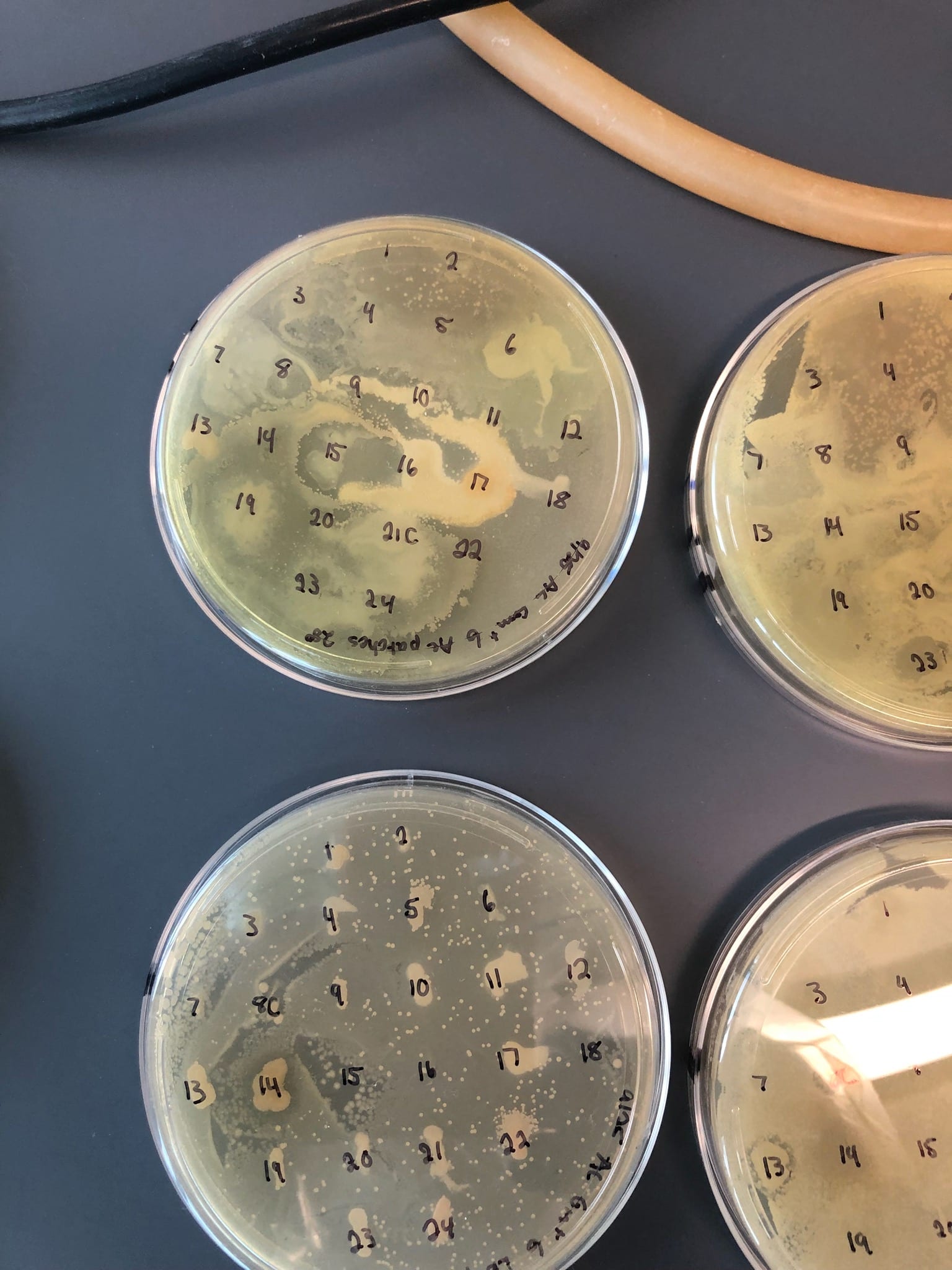
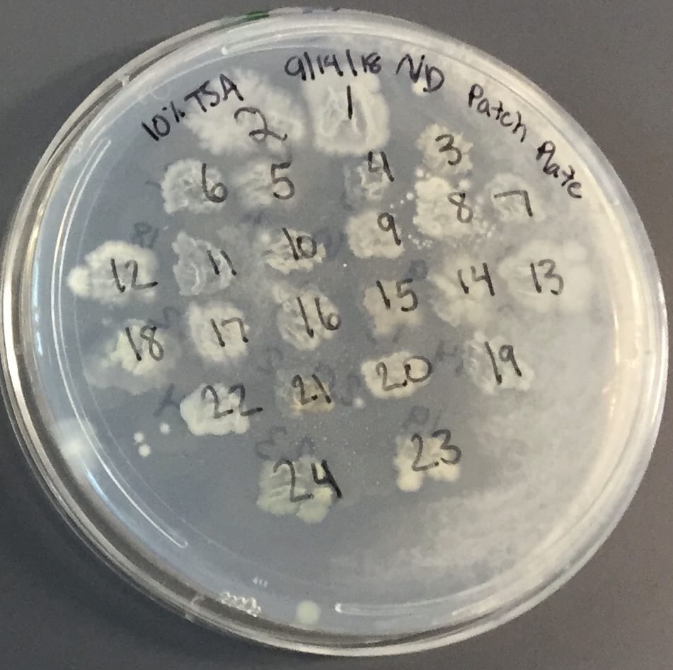
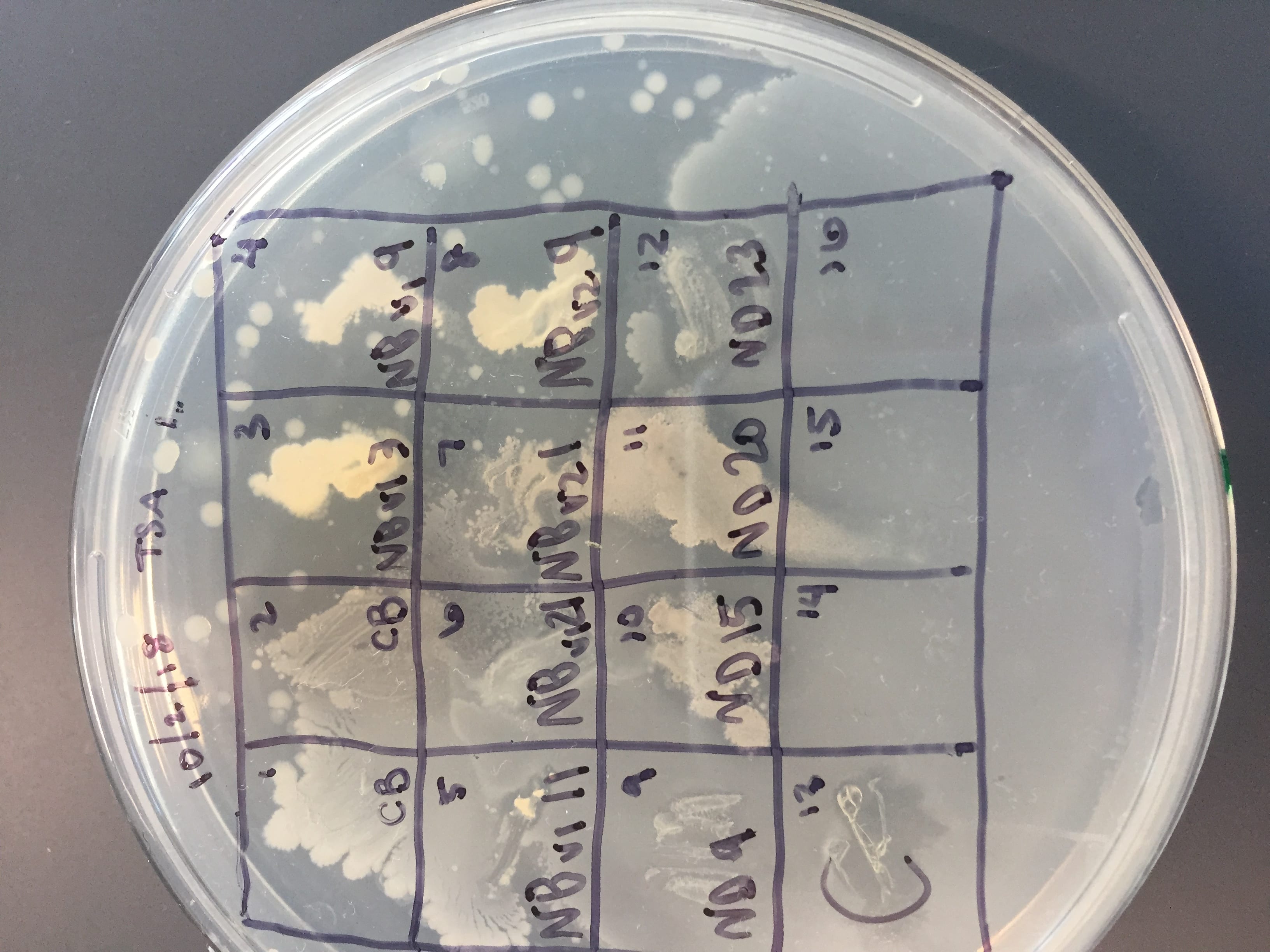
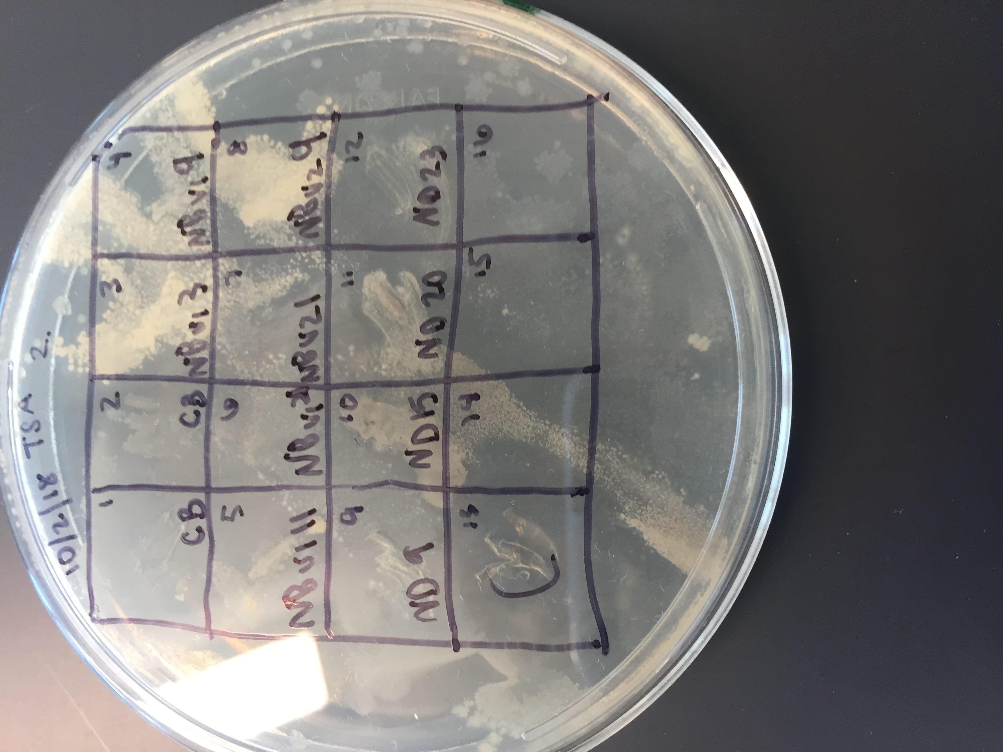
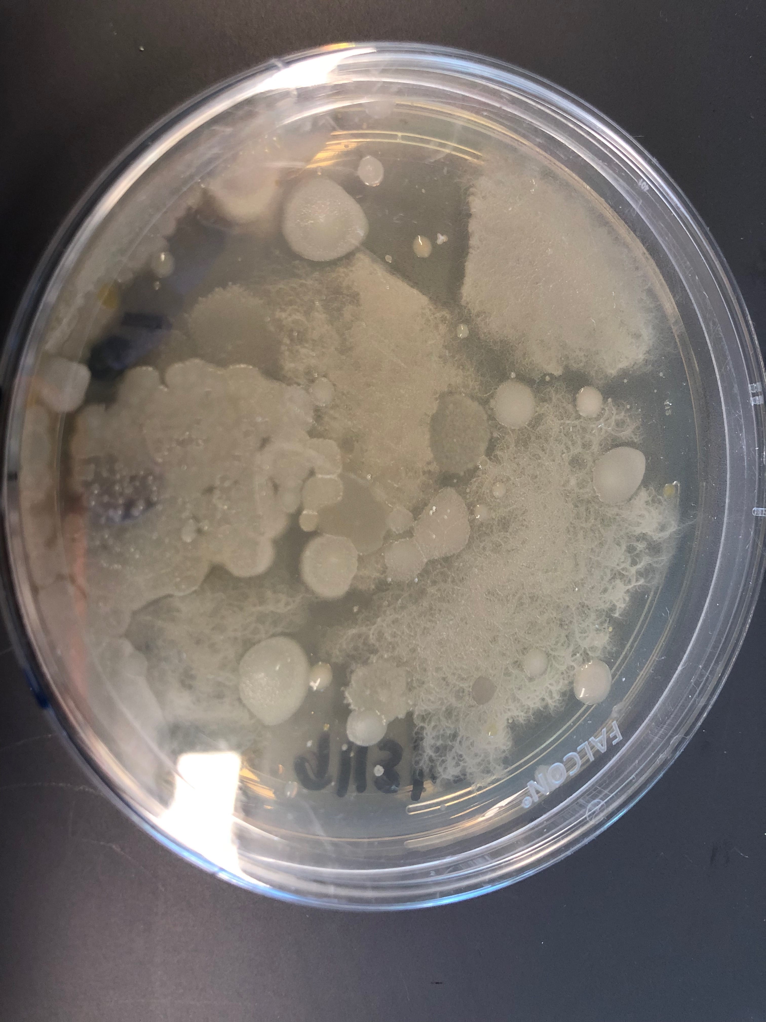
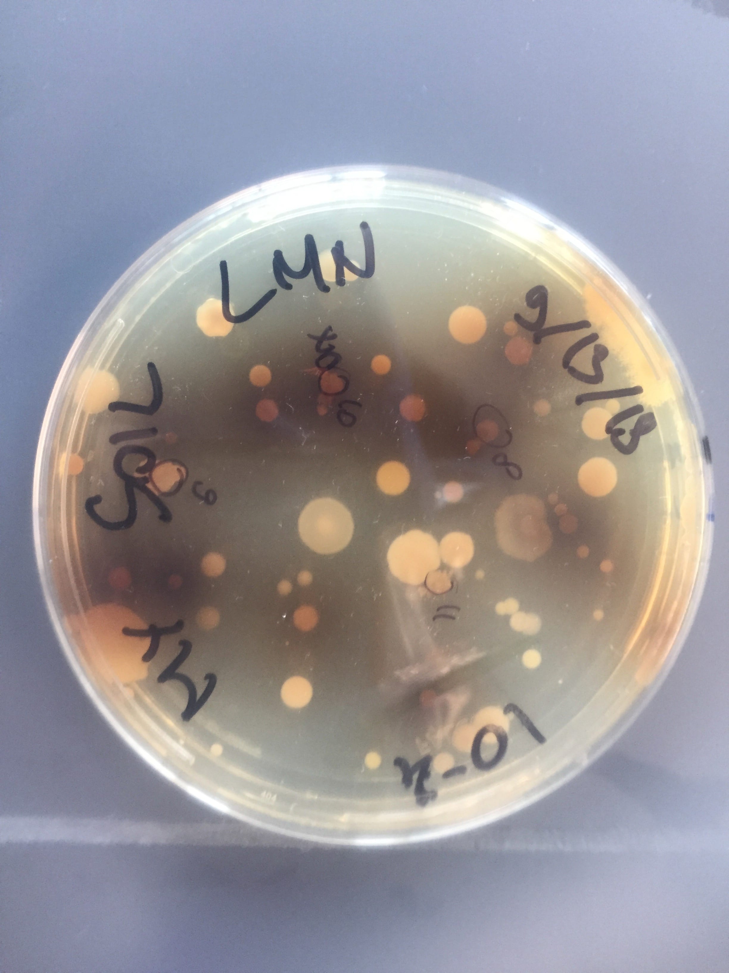
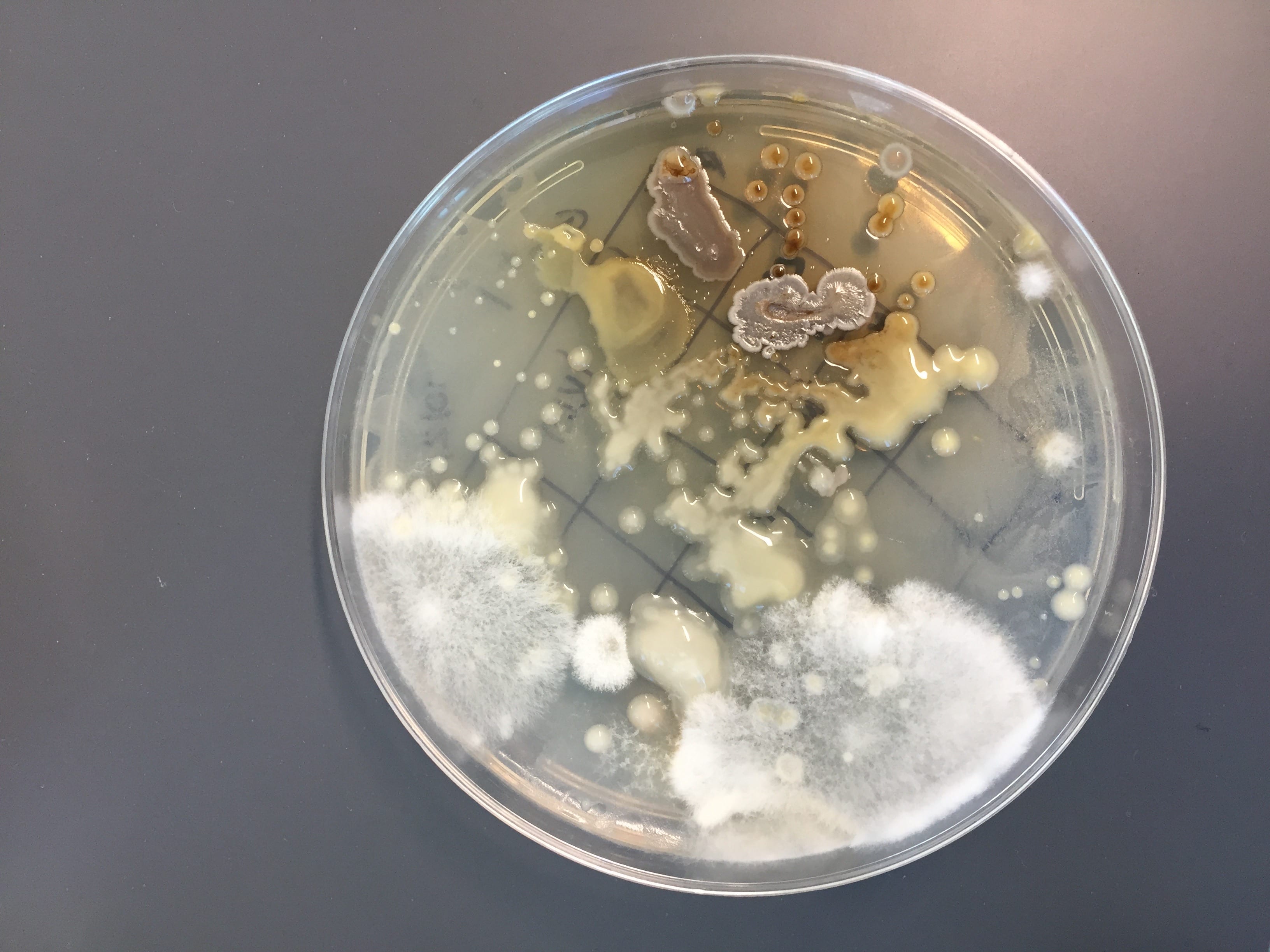
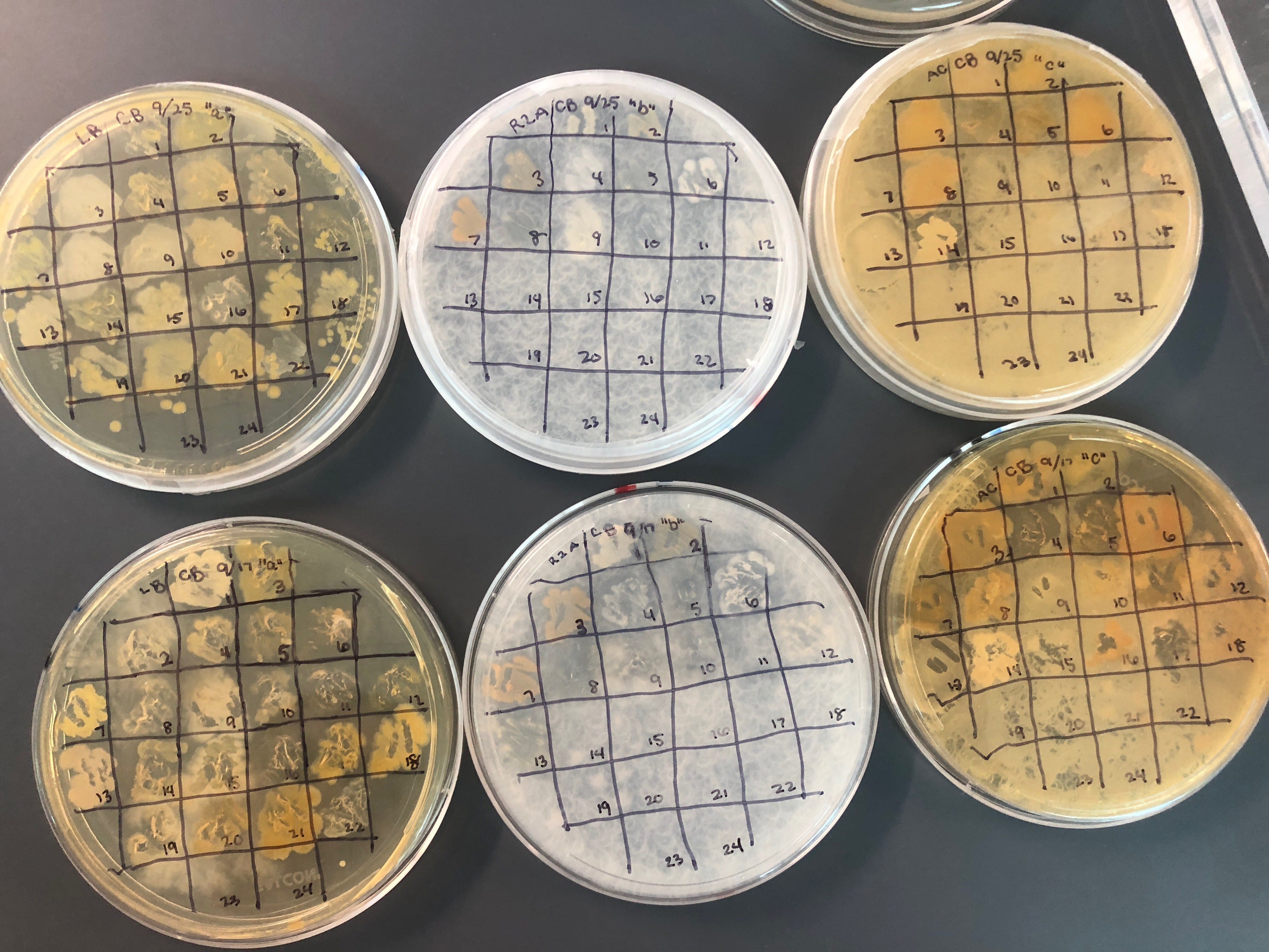
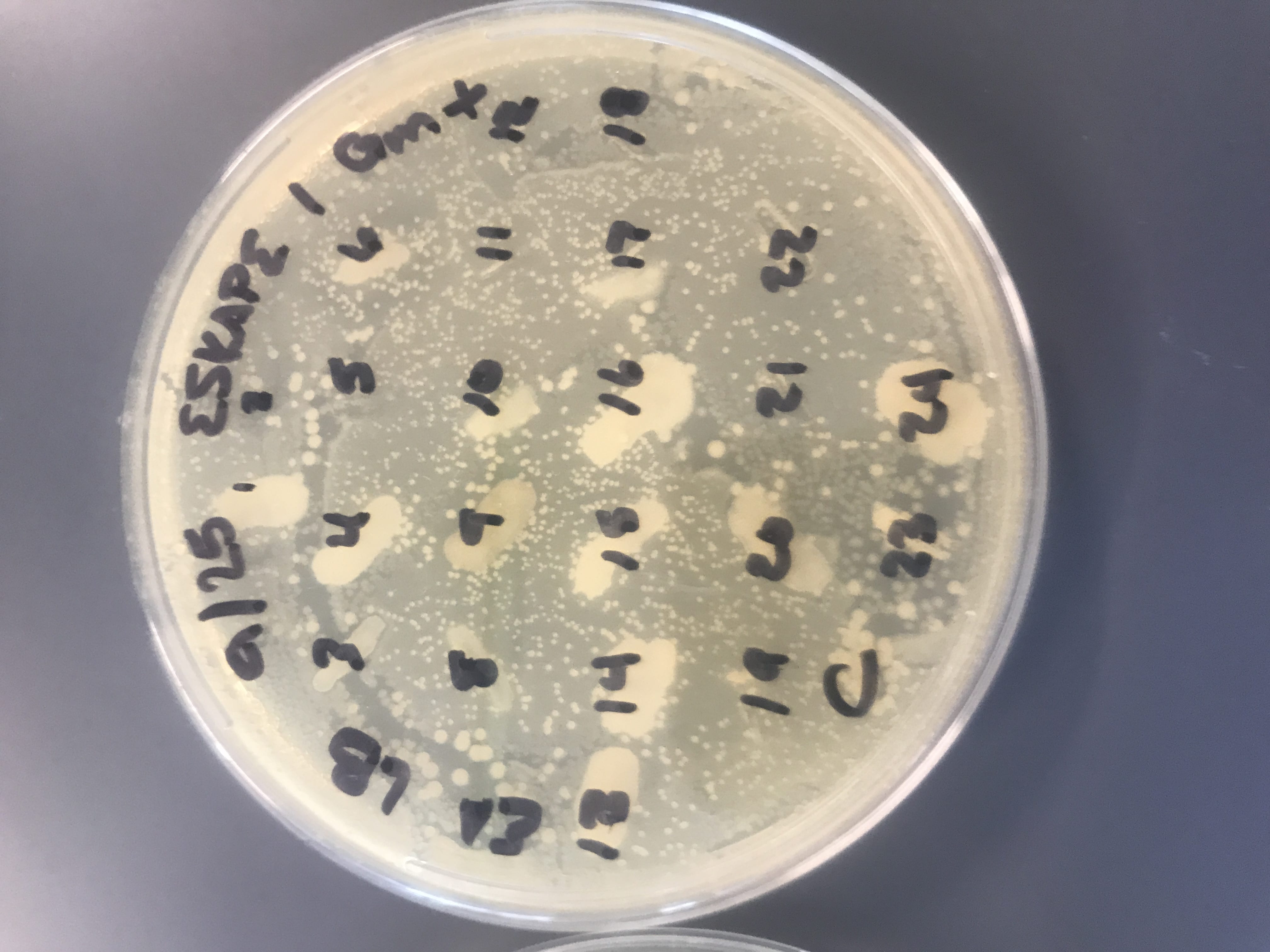
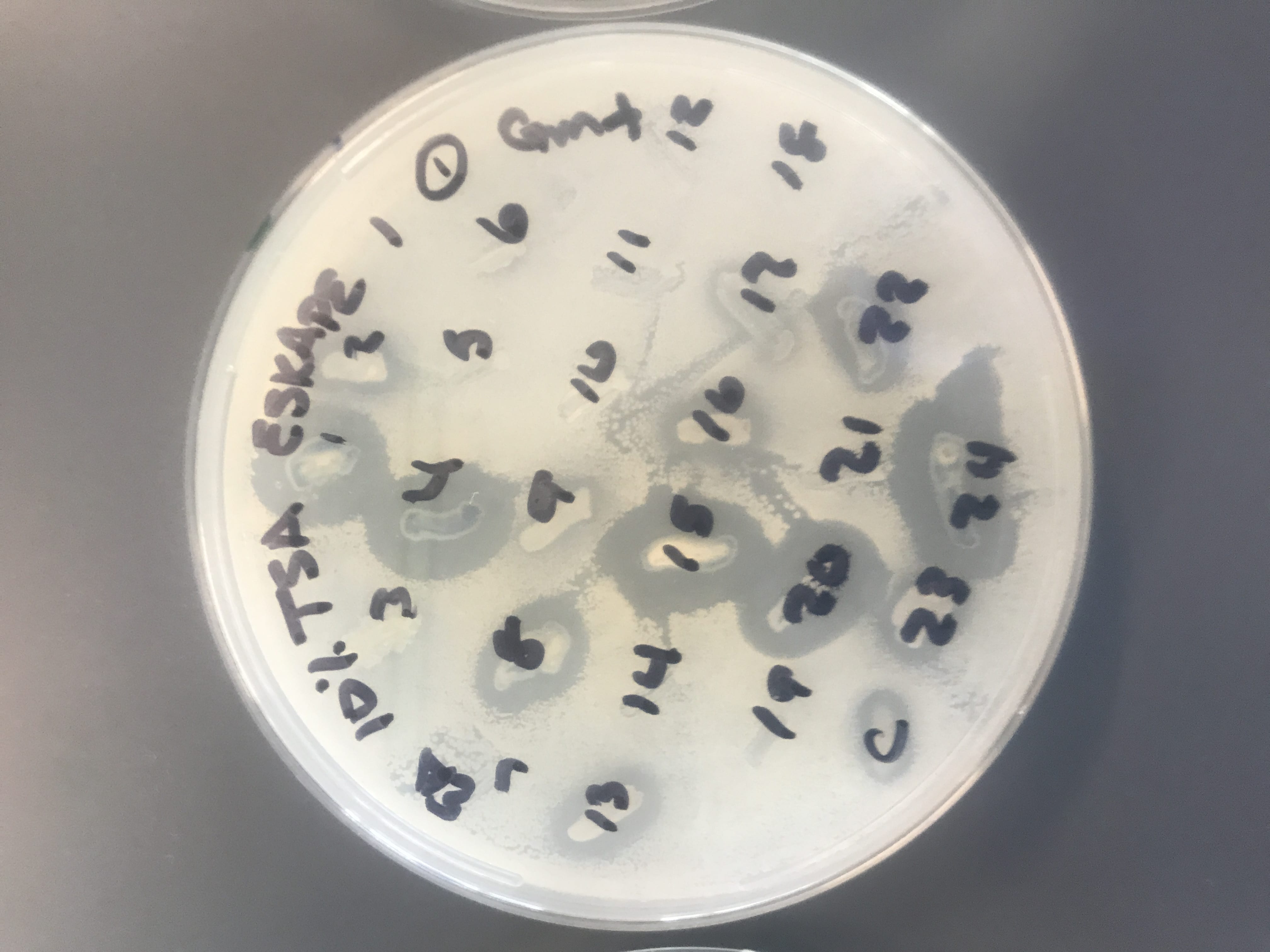
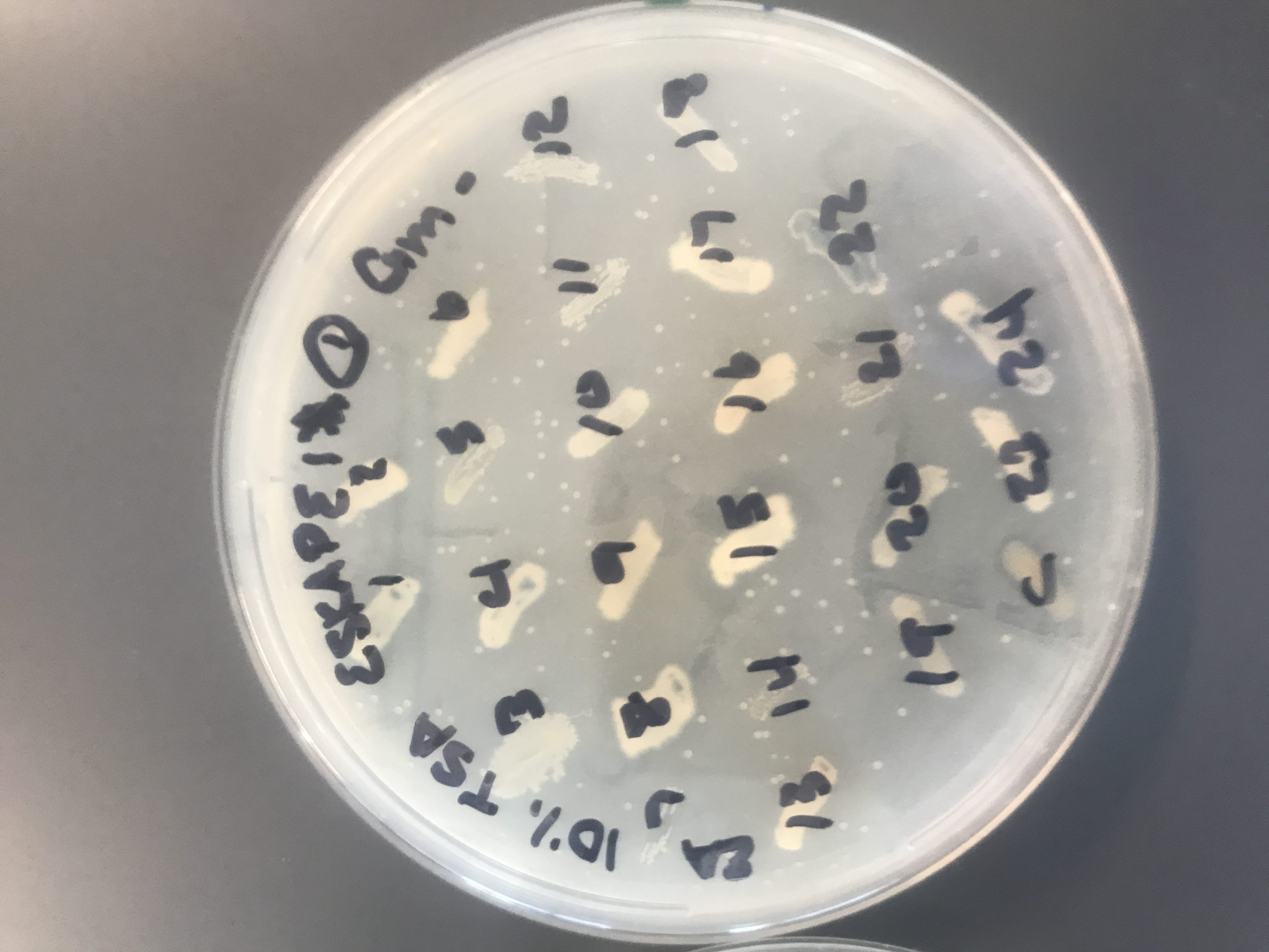
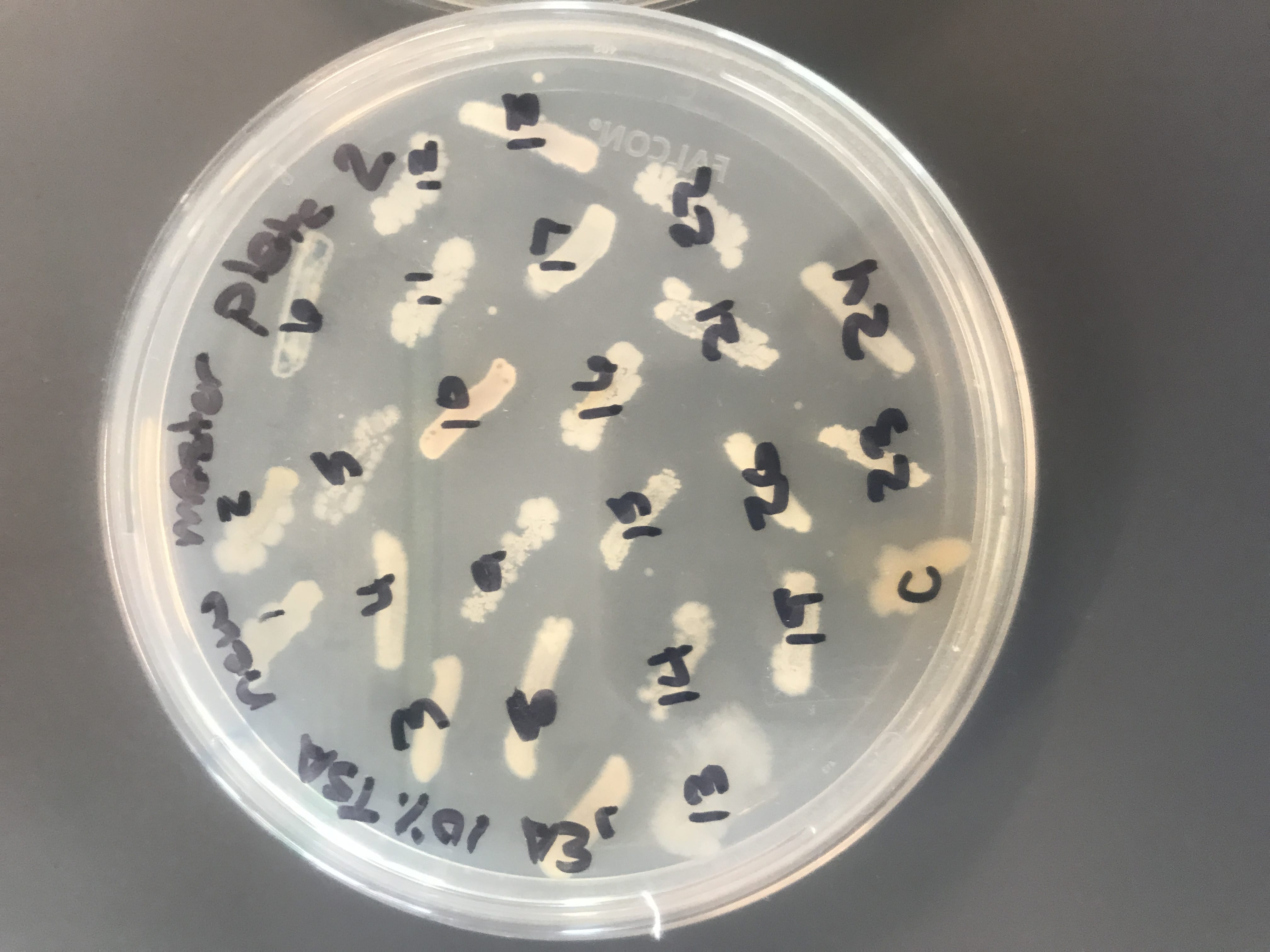
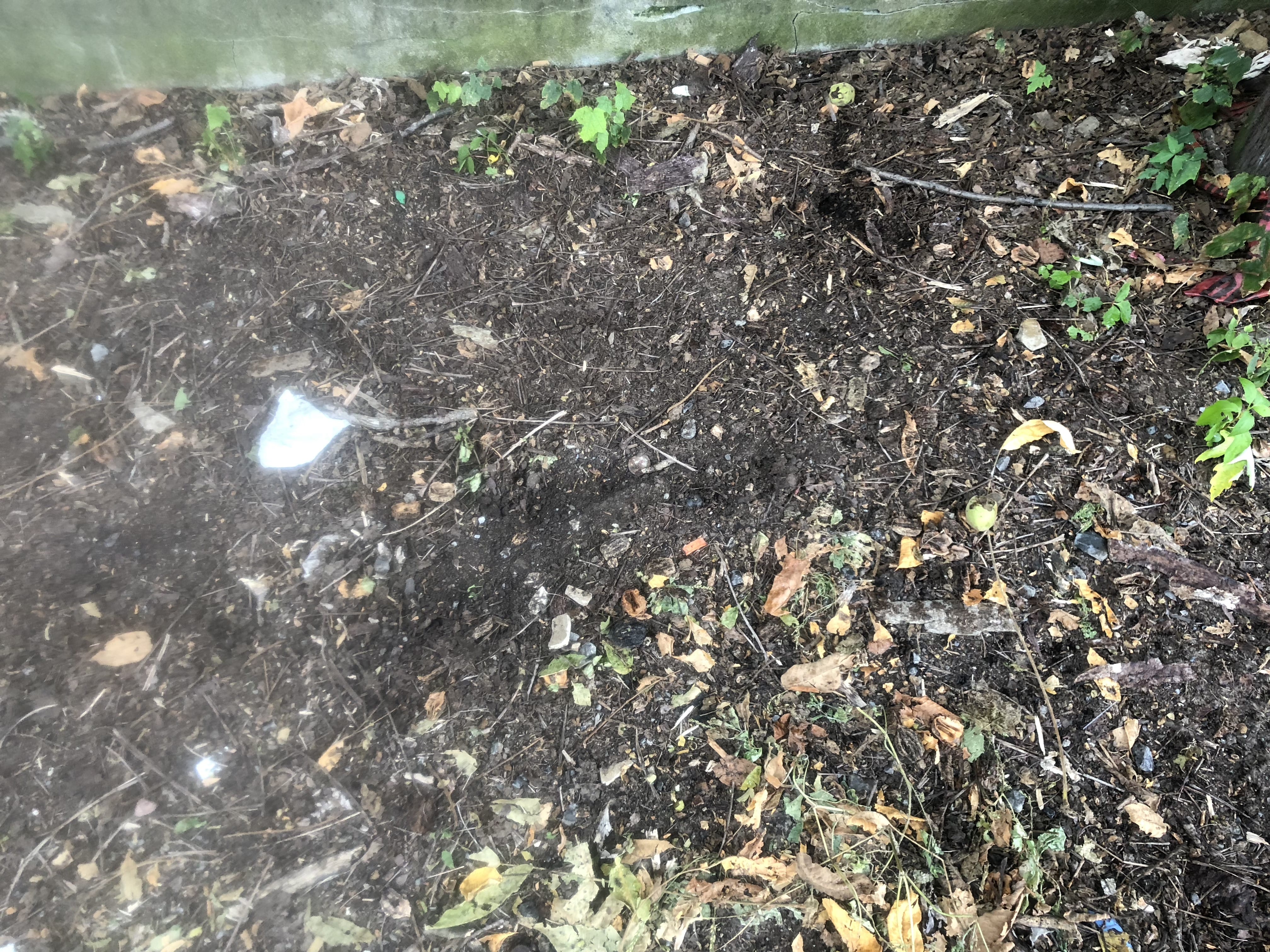
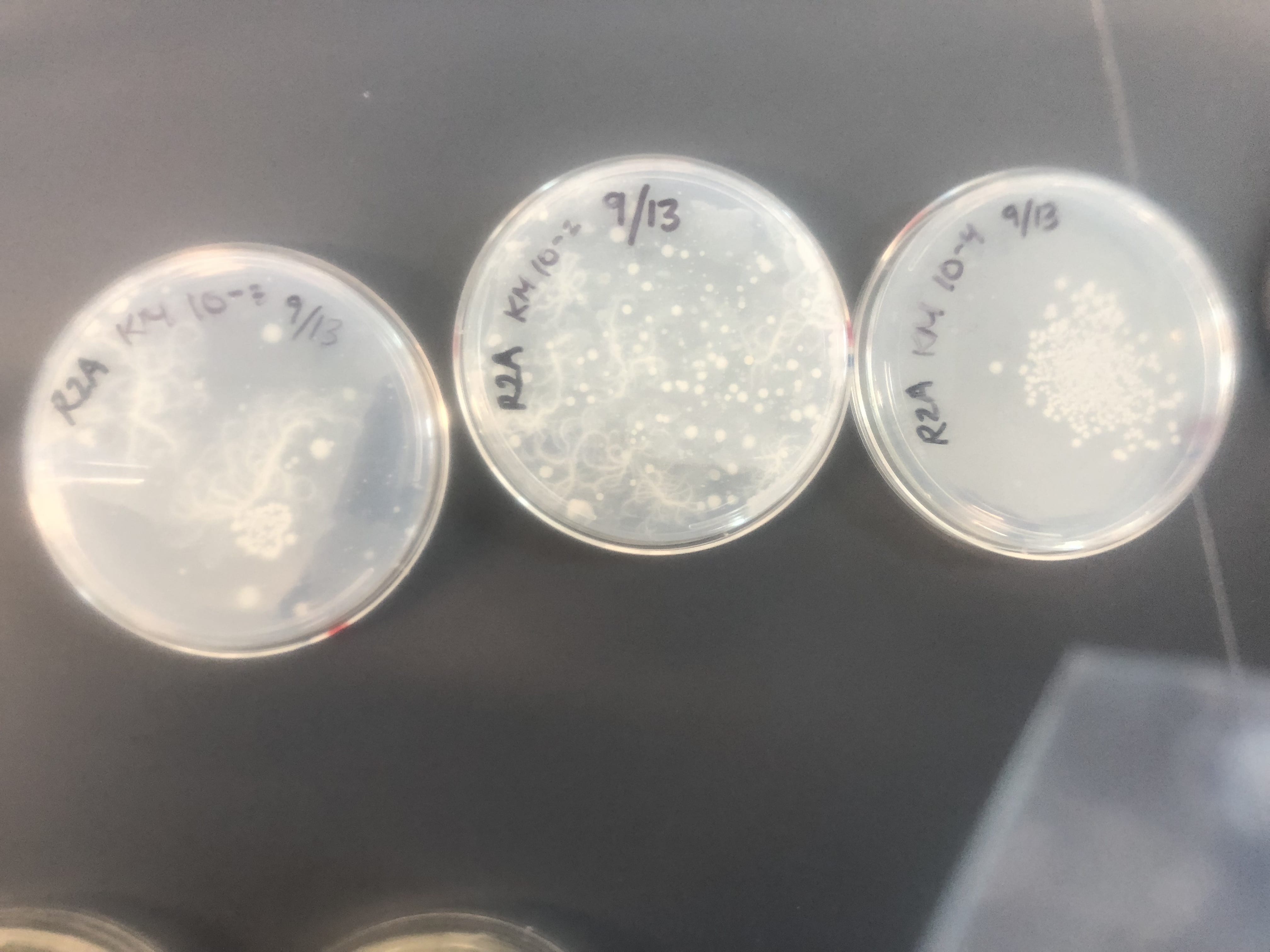
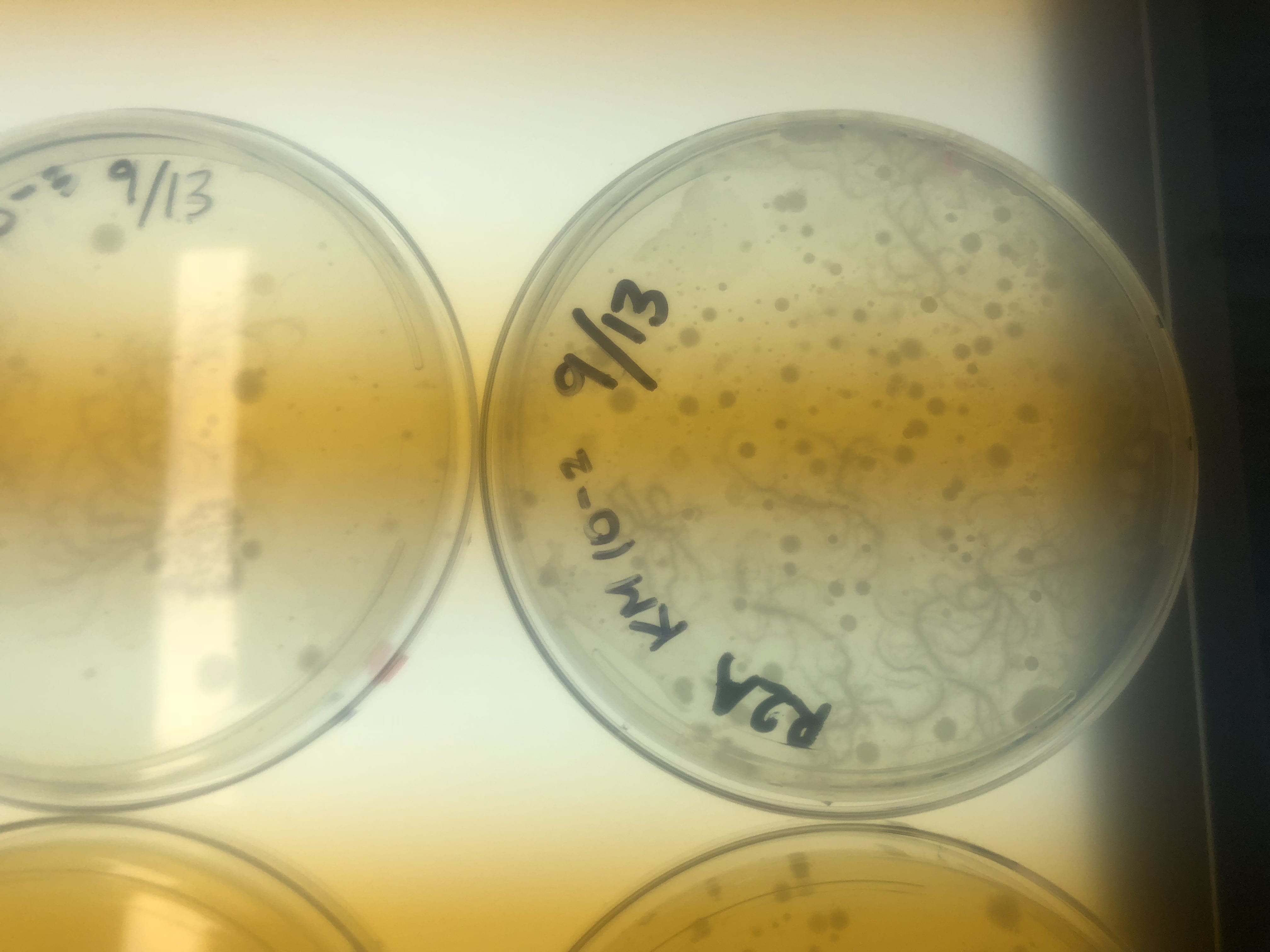
First, bacteria were grown on three different culture media – LB, AC, and R2A – at three different dilution factors.  LB and AC were grown at 10-2, 10-3, and 10-4, while R2A was grown at 10-1, 10-2, and 10-3.  This was done, because they provided three different nutrient levels; Rich, somewhat rich, and somewhat sparse.

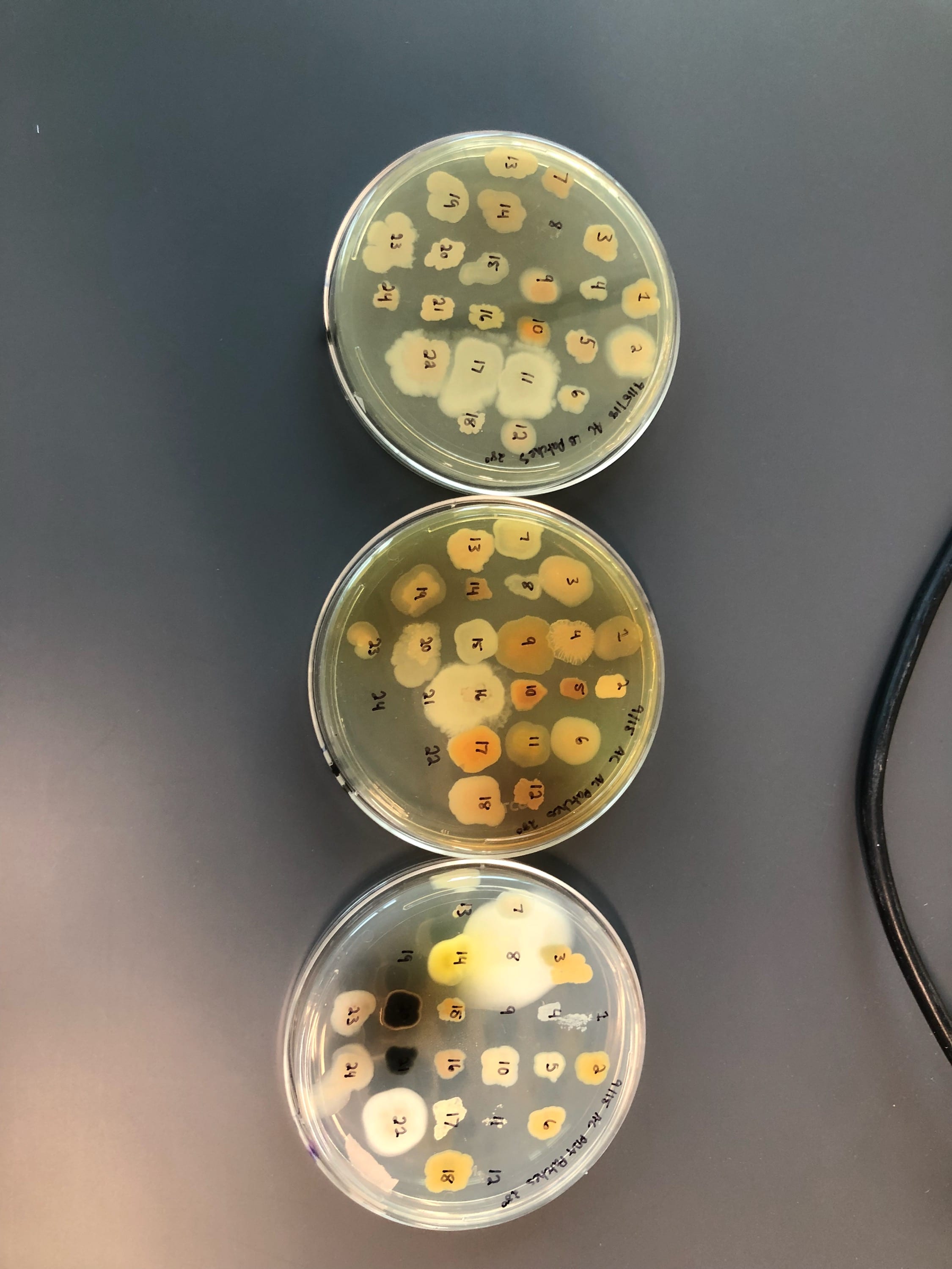
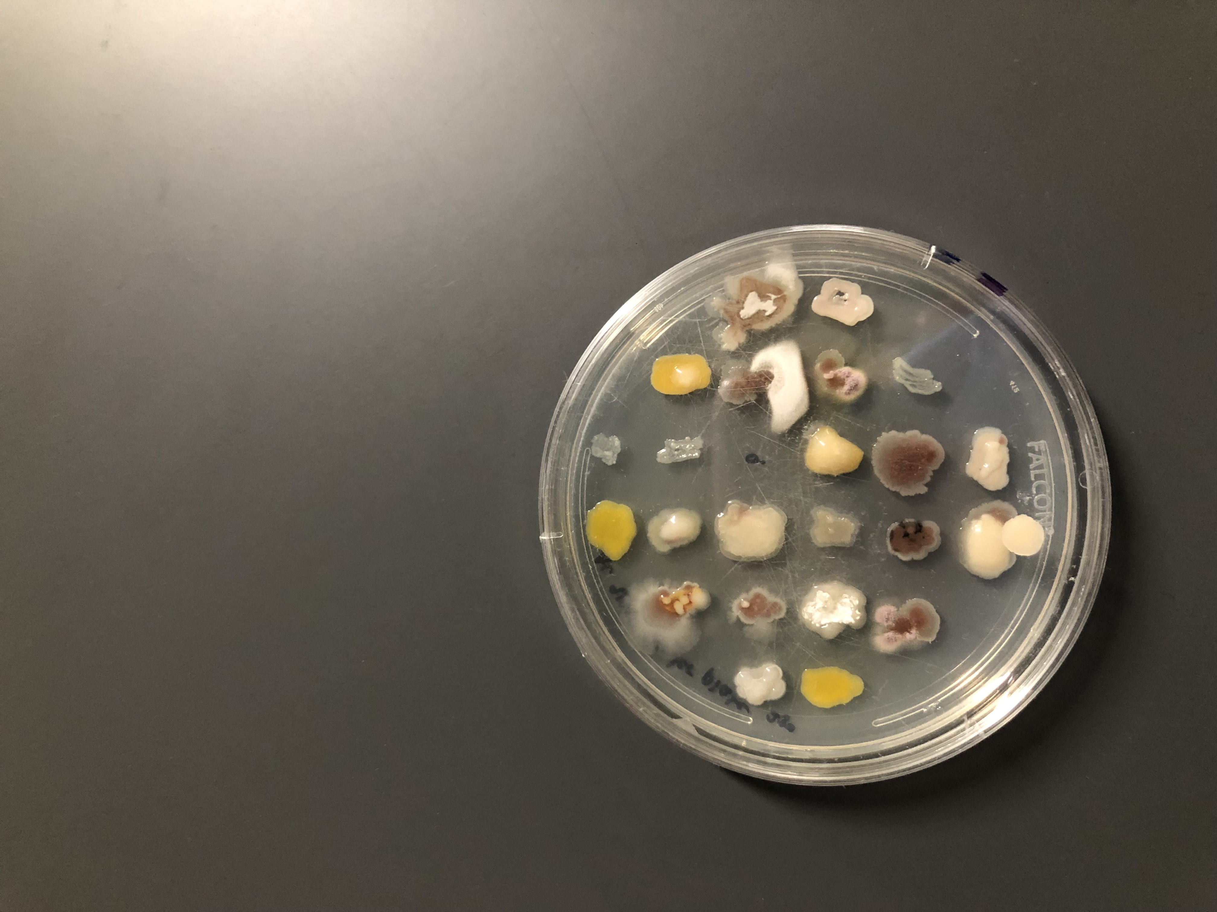
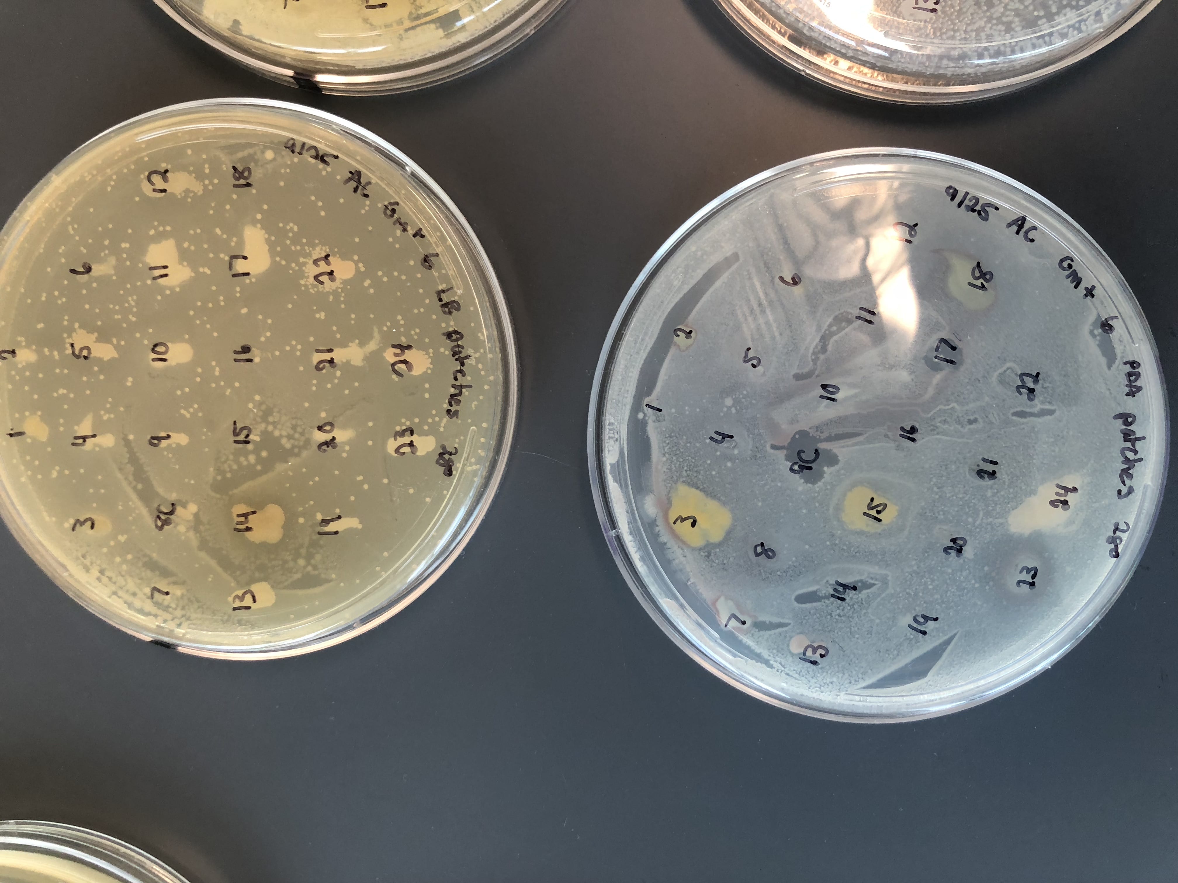
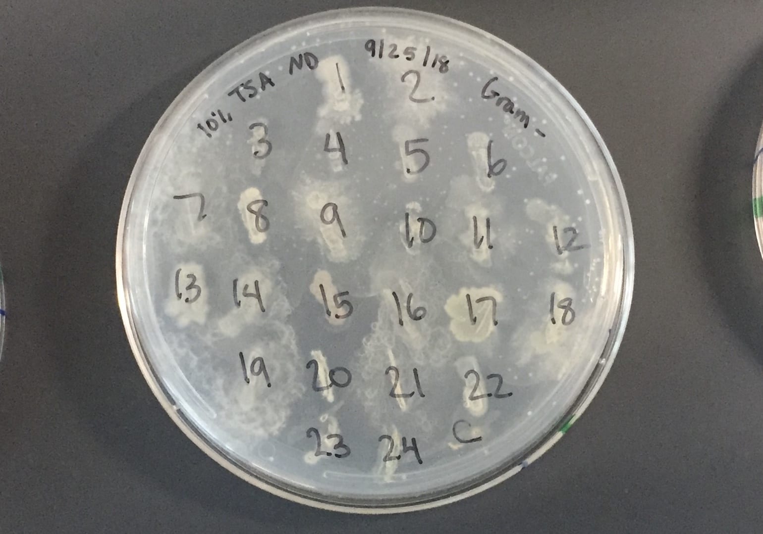
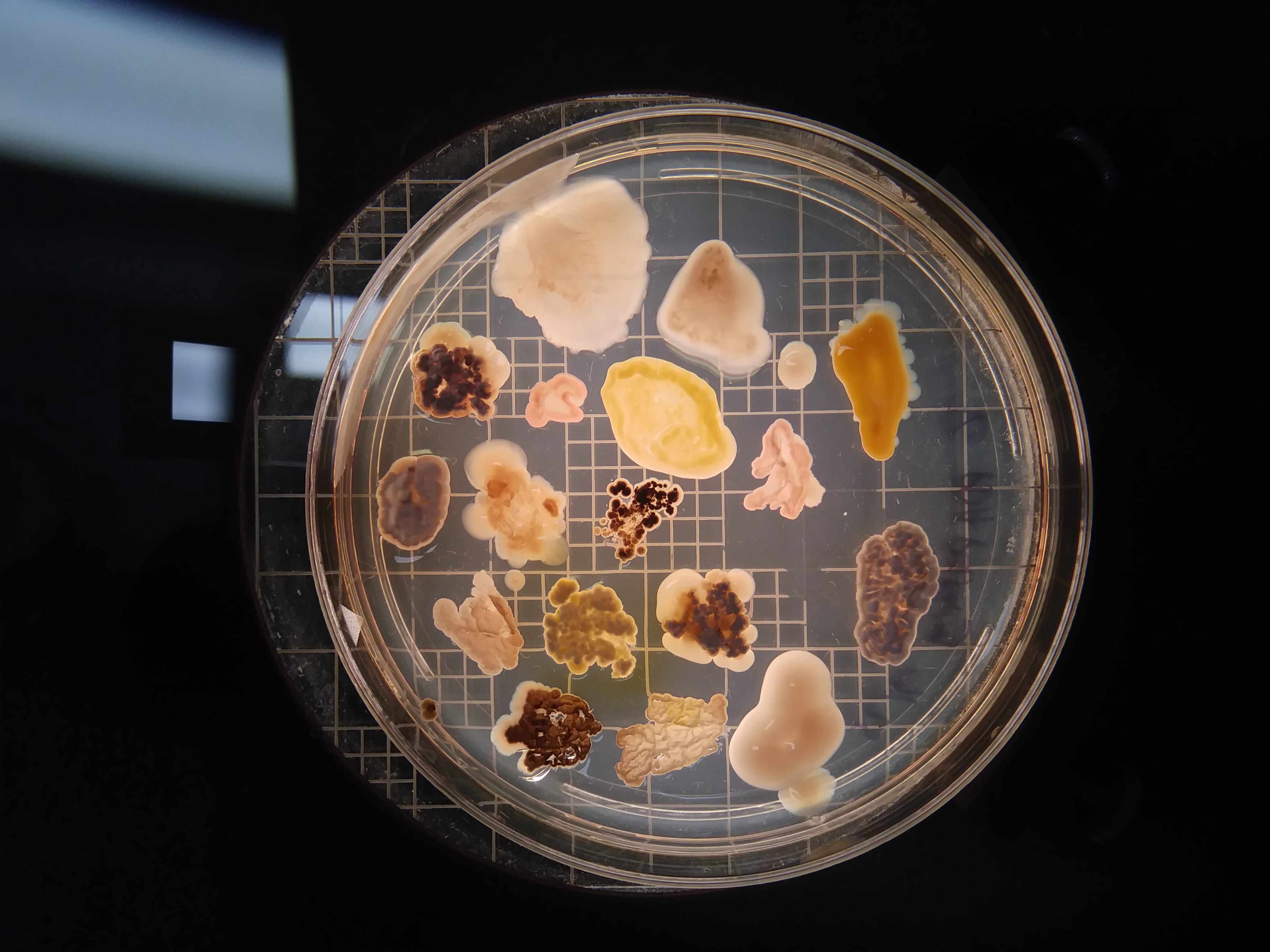
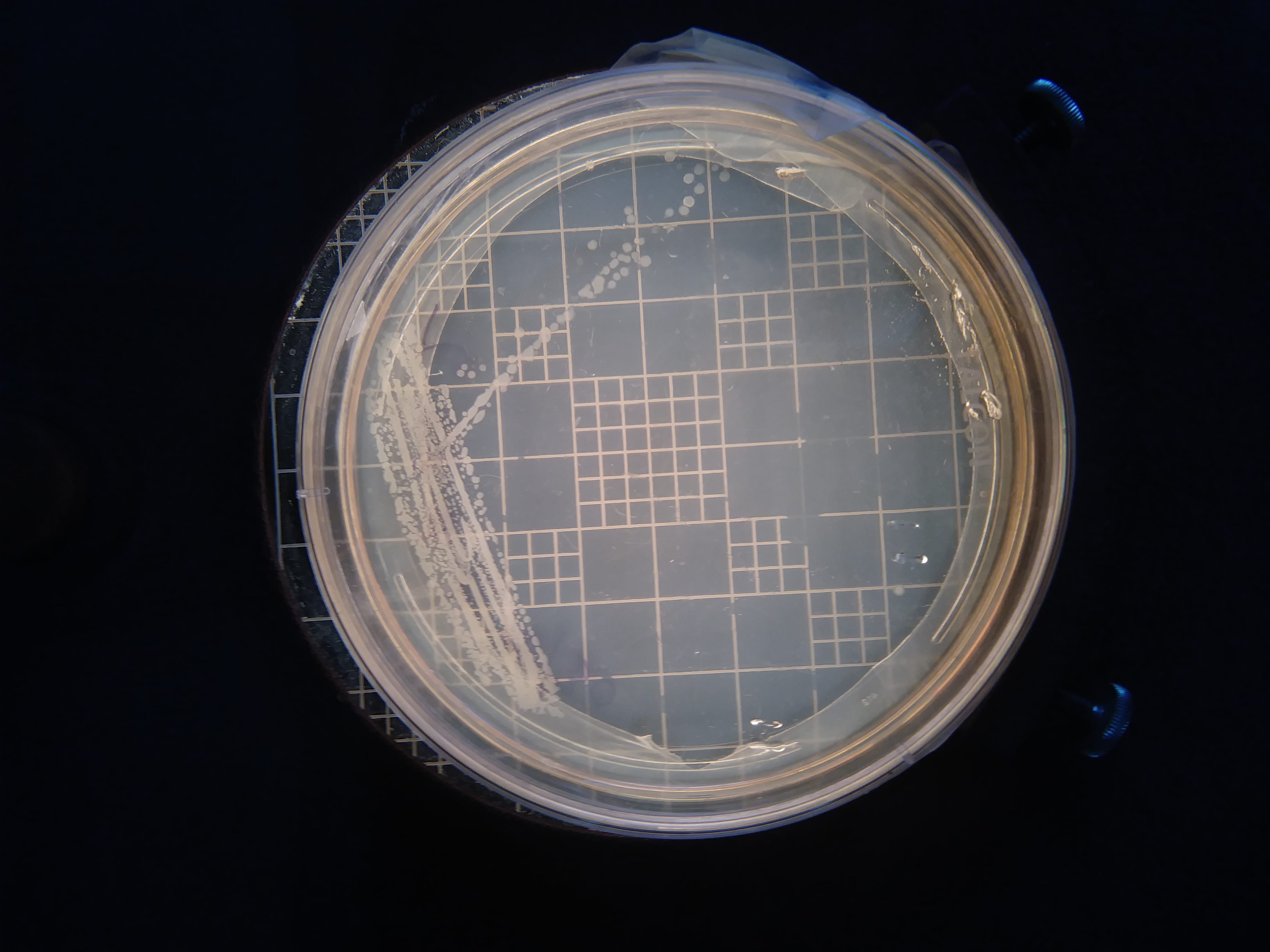
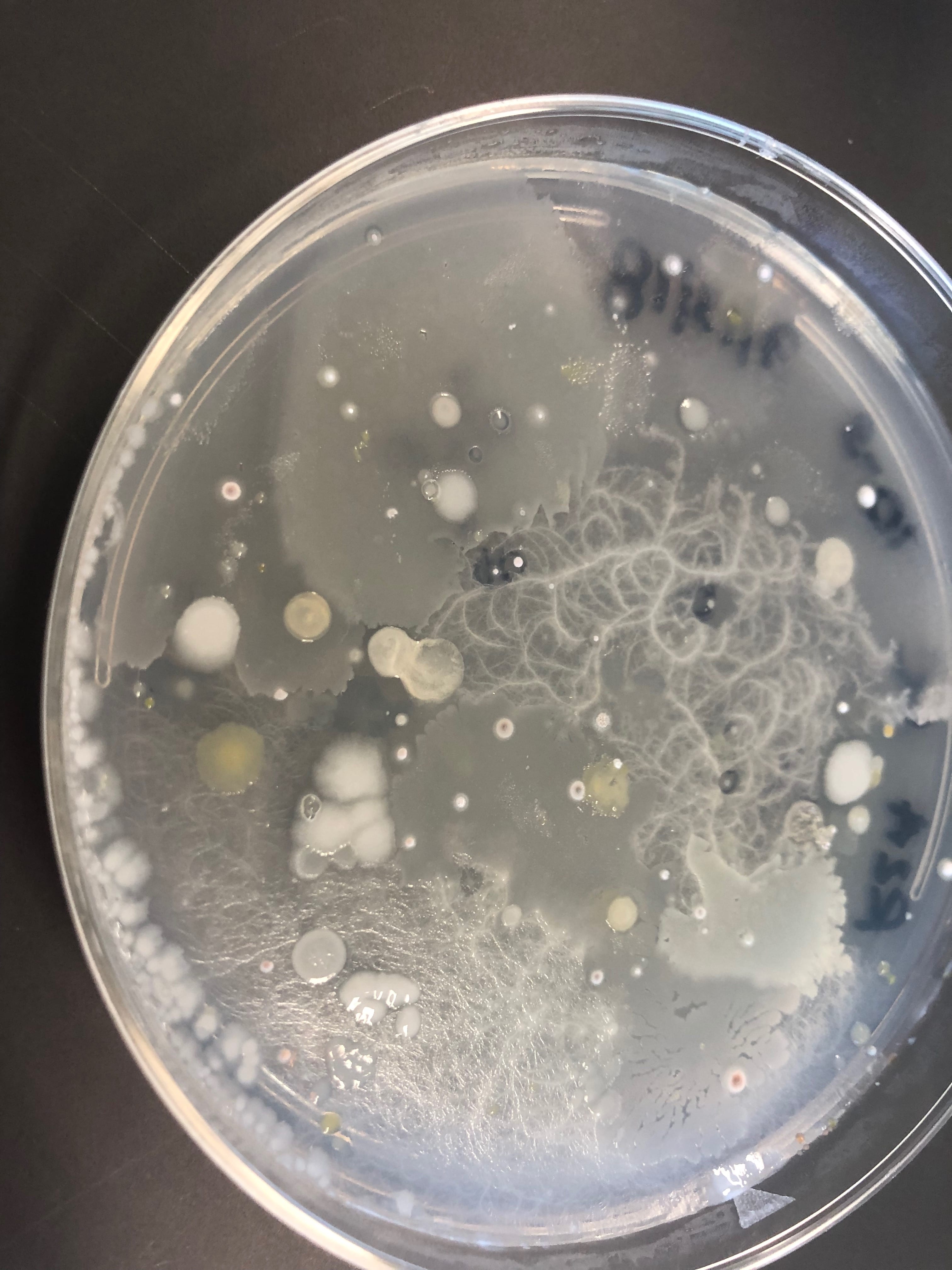
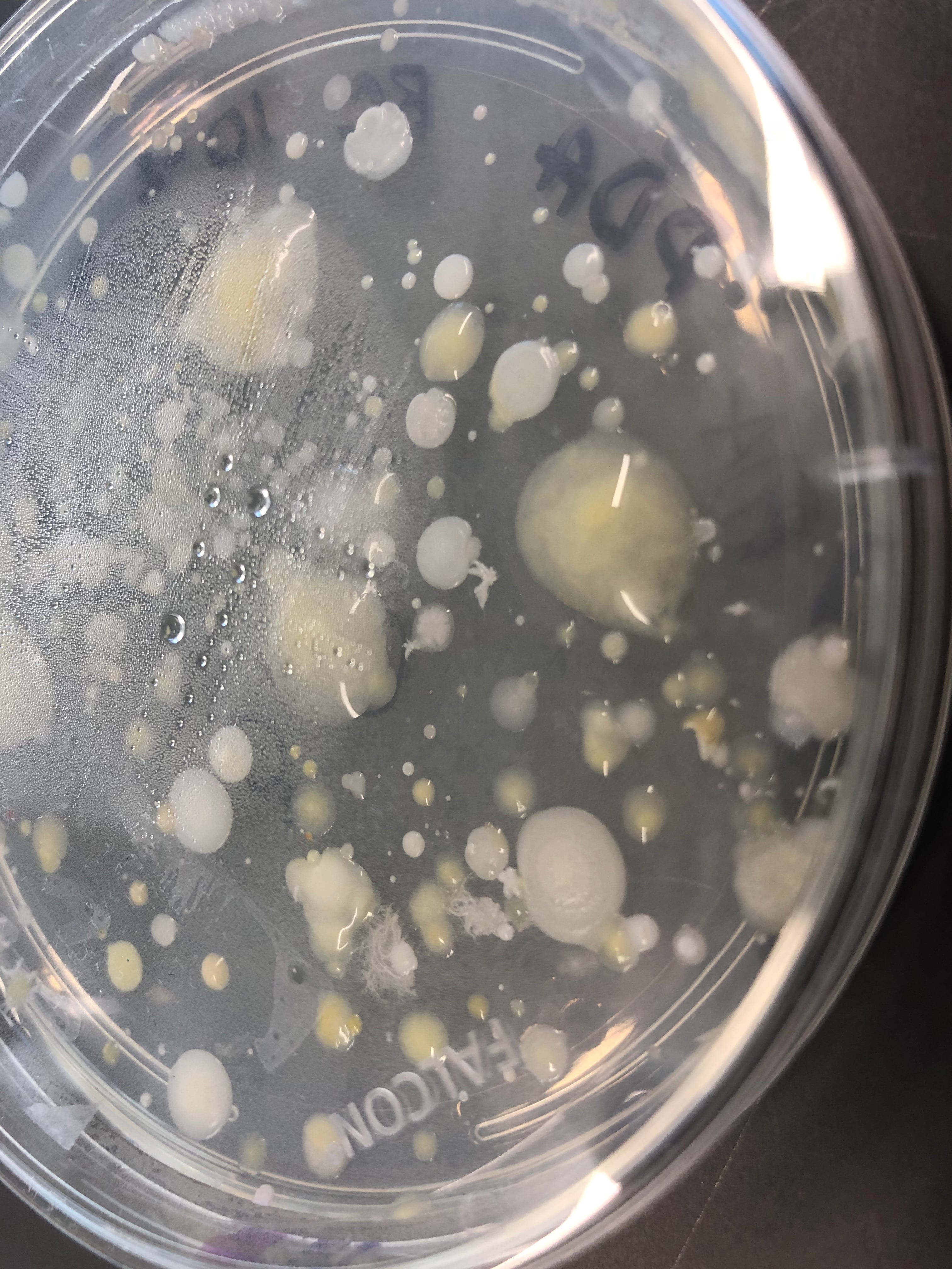
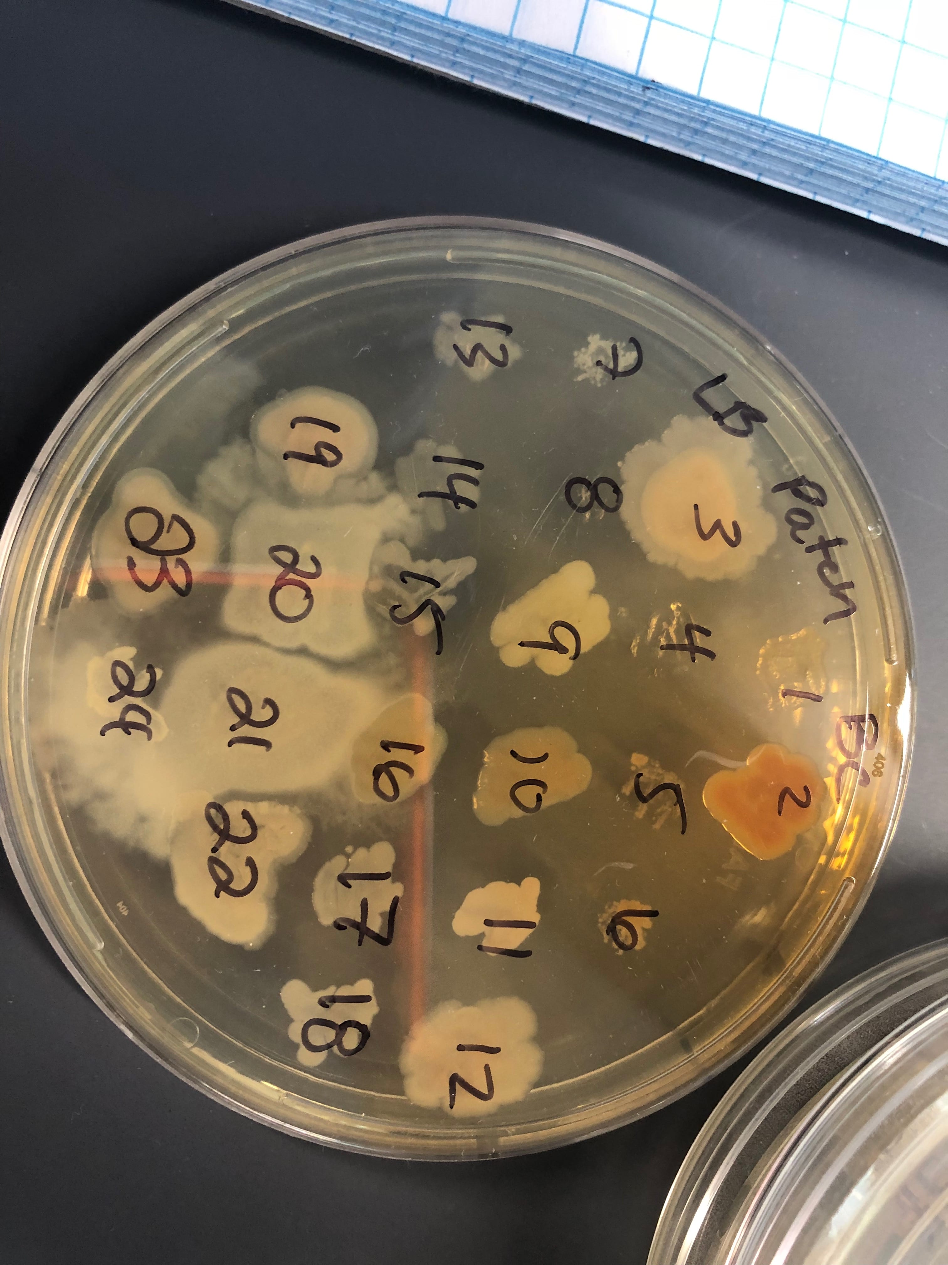
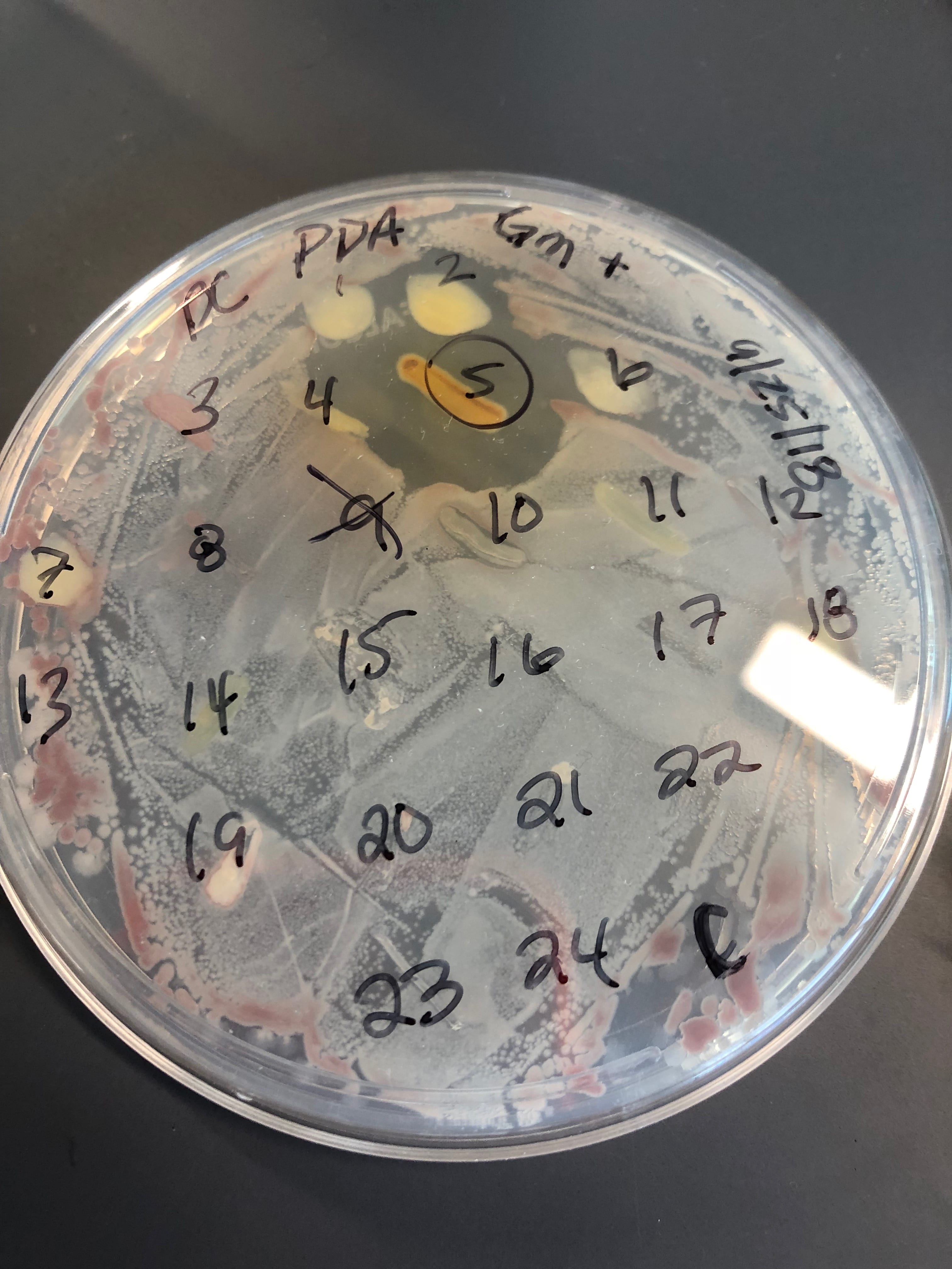
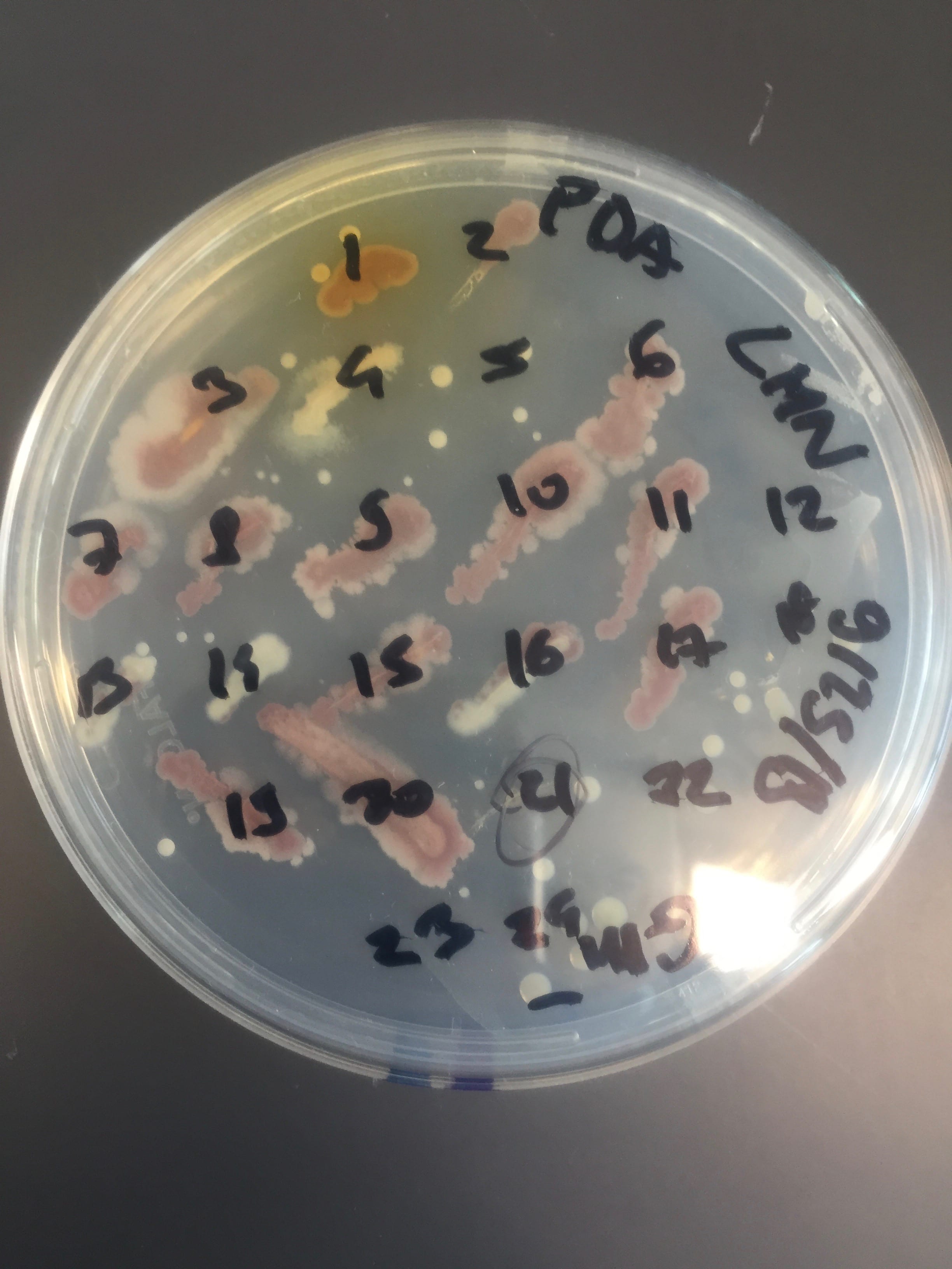
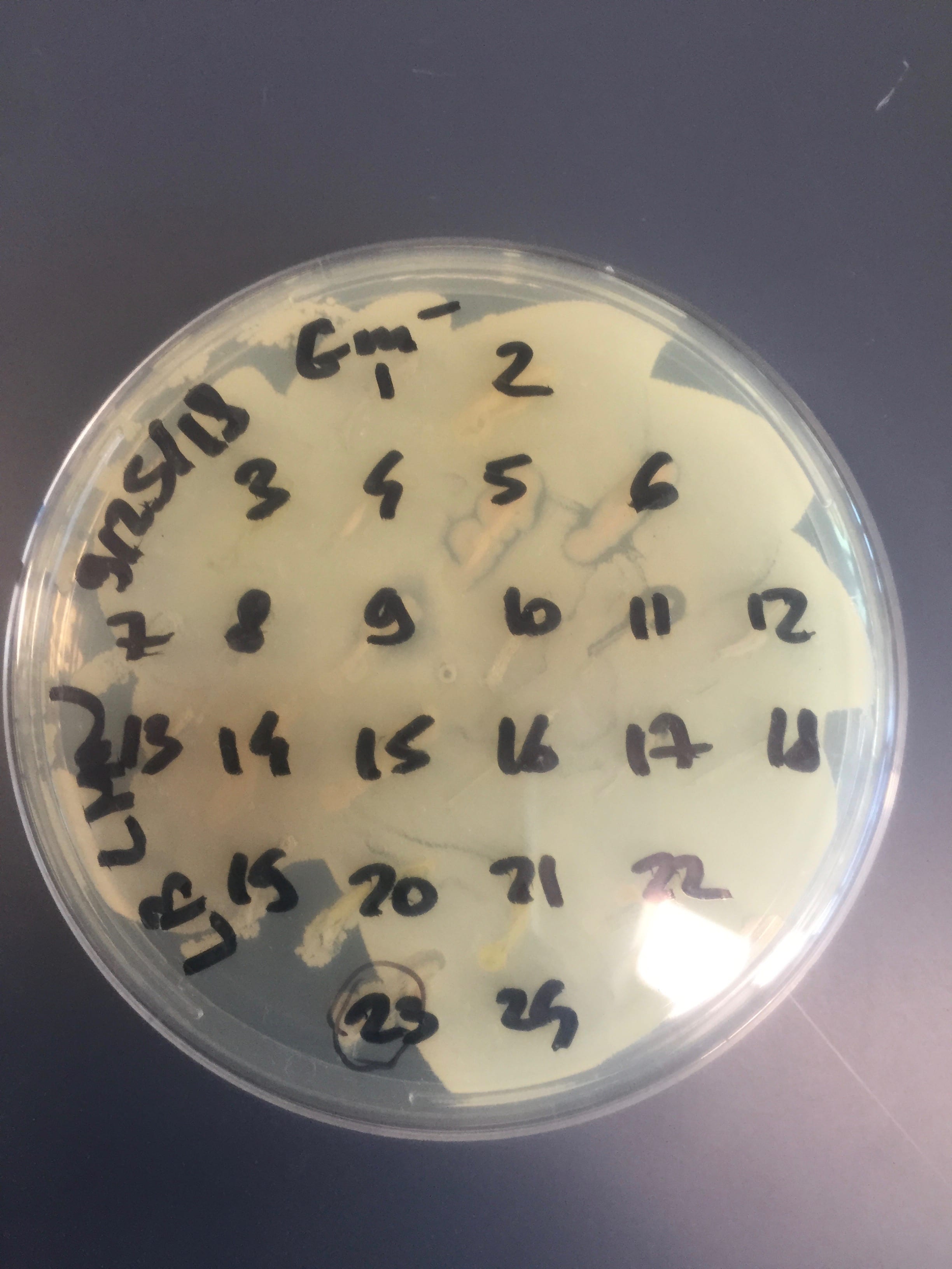
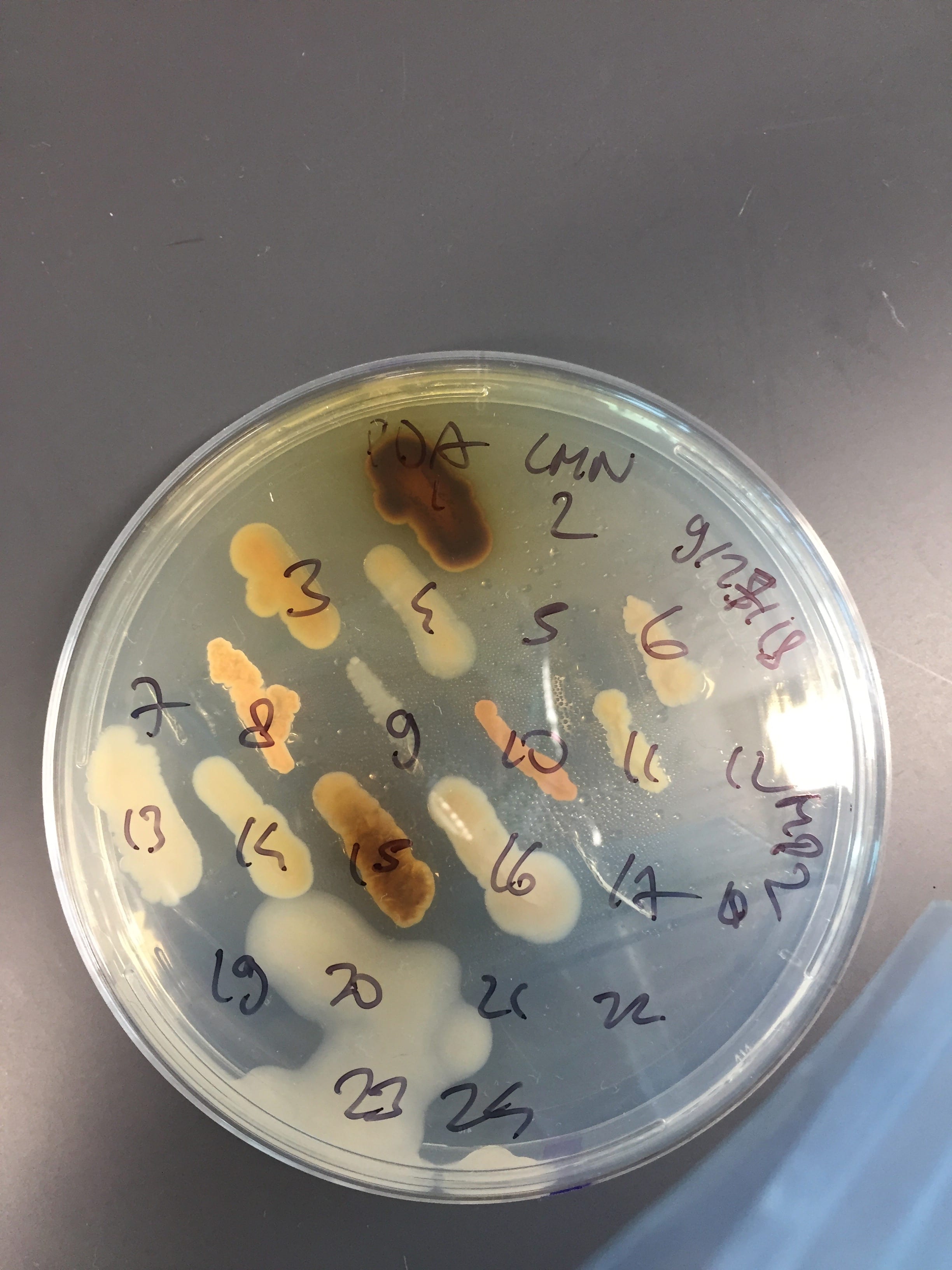
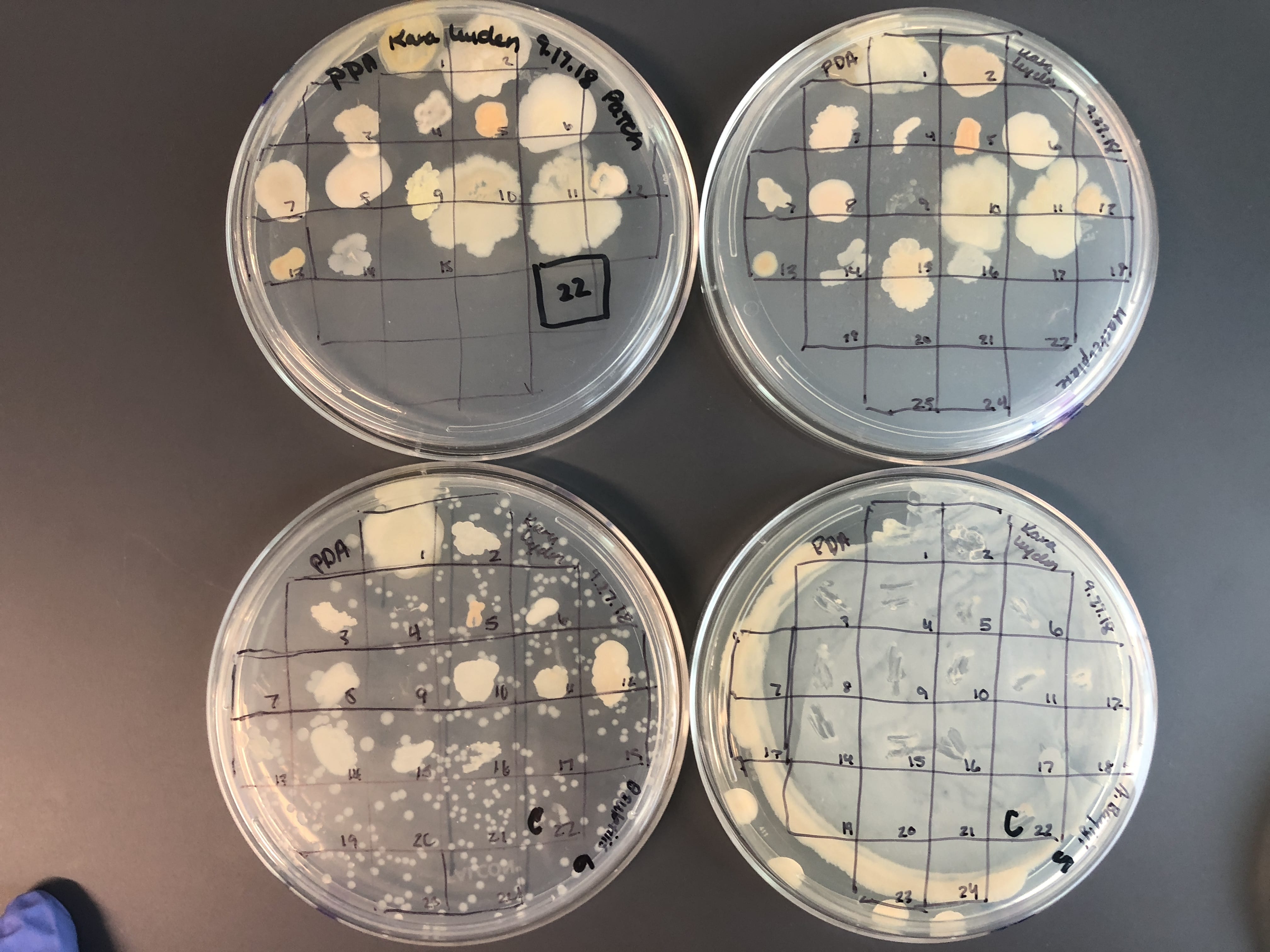
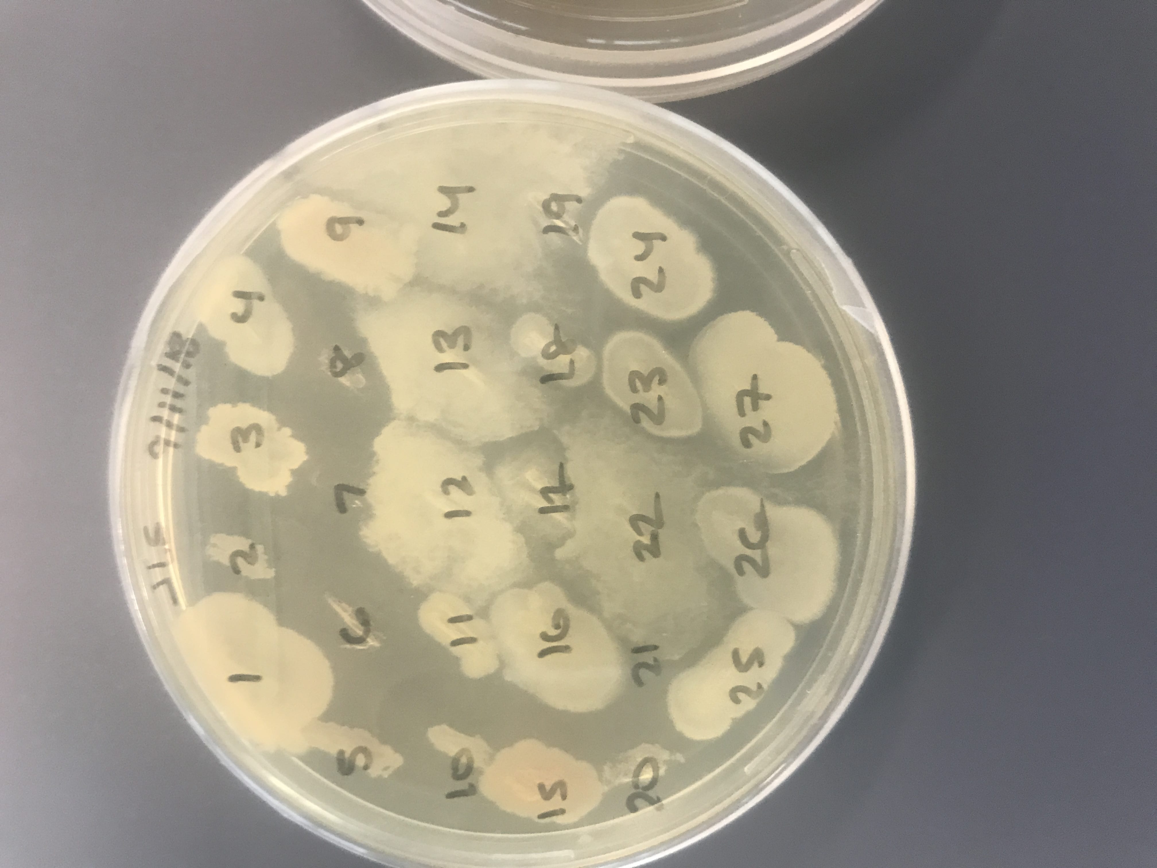
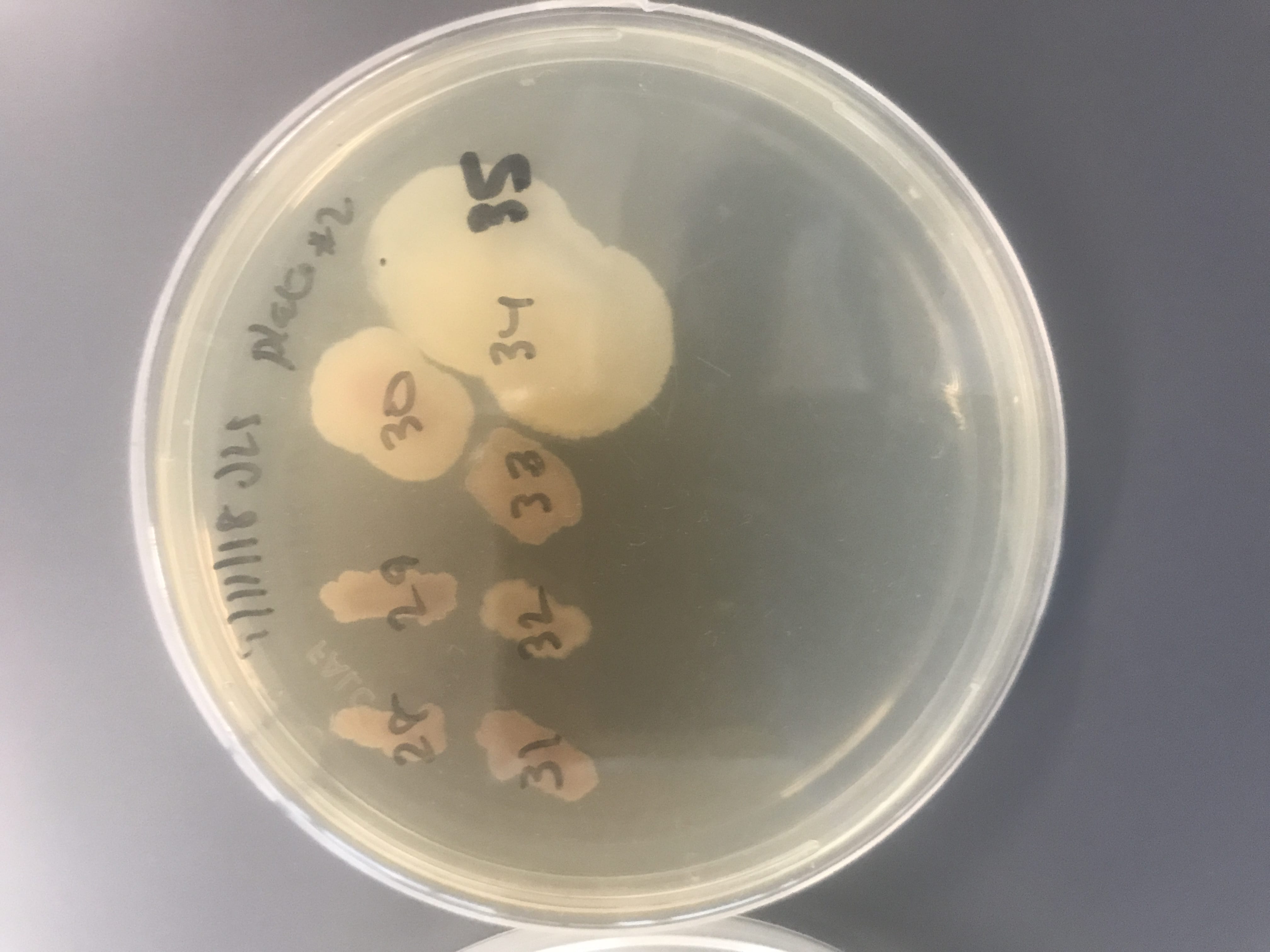
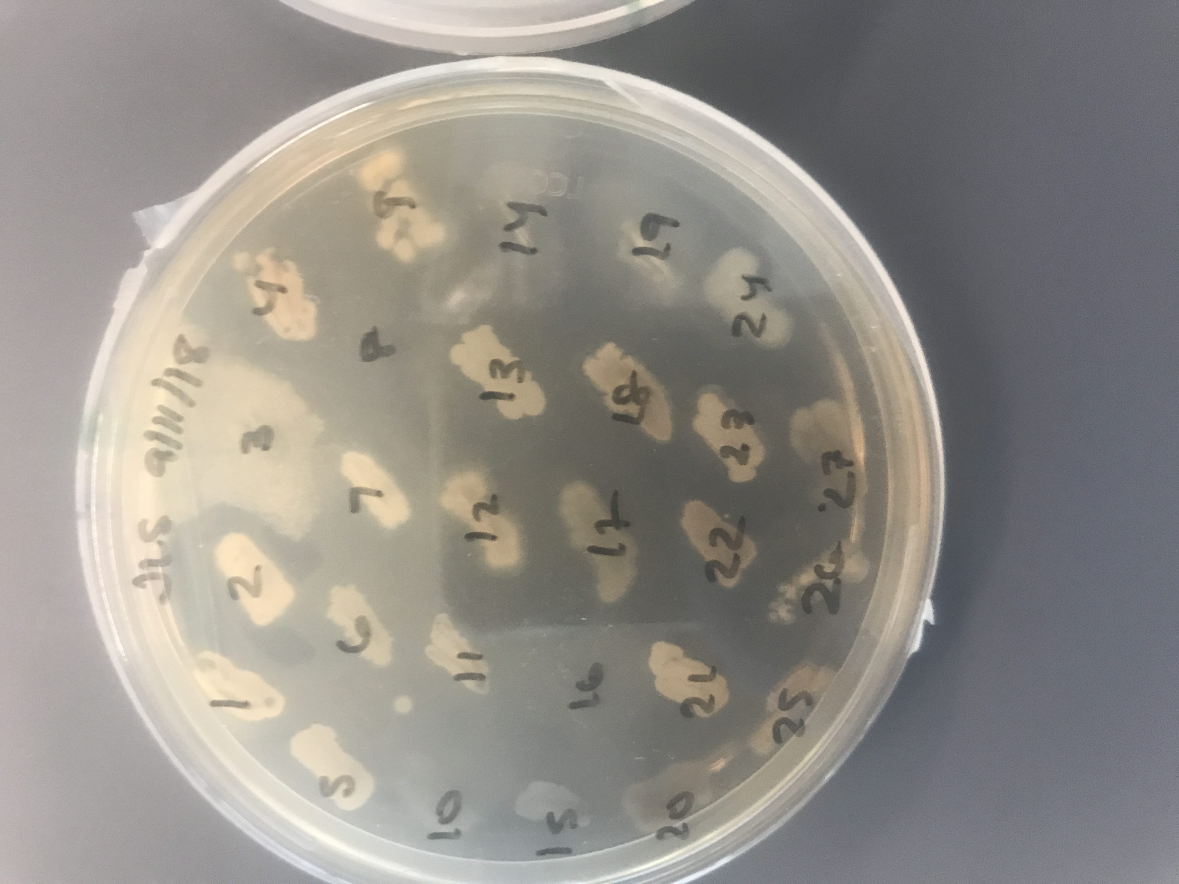
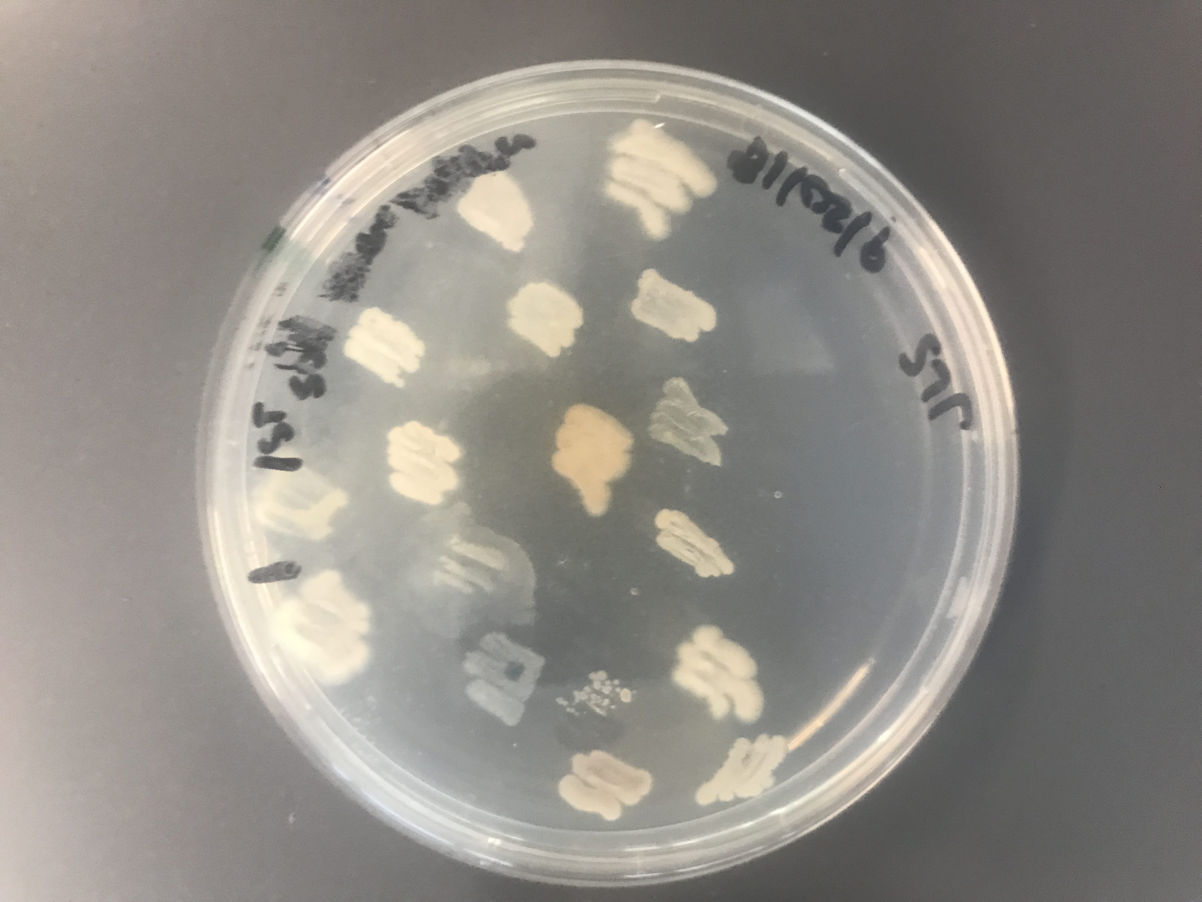
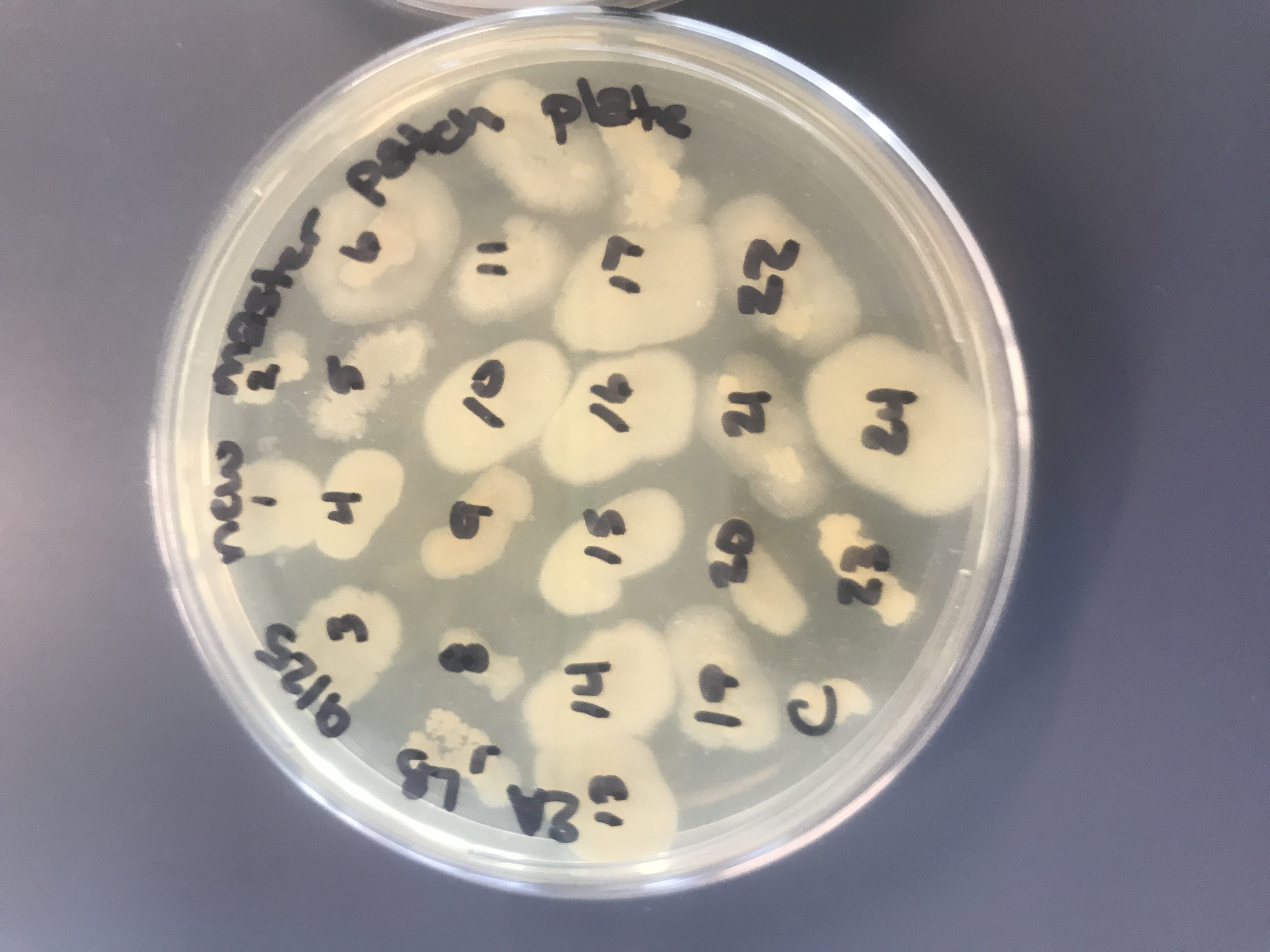
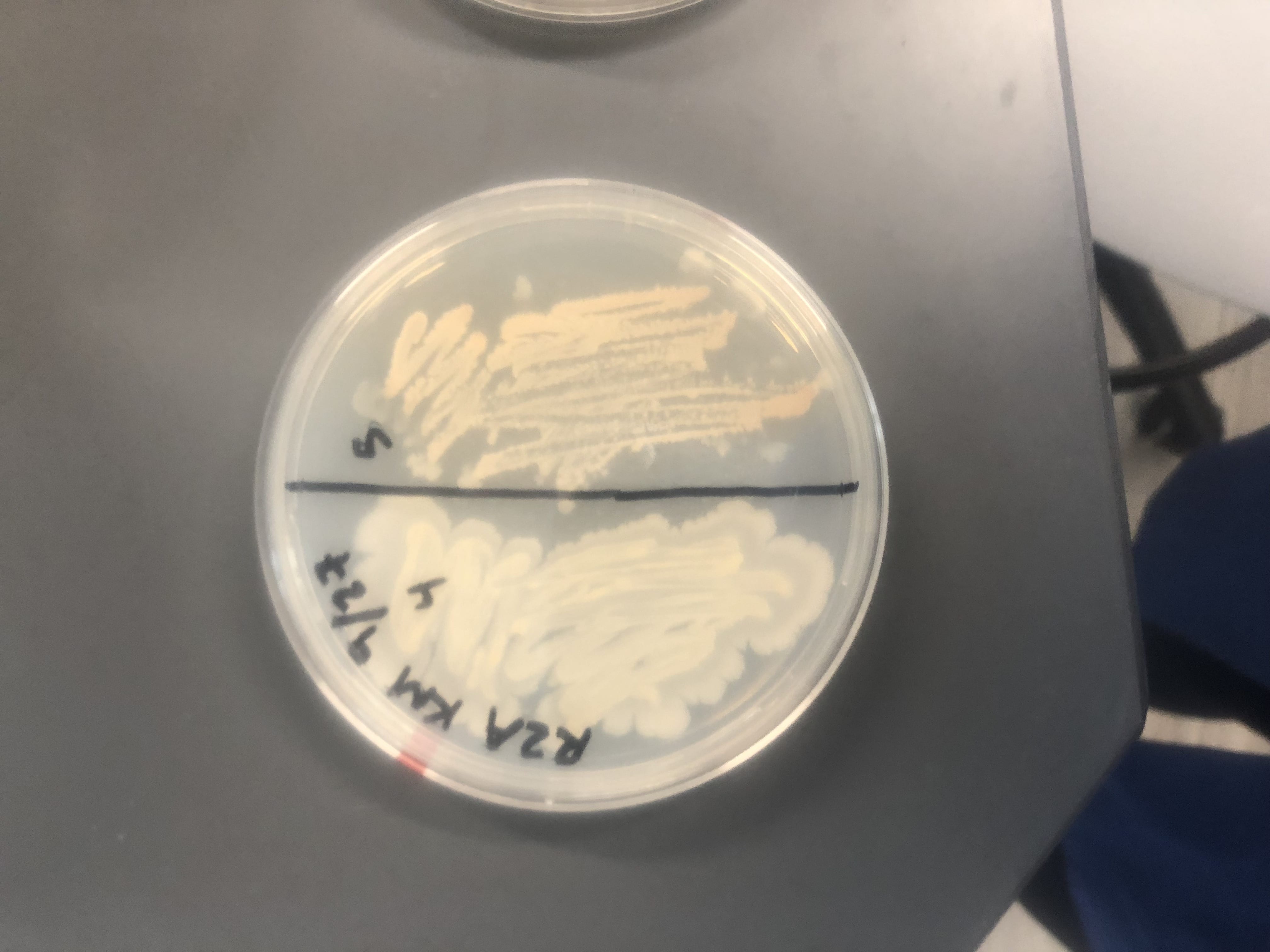
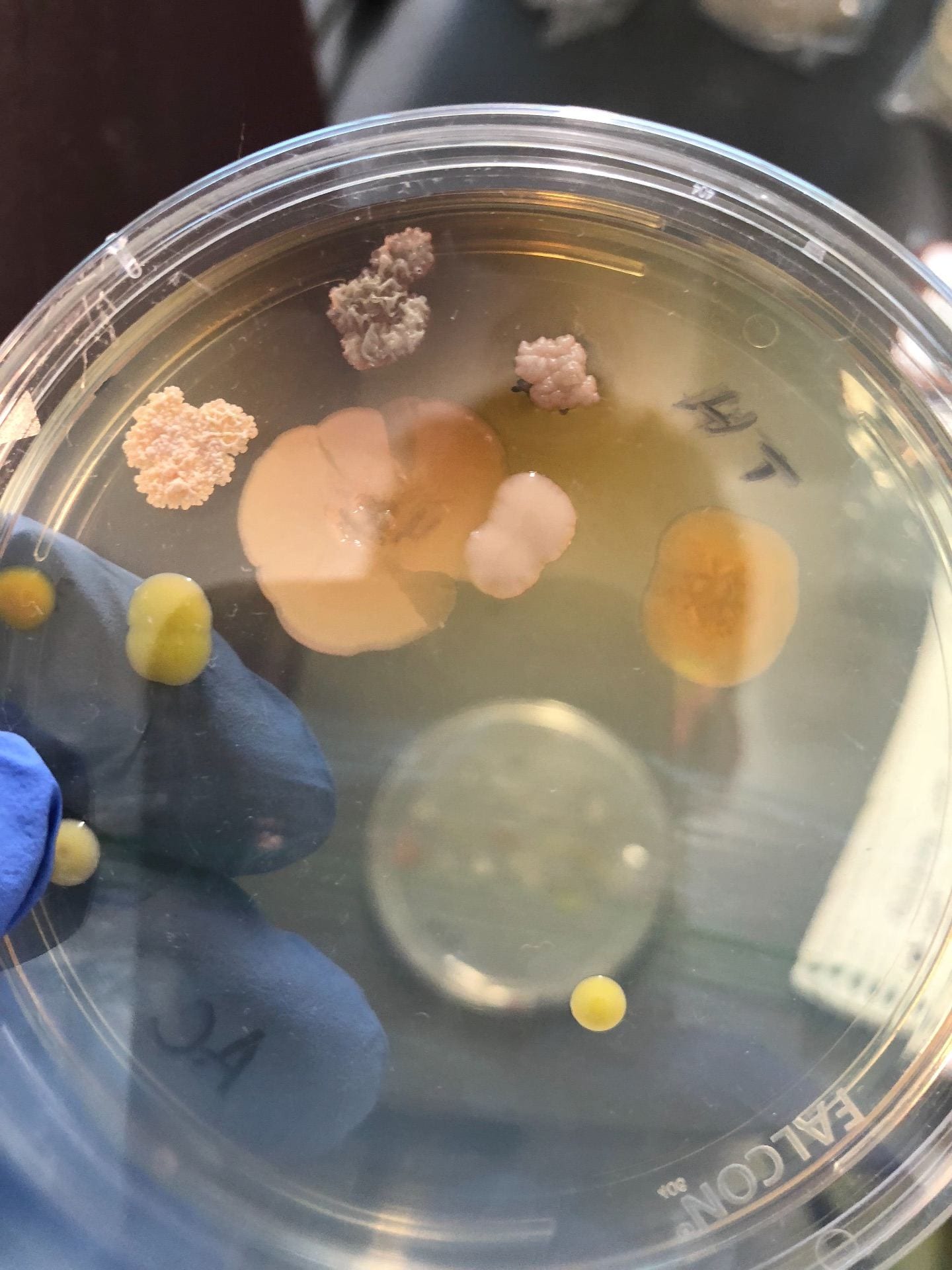
After the bacteria were grown they were selected for different traits that set them apart.  They were then picked and patched onto individual media corresponding to the media they were picked from.  12 cultures were picked from LB, 8 were picked from AC, and 16 were picked from R2A.

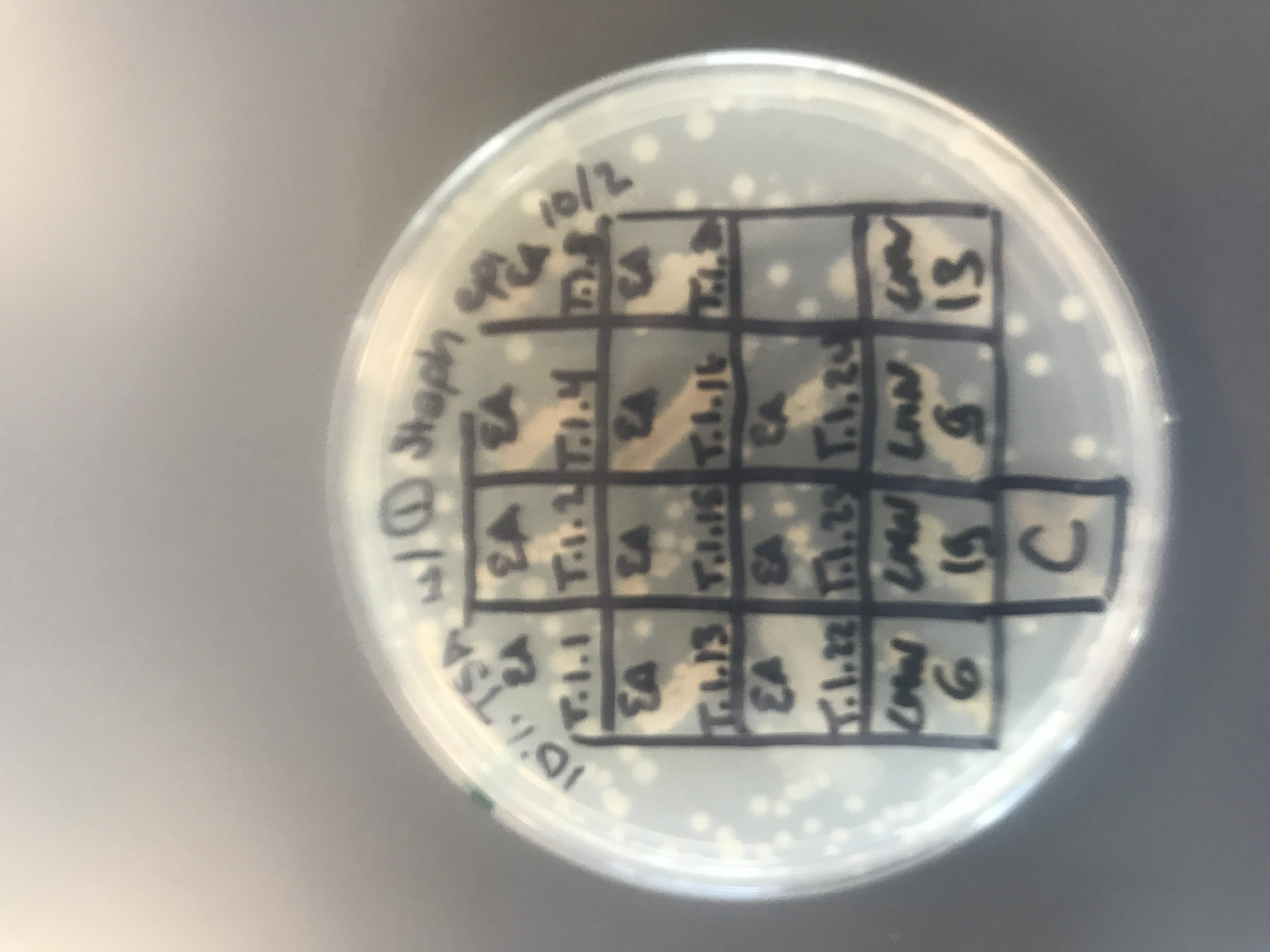
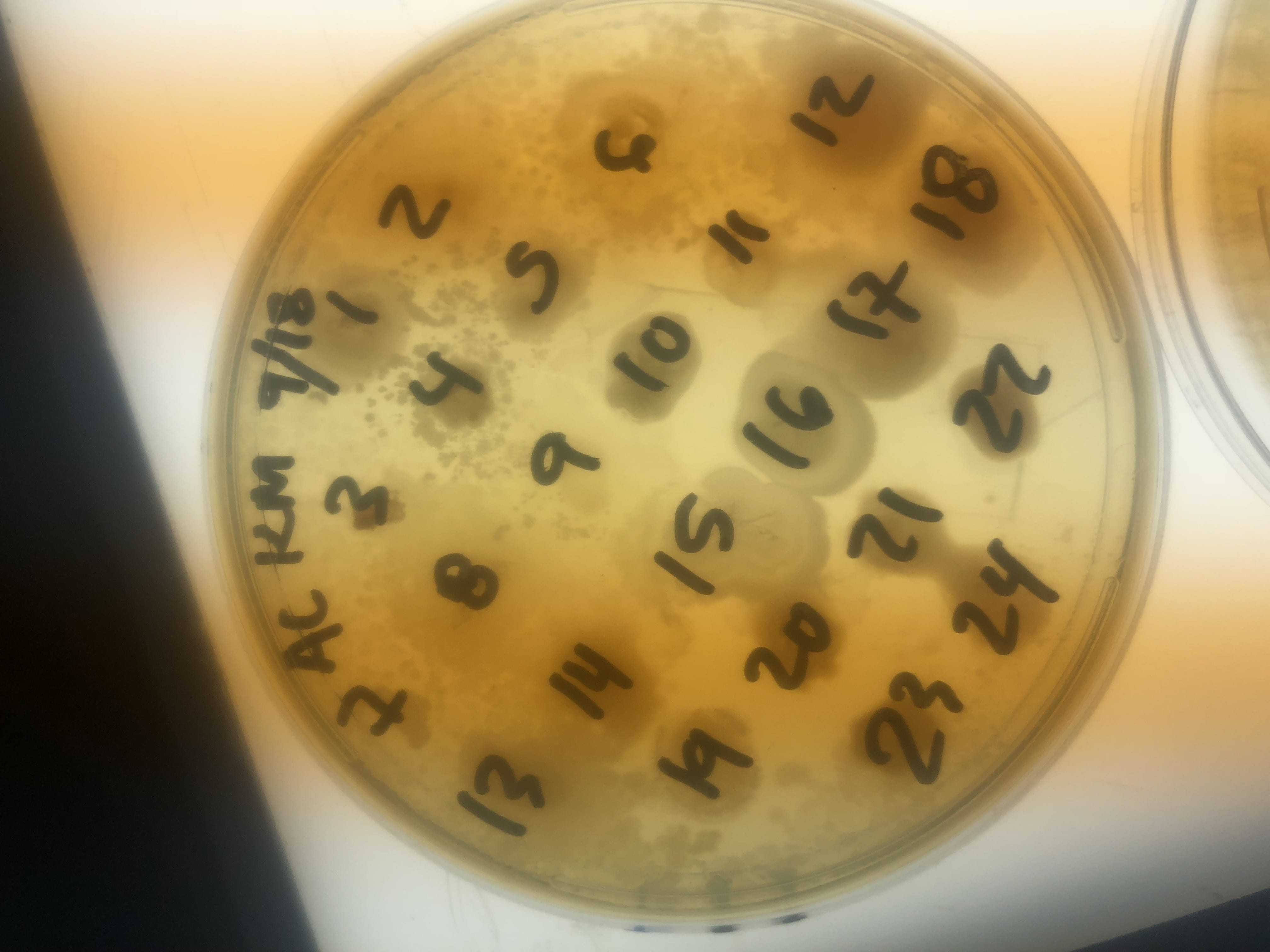
LB patch plate after selection for unique bacteria.

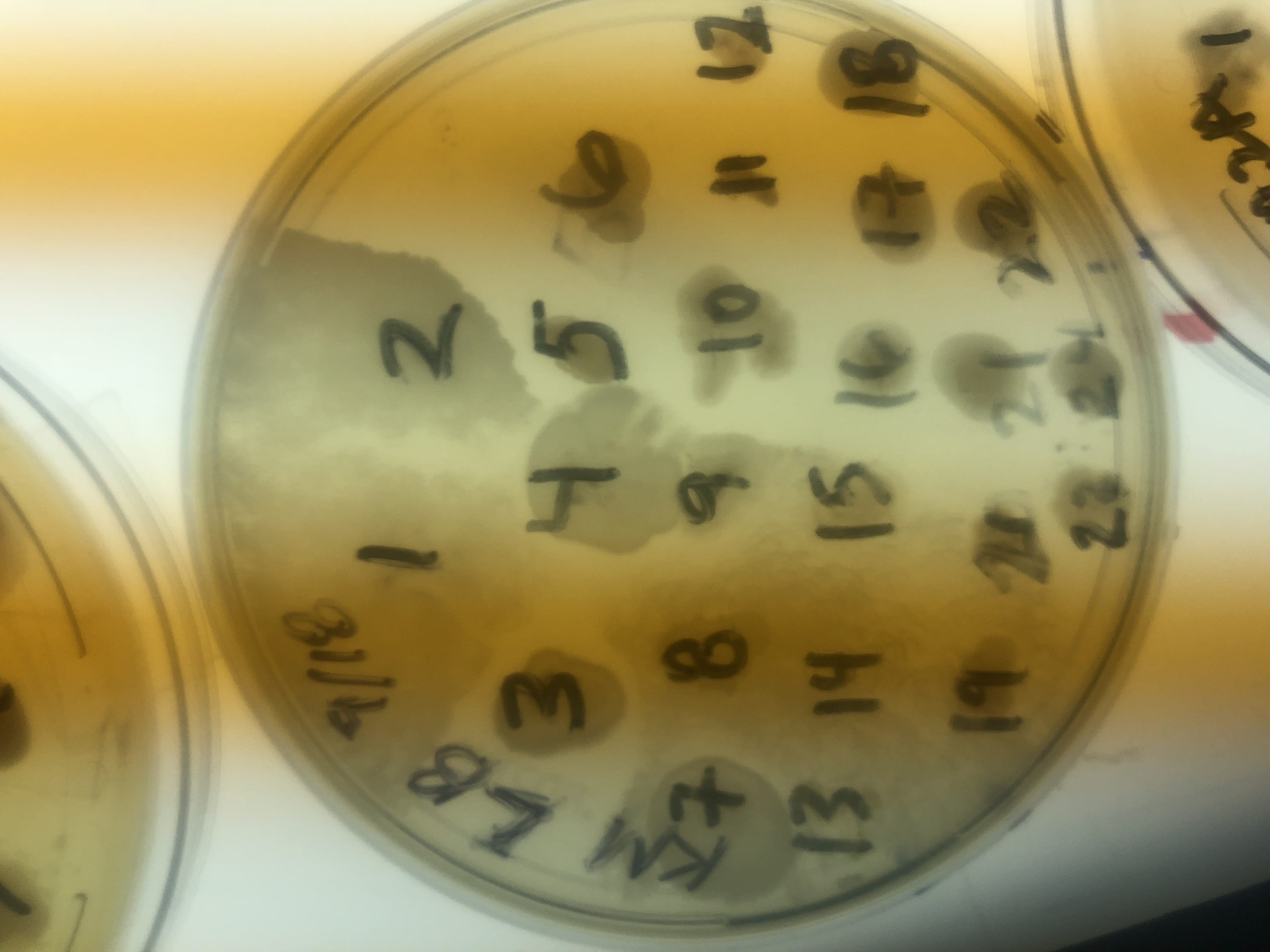
AC patch plate after selection for unique bacteria.

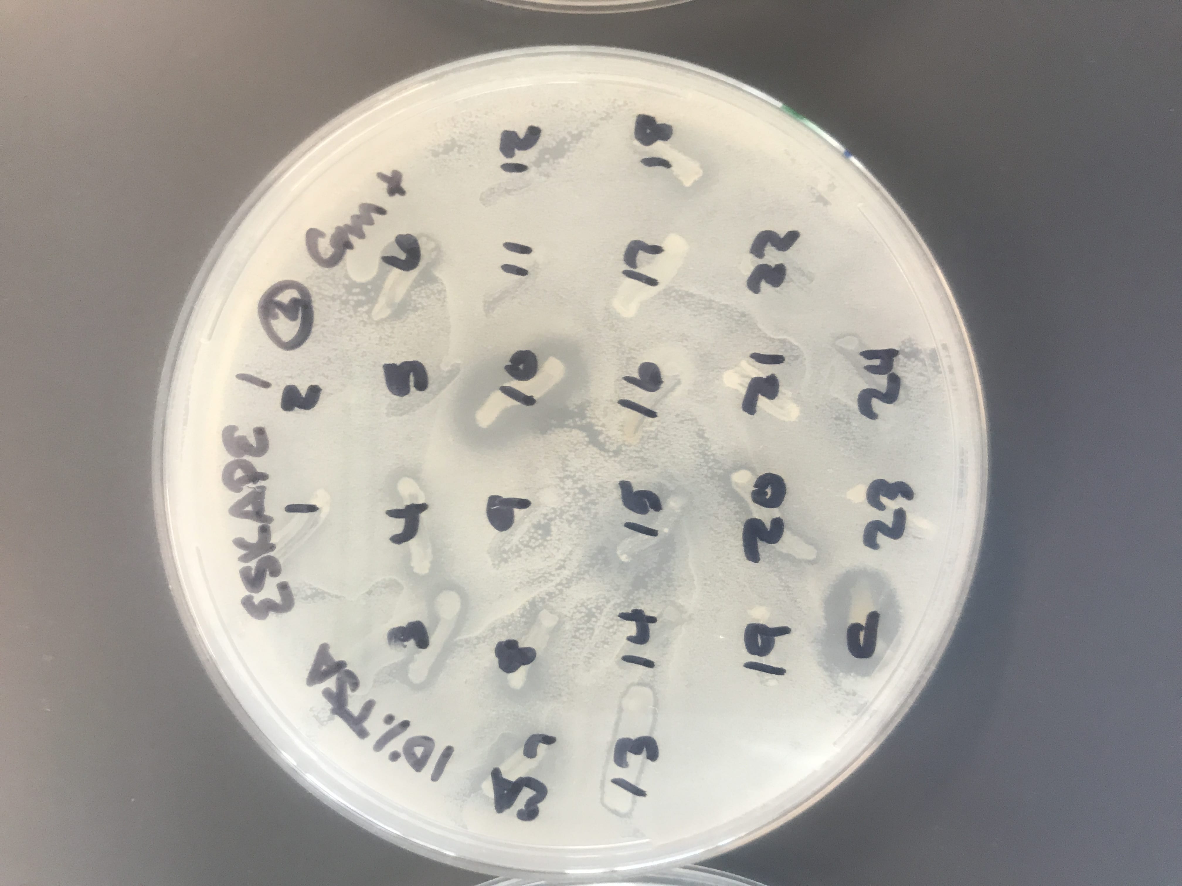
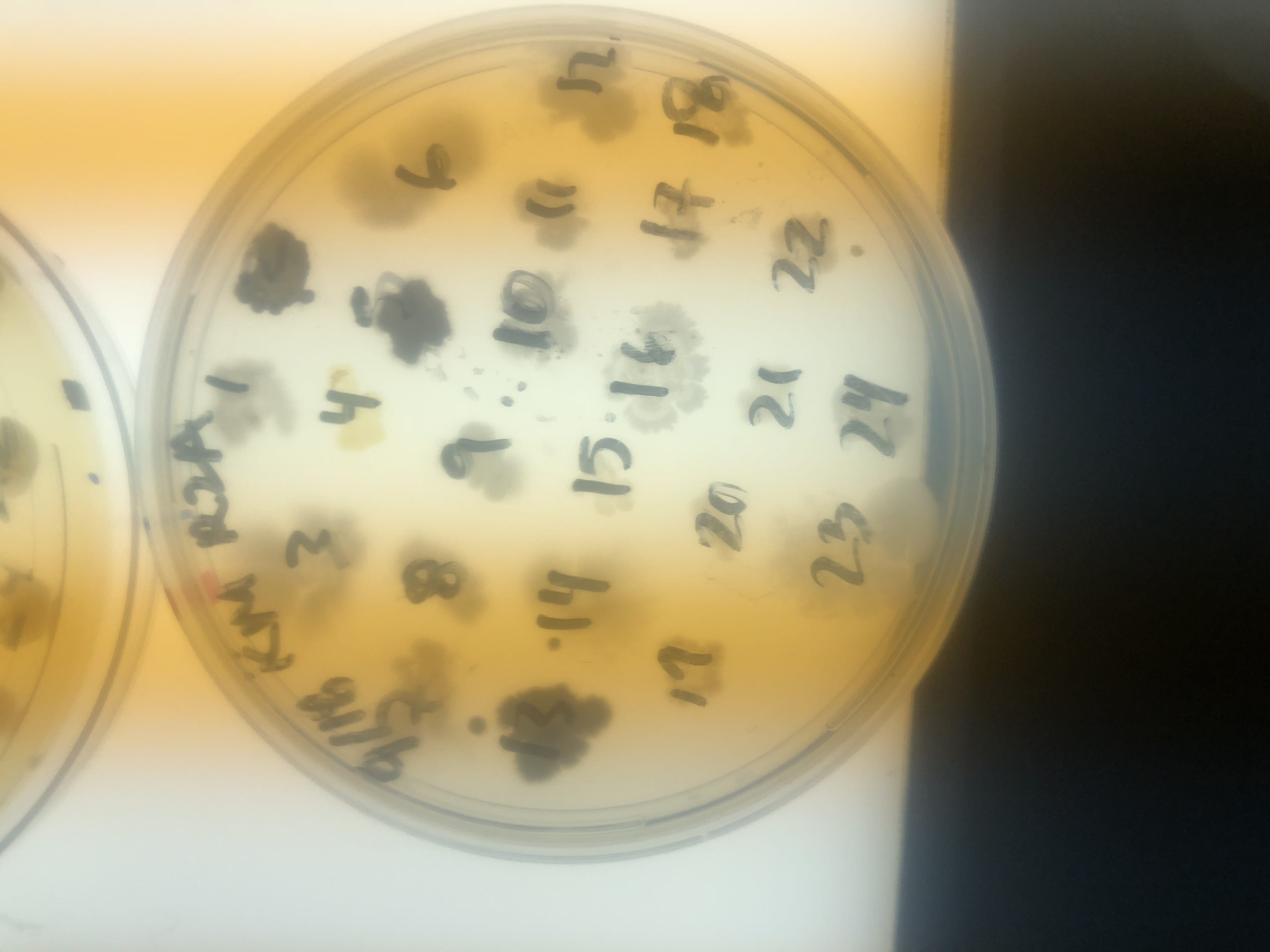
R2A patch plate after selection for unique bacteria.

I thought it was so cool how many different colors and textures were produced by these bacteria!  It’s safe to say that Professor Salvo was right – this soil is pretty diverse!

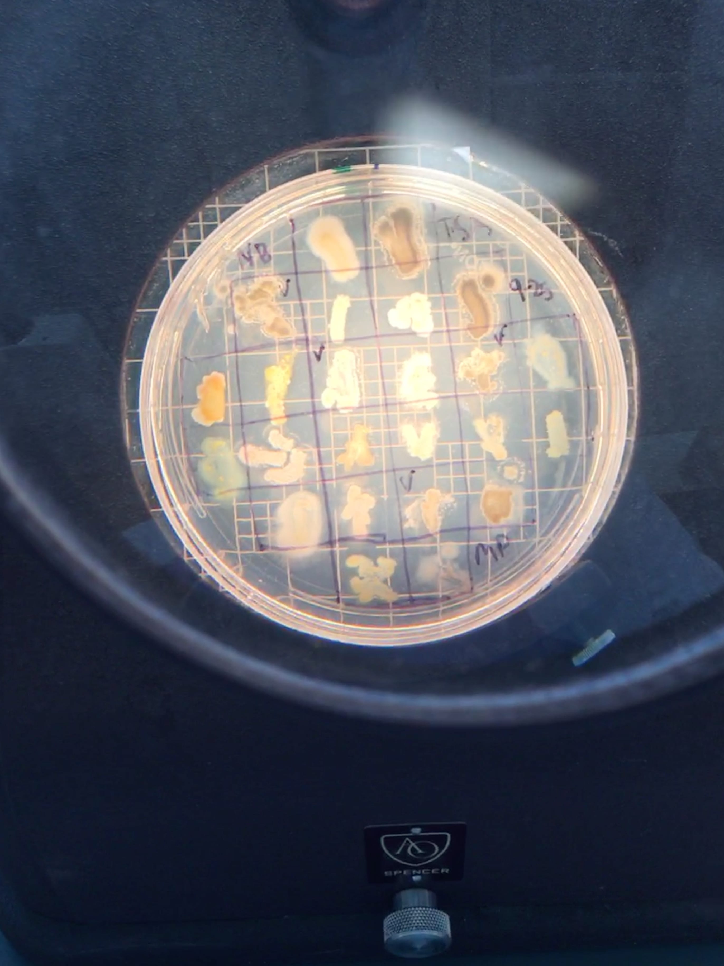
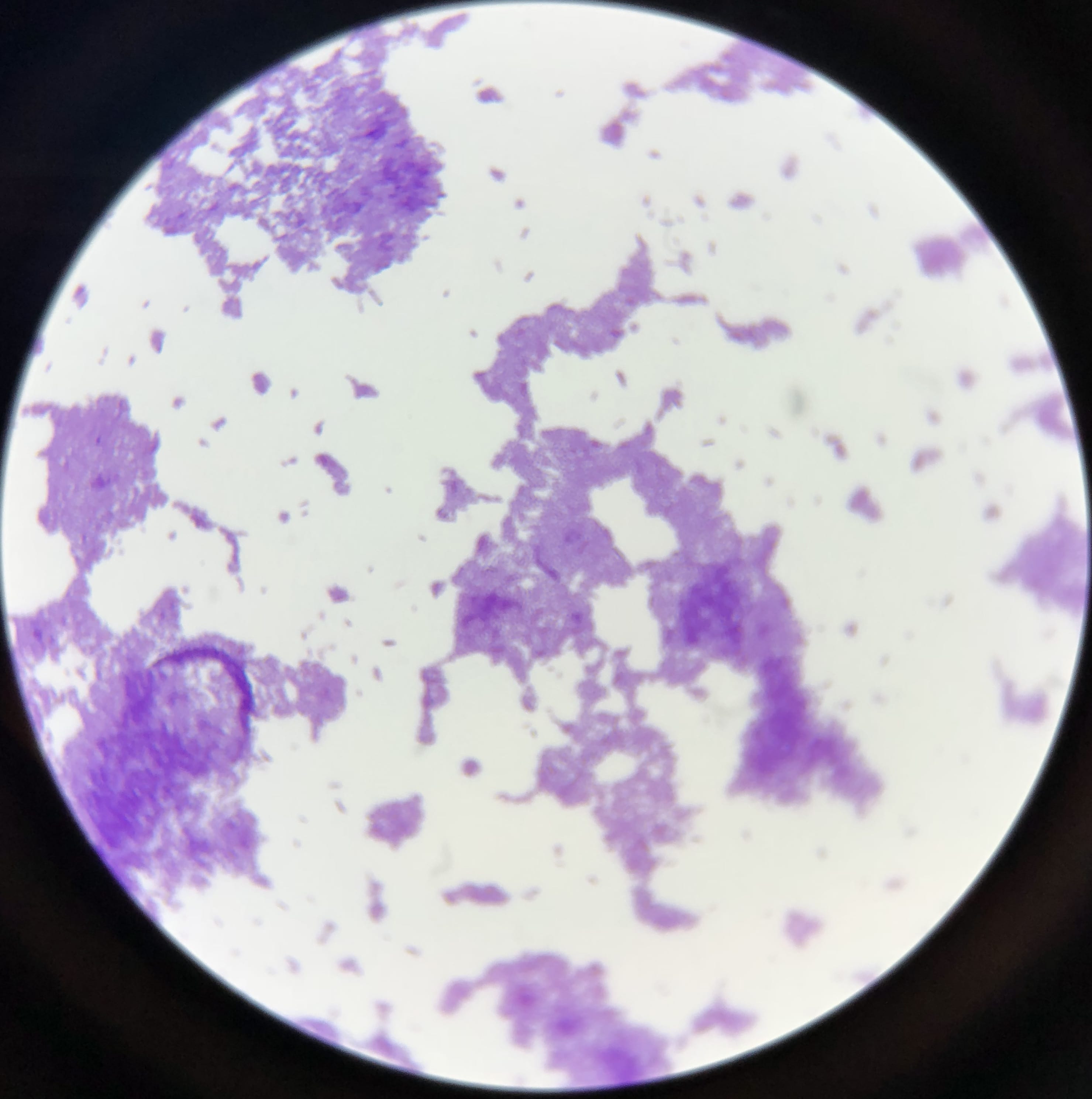
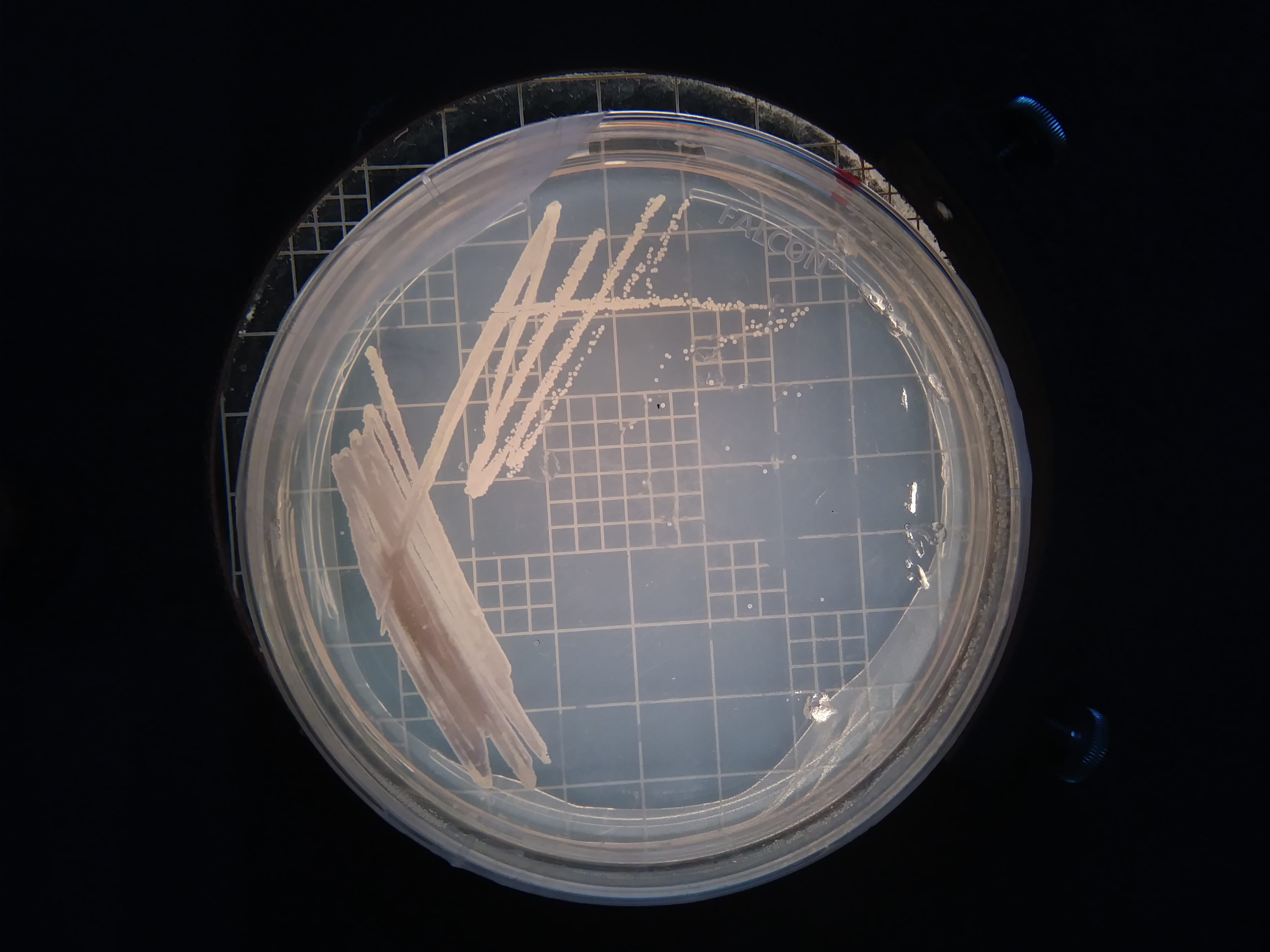
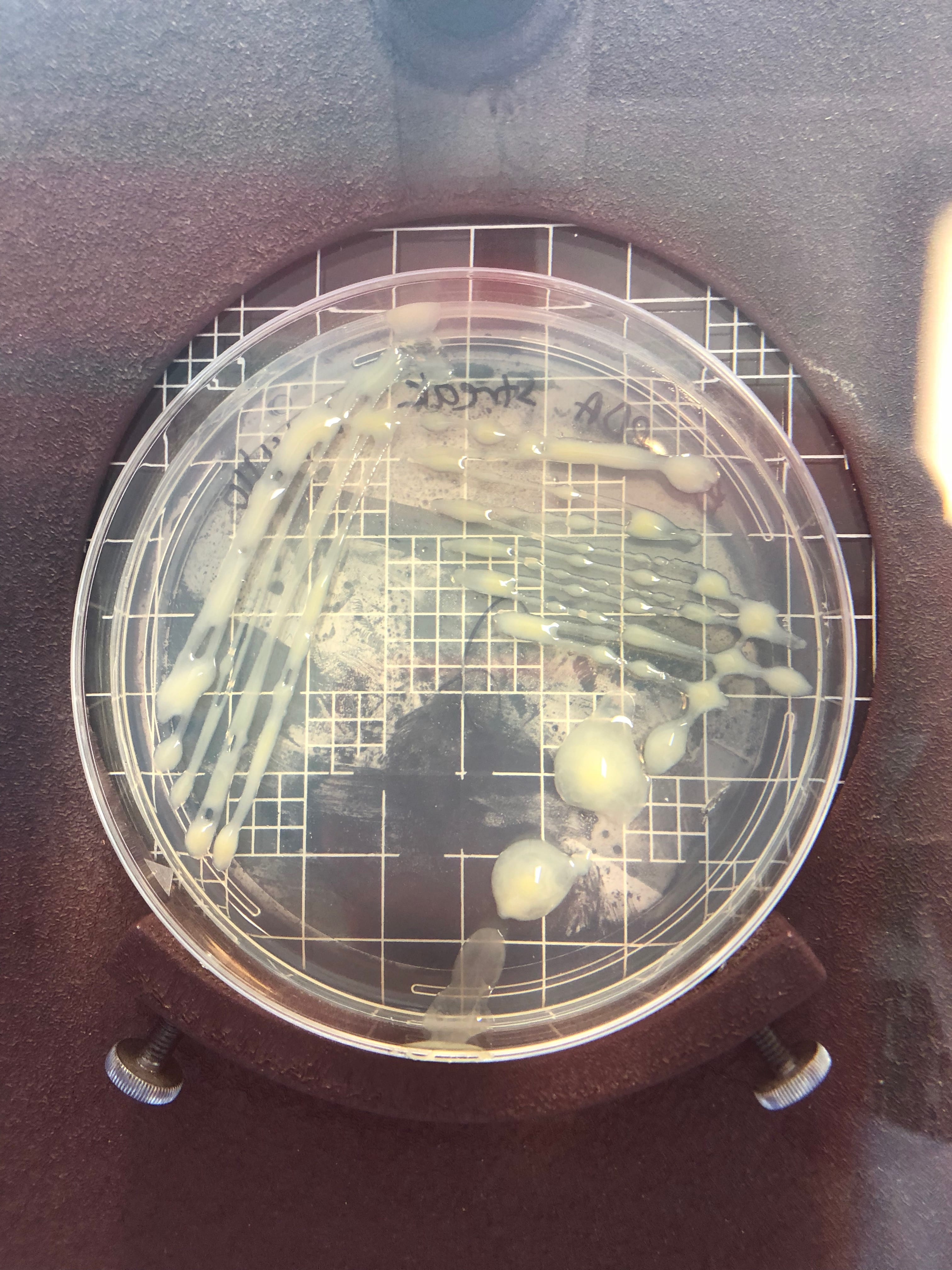
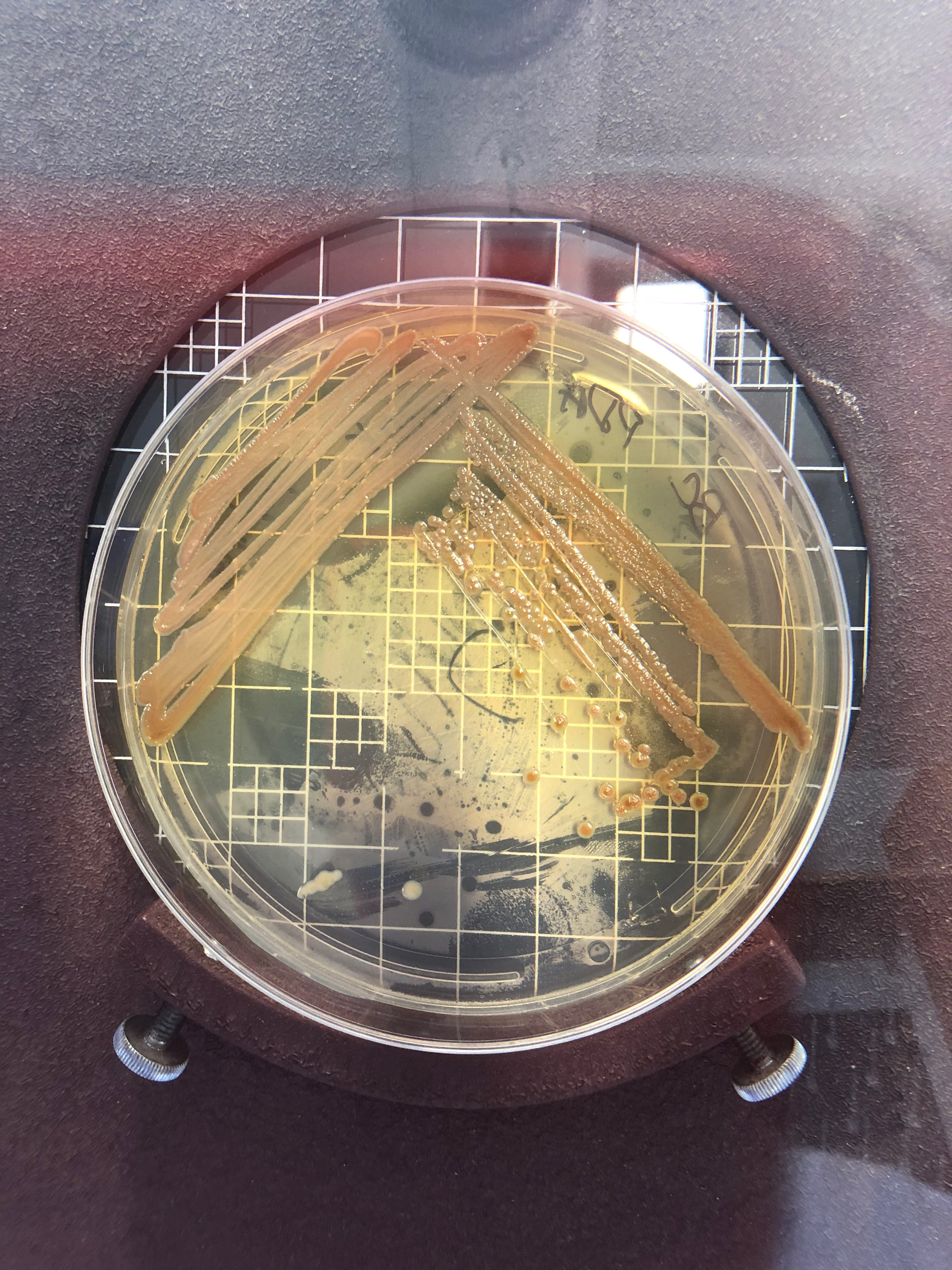
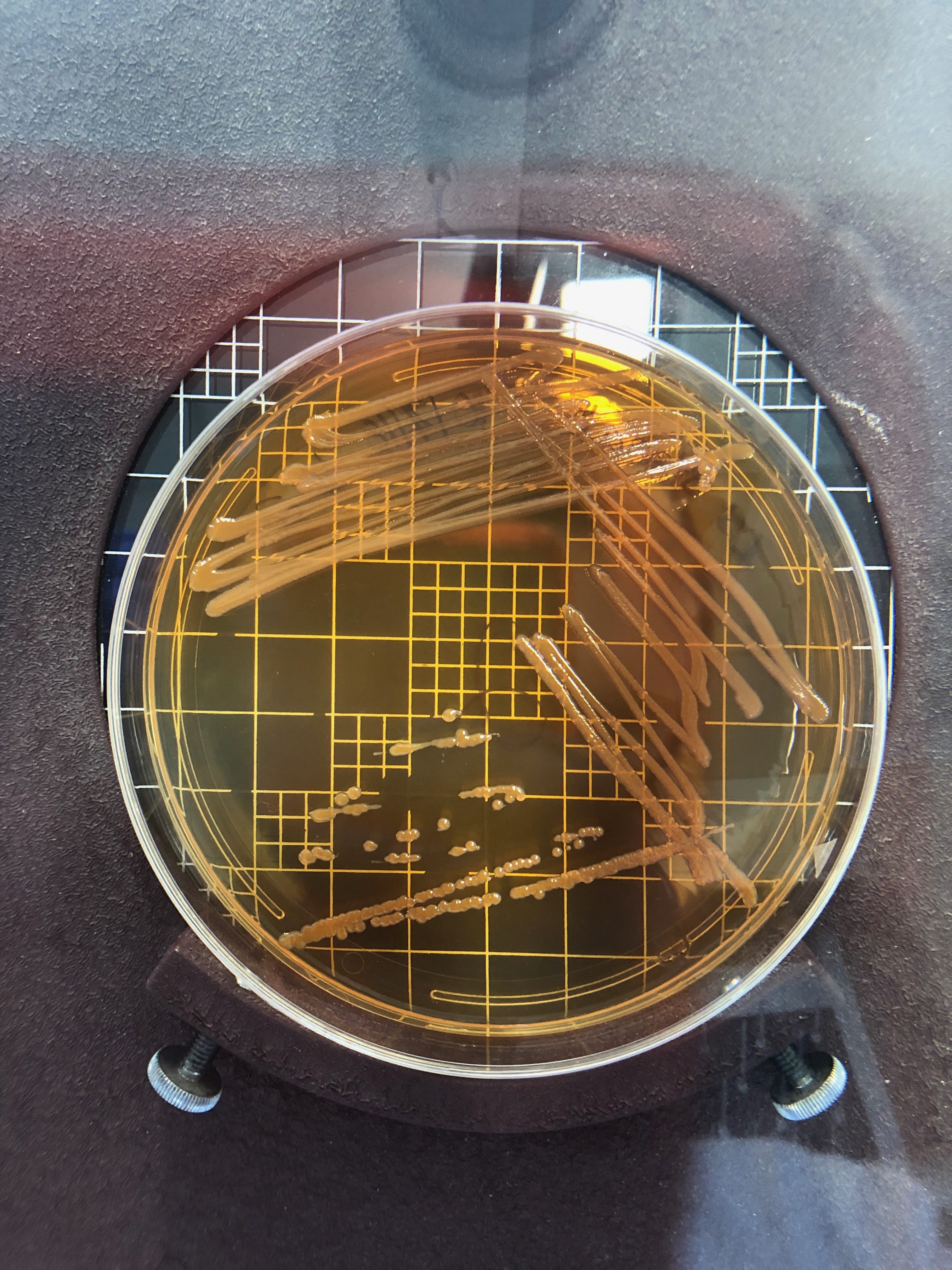
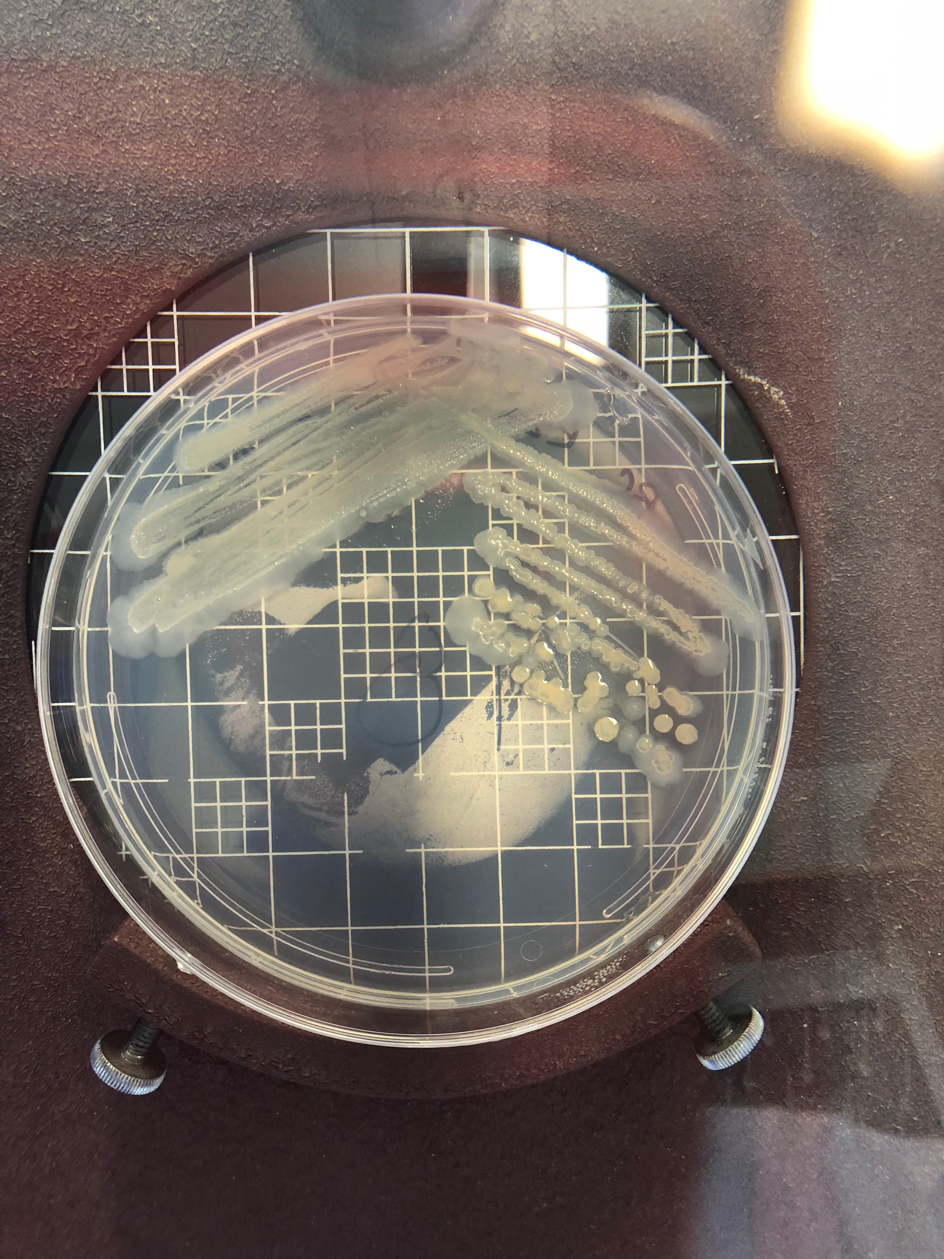
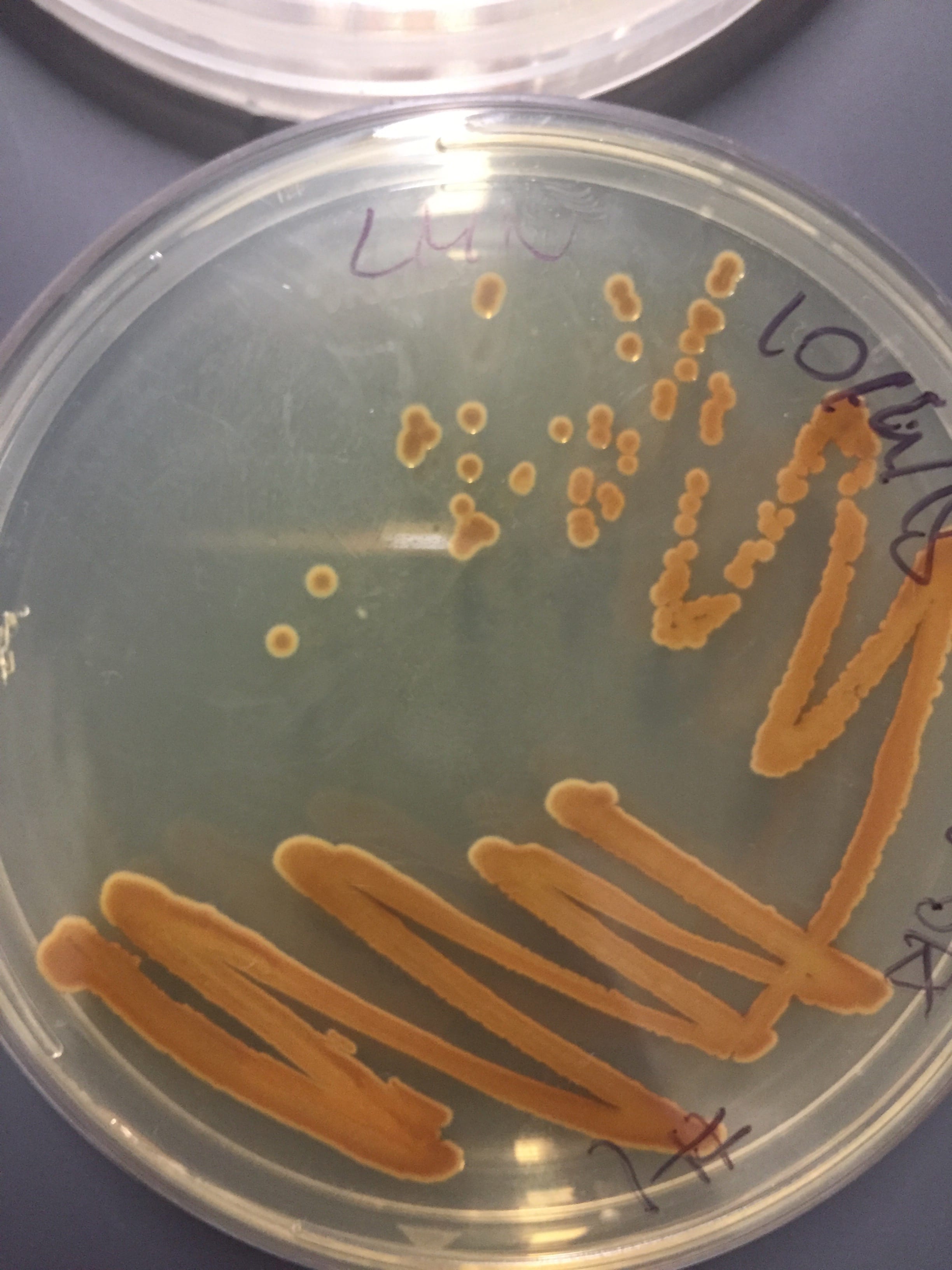
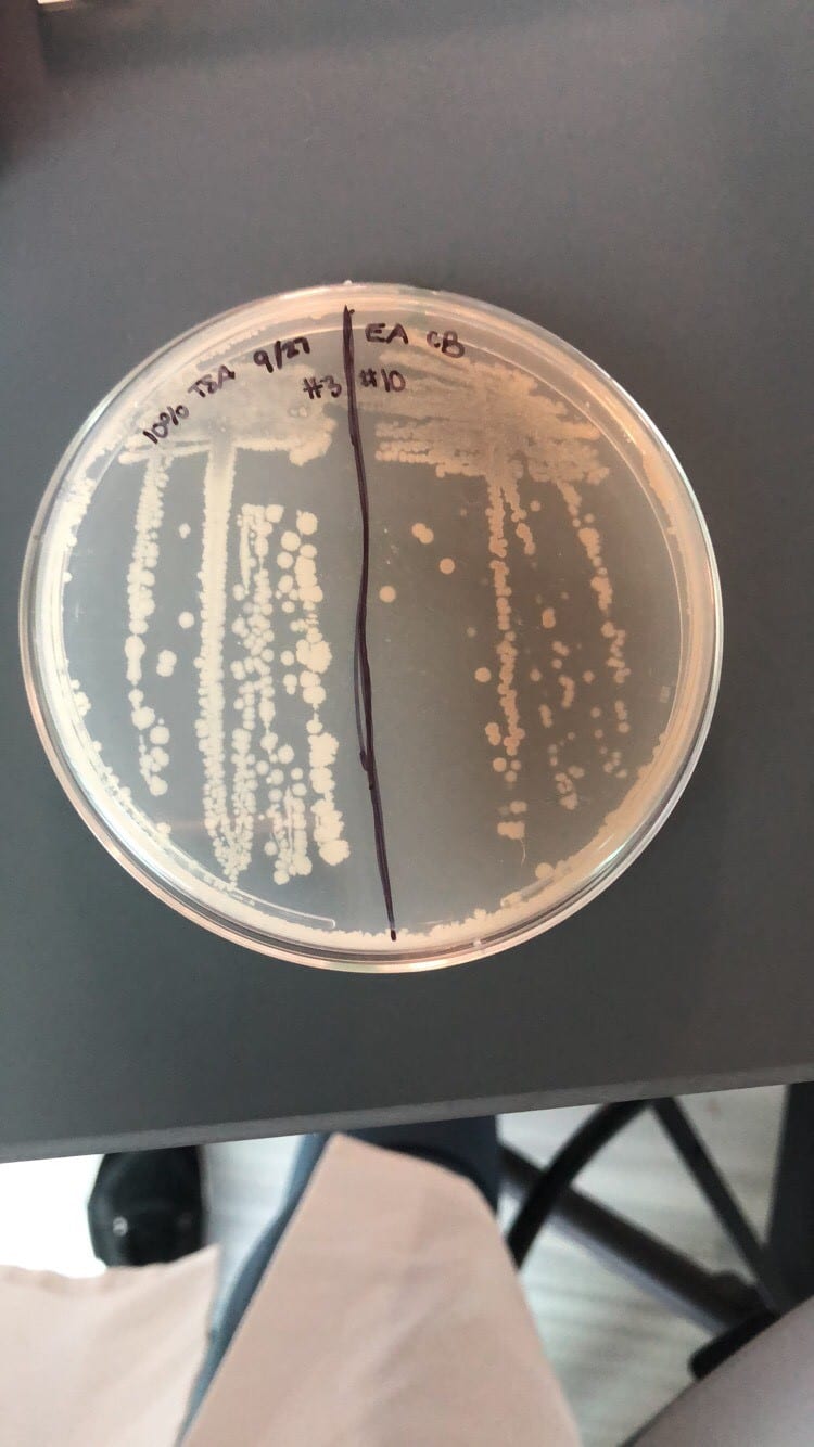
After testing all of these strains against different tester bacteria, the only colonies that actually exhibited any inhibition pattern were strains 1A, 3A, and 2B.  These colonies were isolated after they were tested, and even though I started off a little rough with streak plates I think it’s safe to say I got the hang of them by the end!

Strain 1A isolated streak plate

Strain 3A isolated streak plate

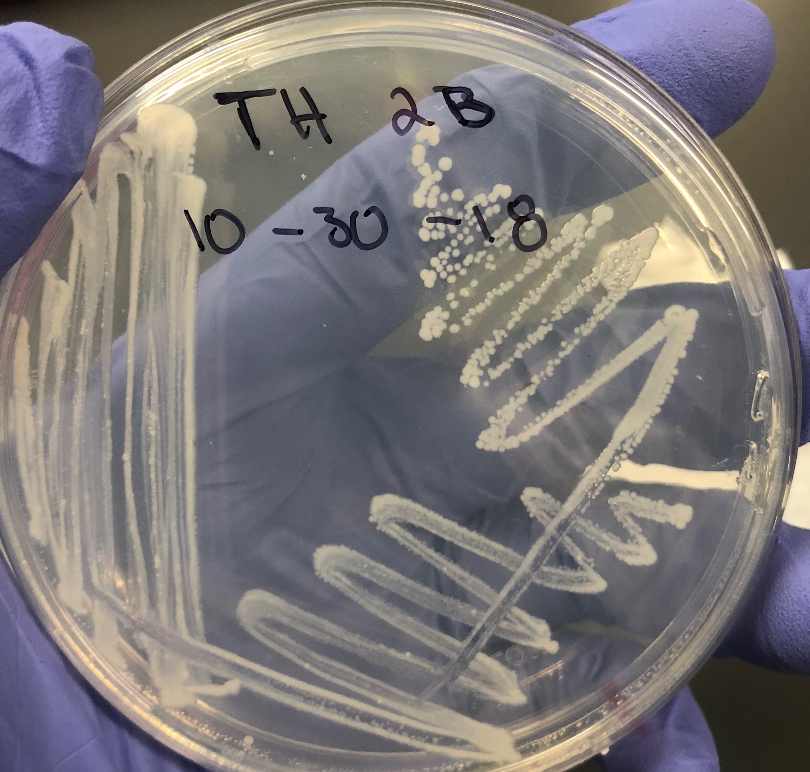
Strain 2B isolated streak plate

Just look at those beautiful streaks! Gotta bring these bacteria over to the Nott… (insert laughs here)

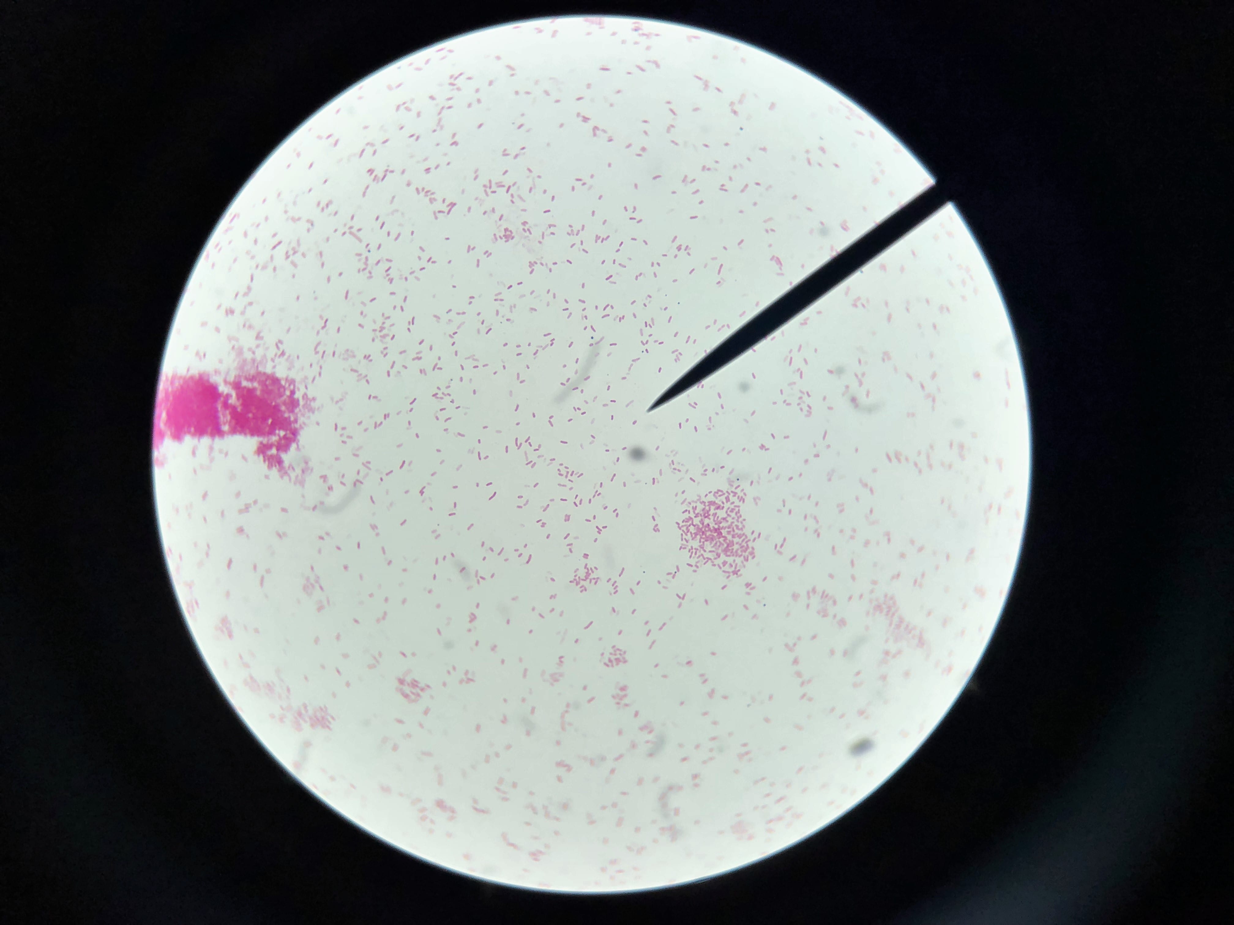
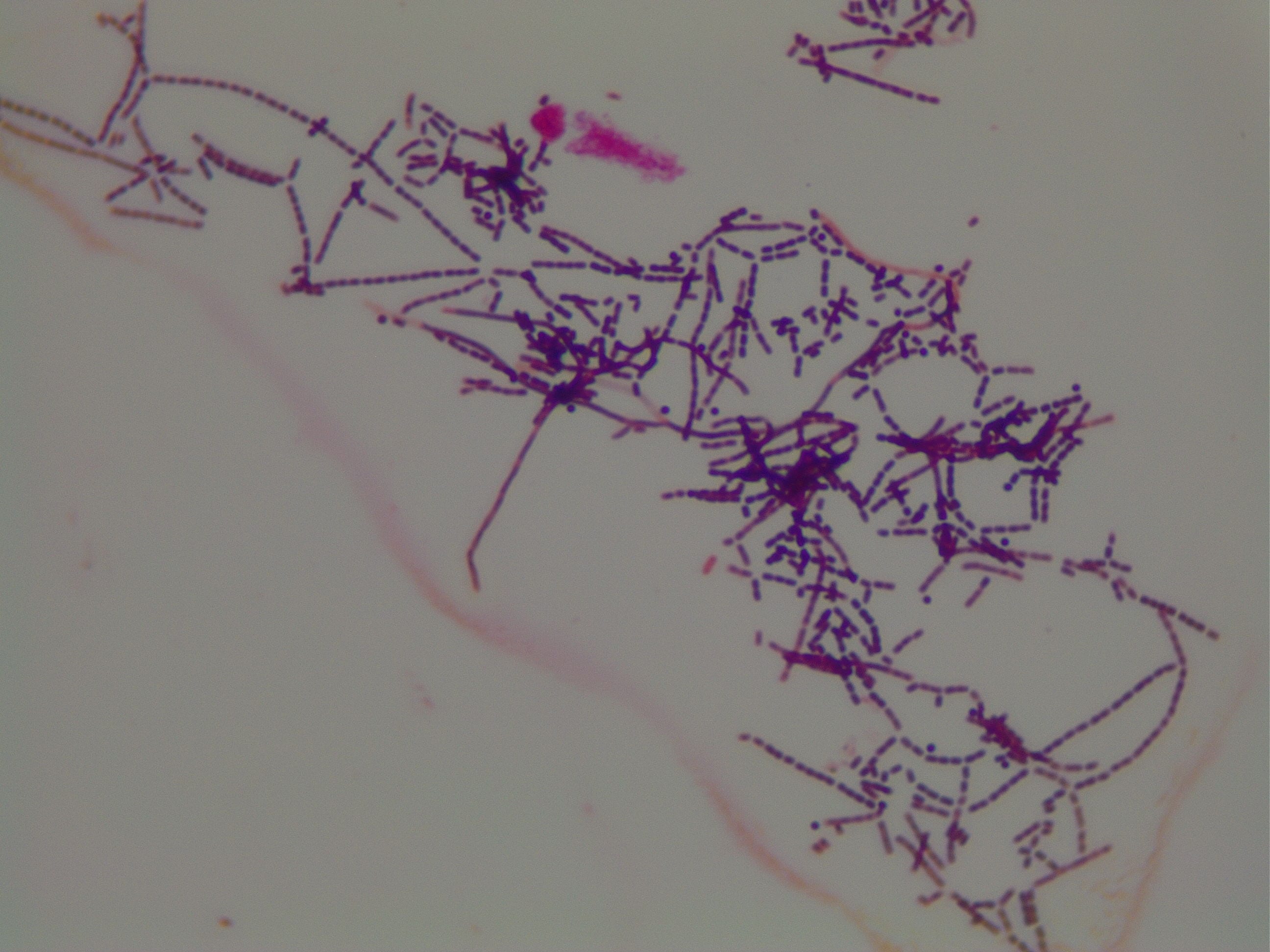
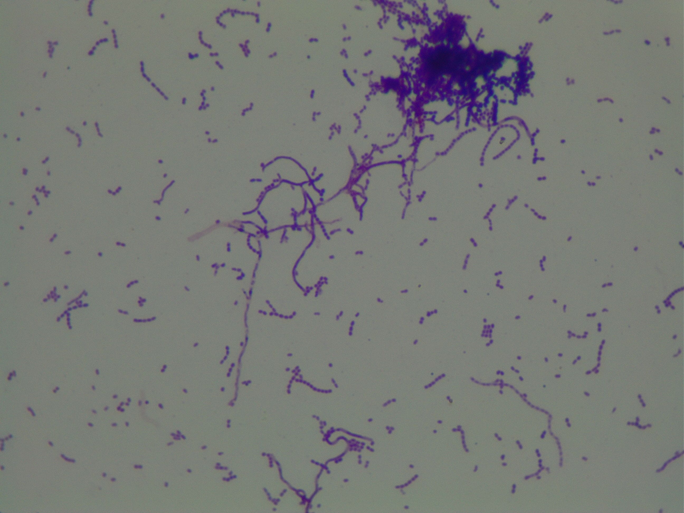
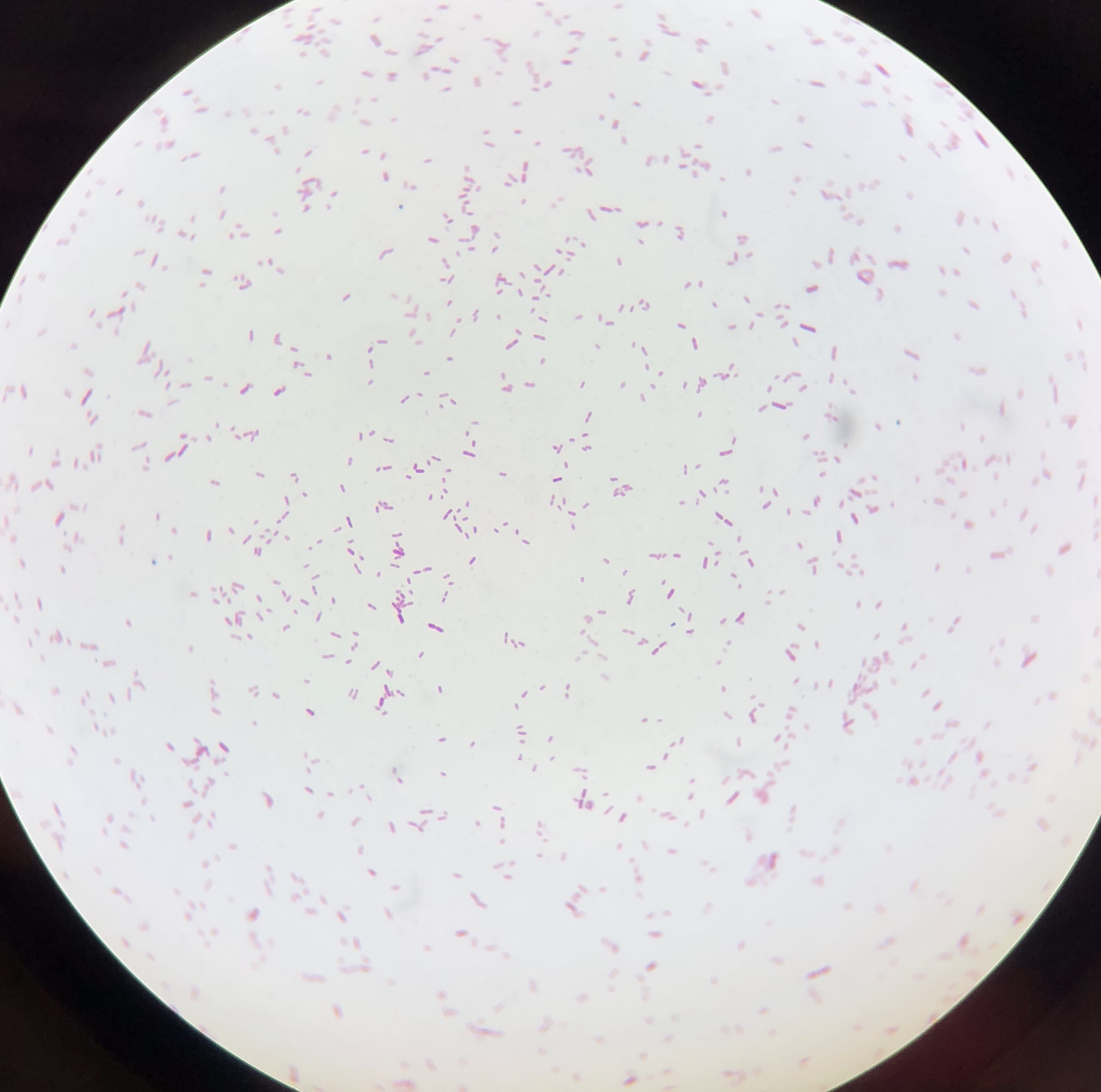
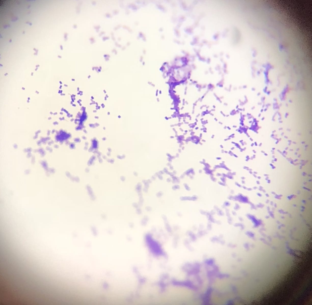
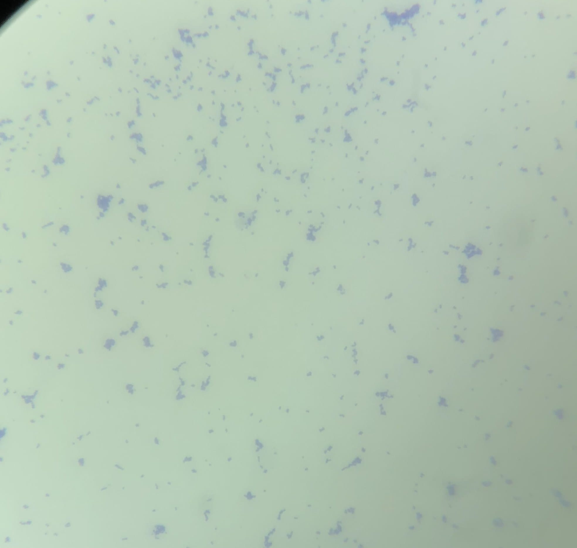
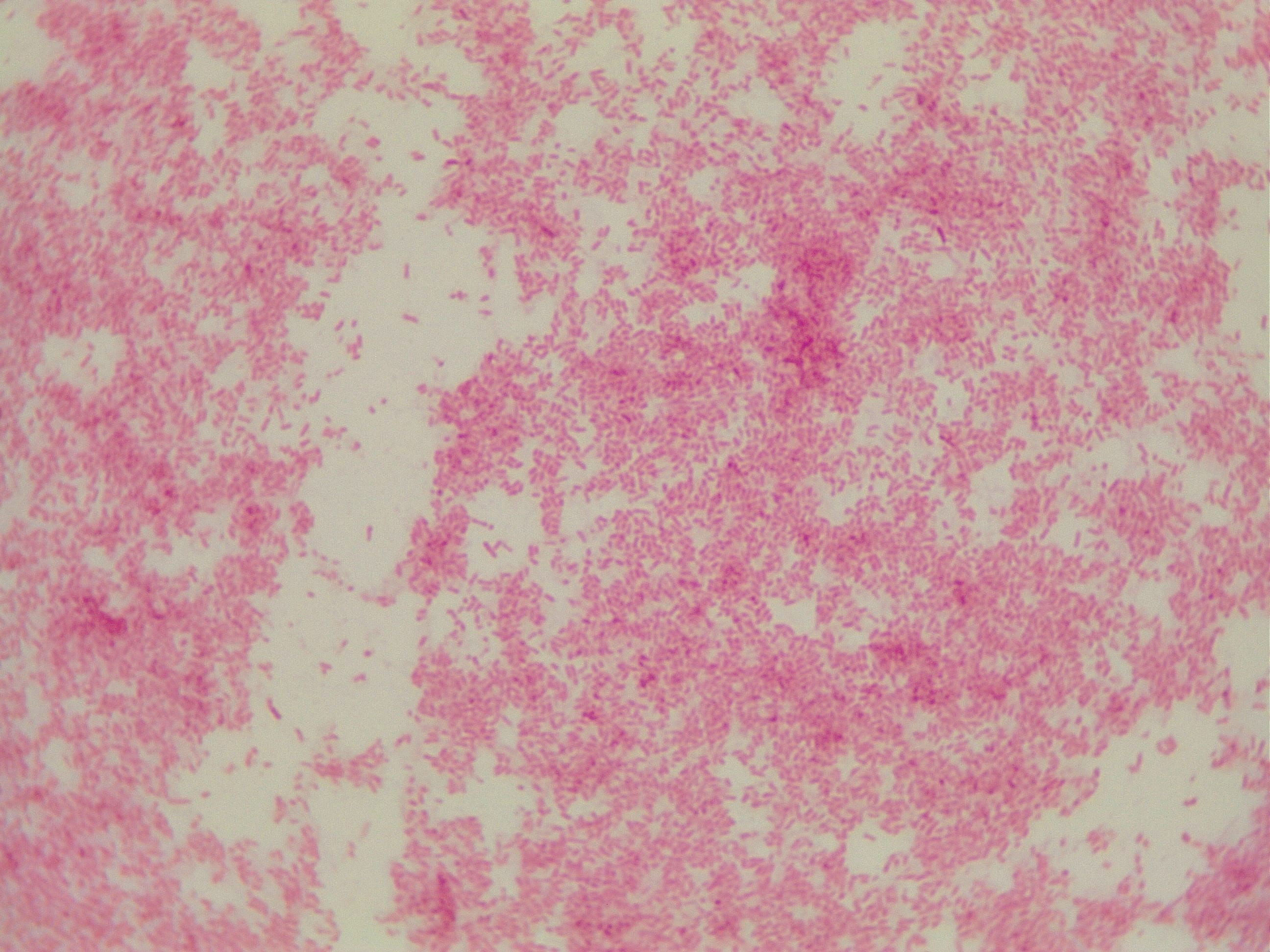
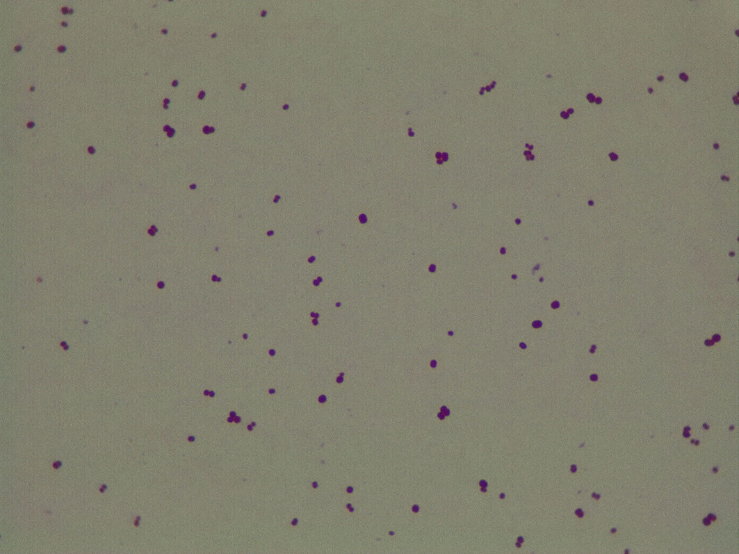
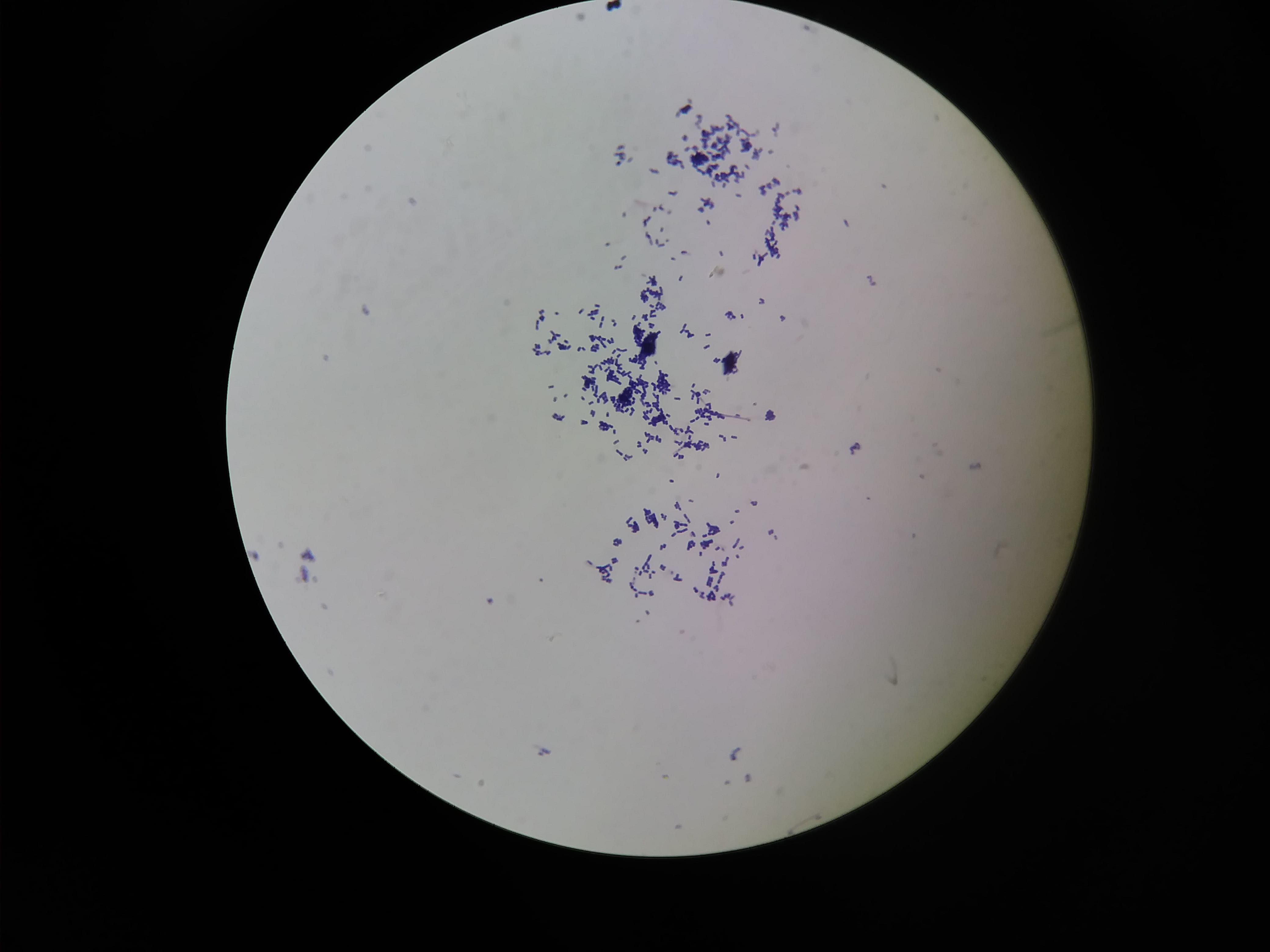
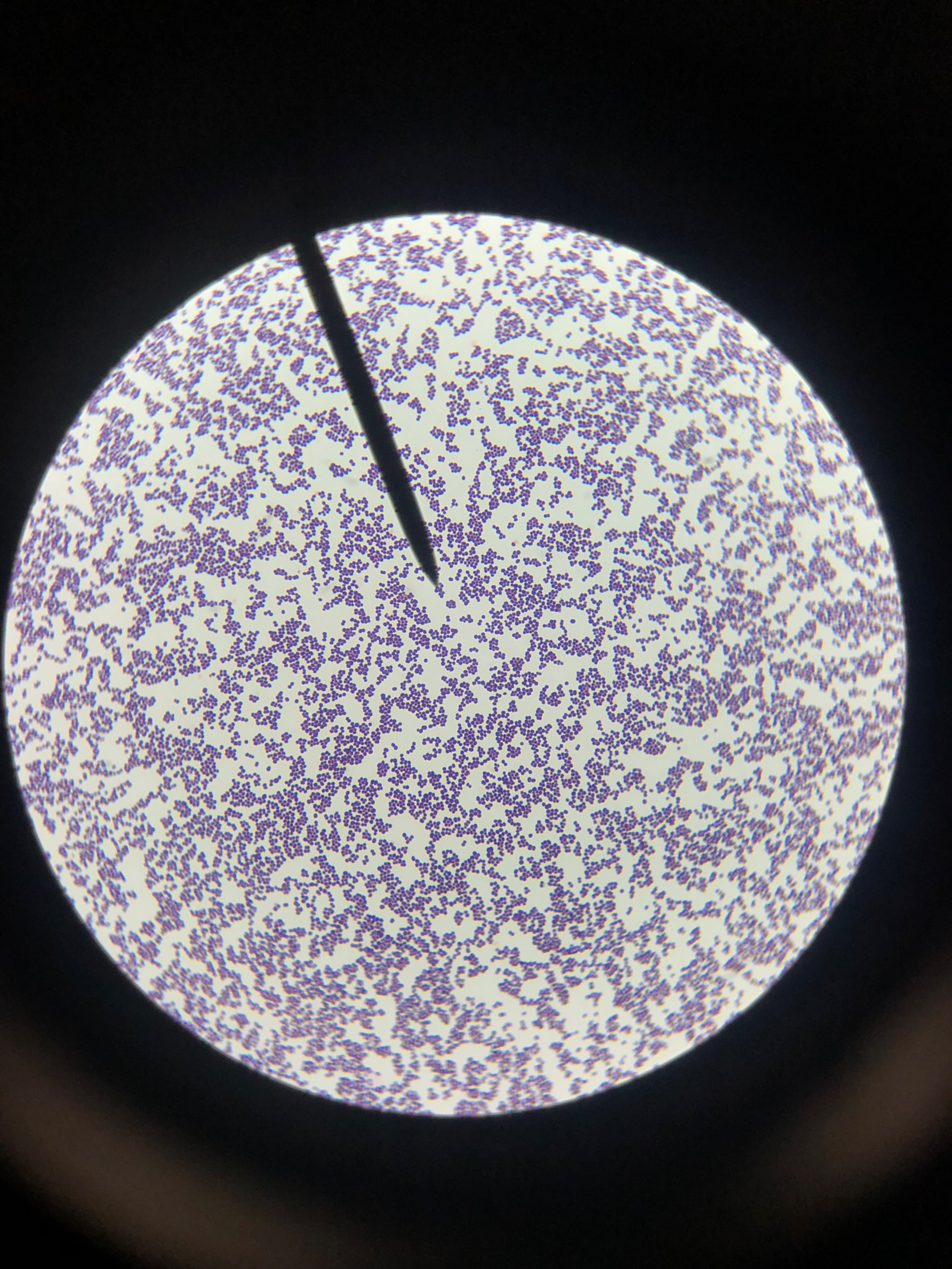
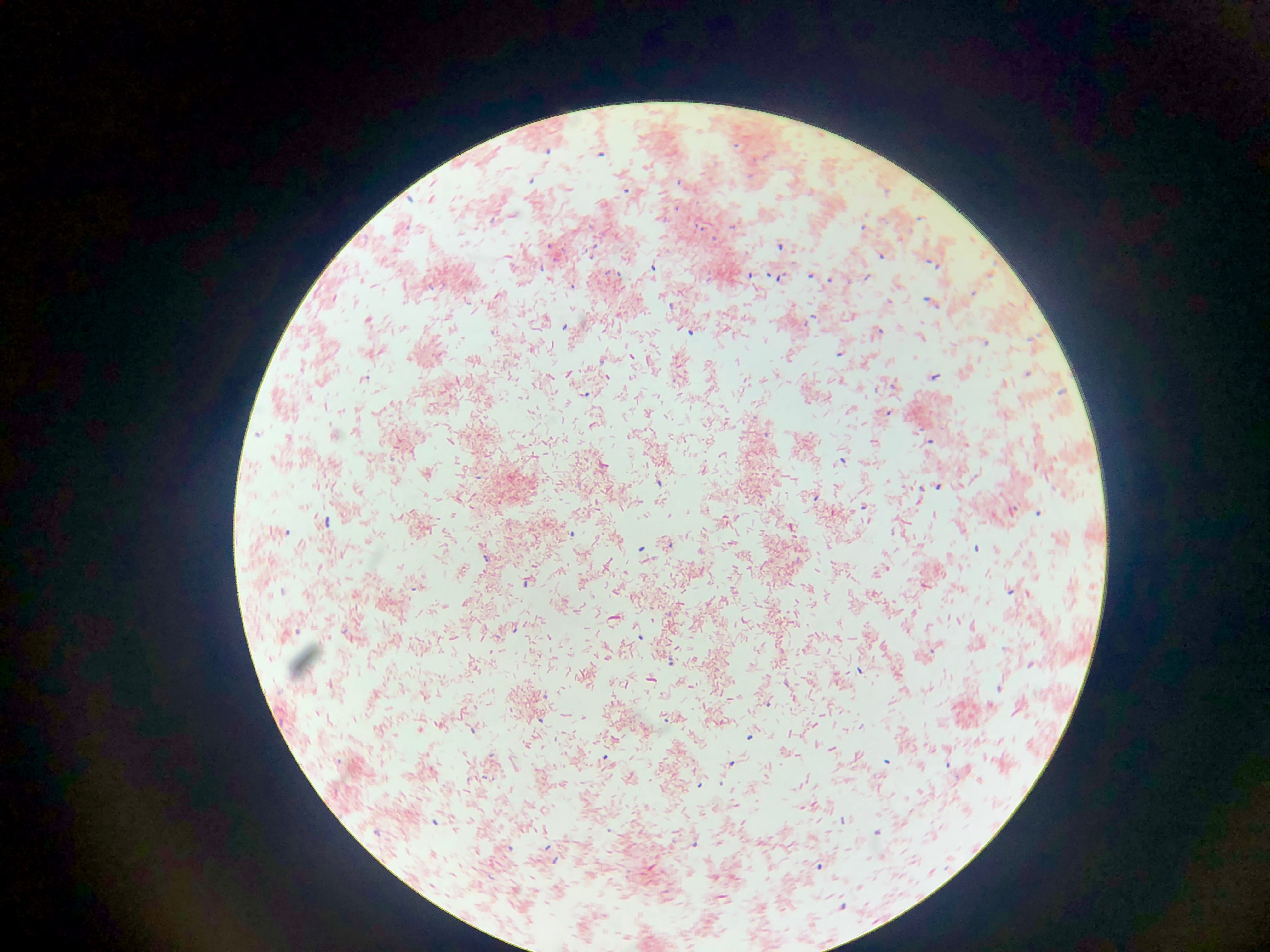
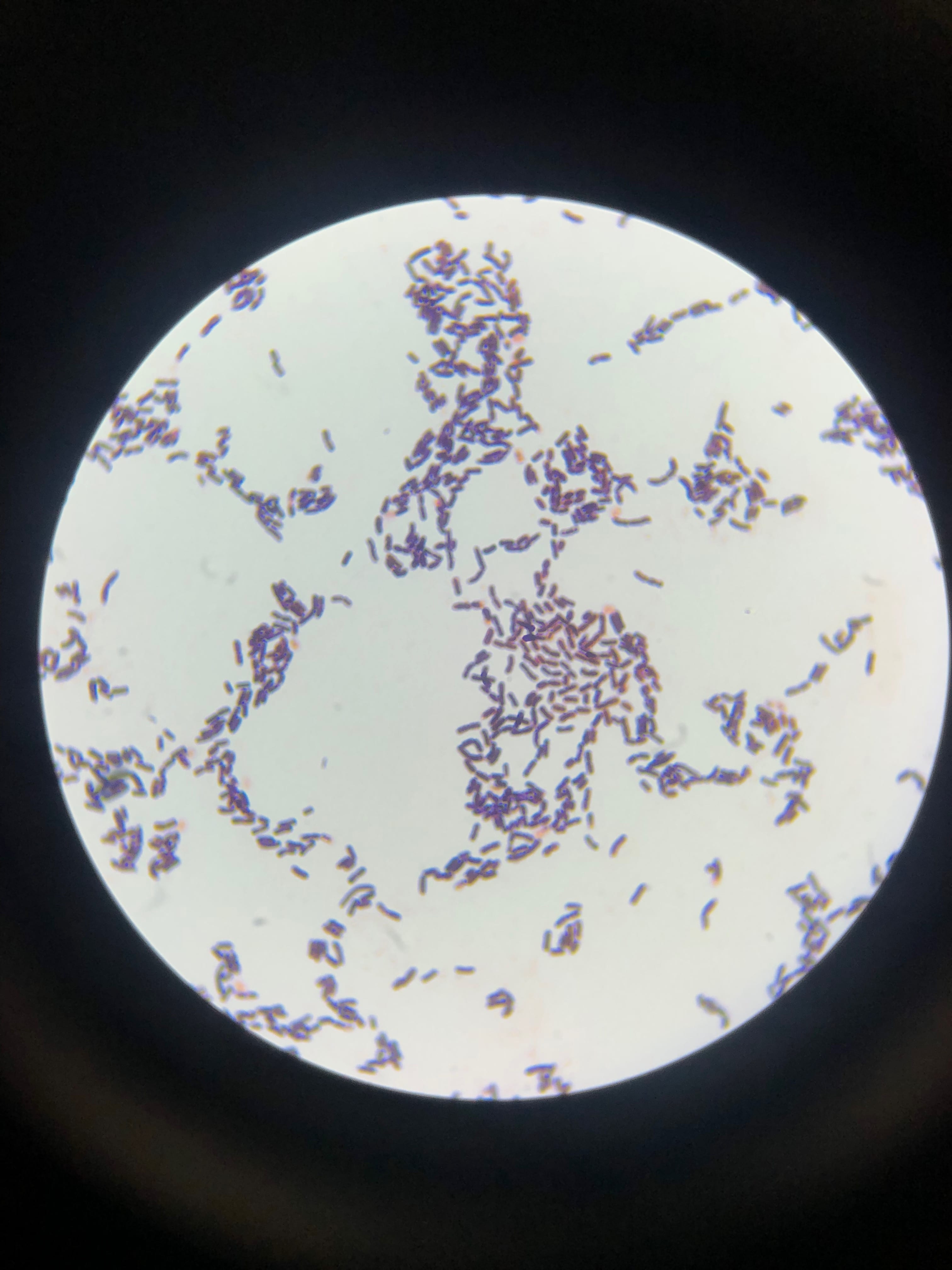
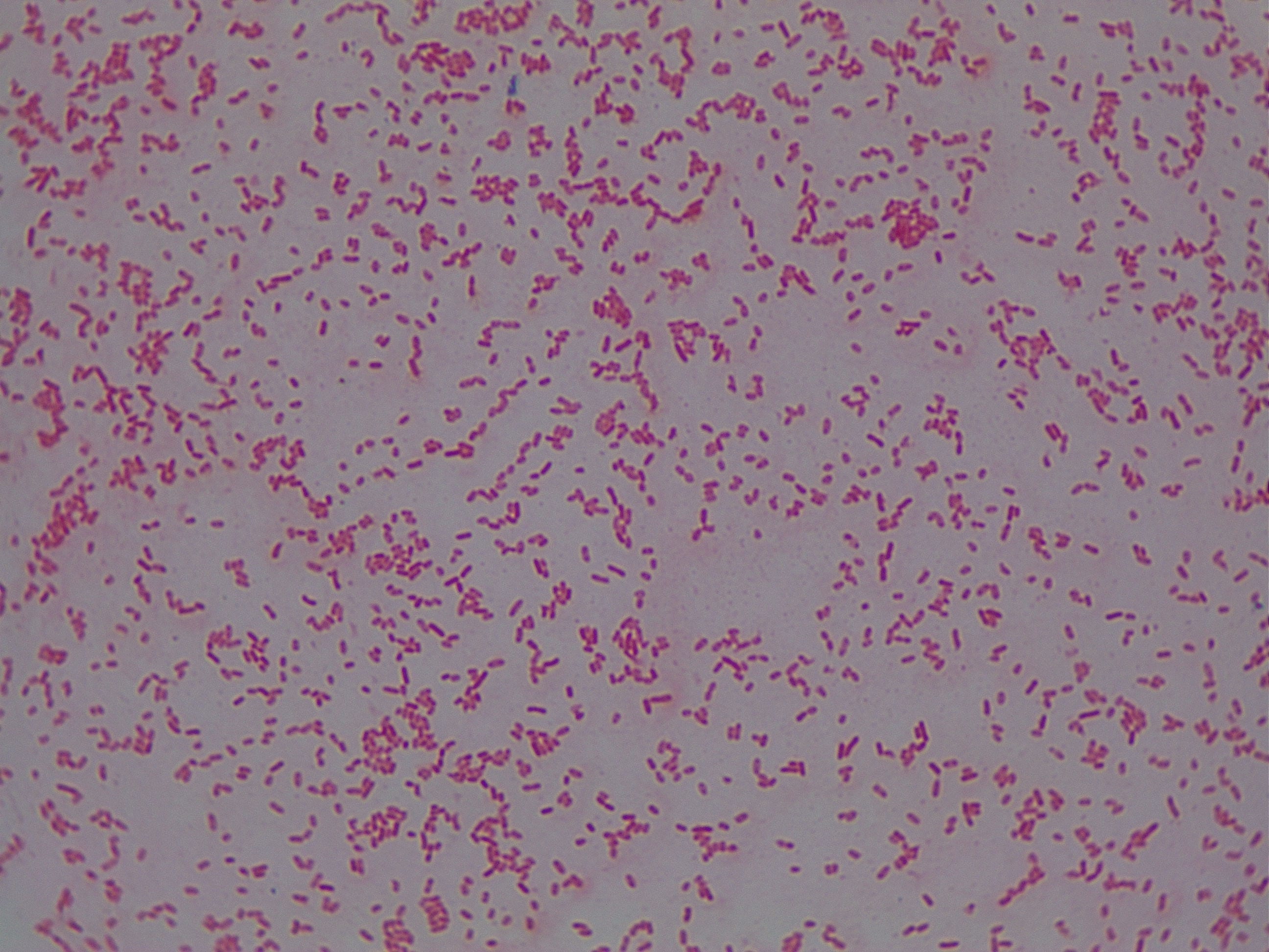
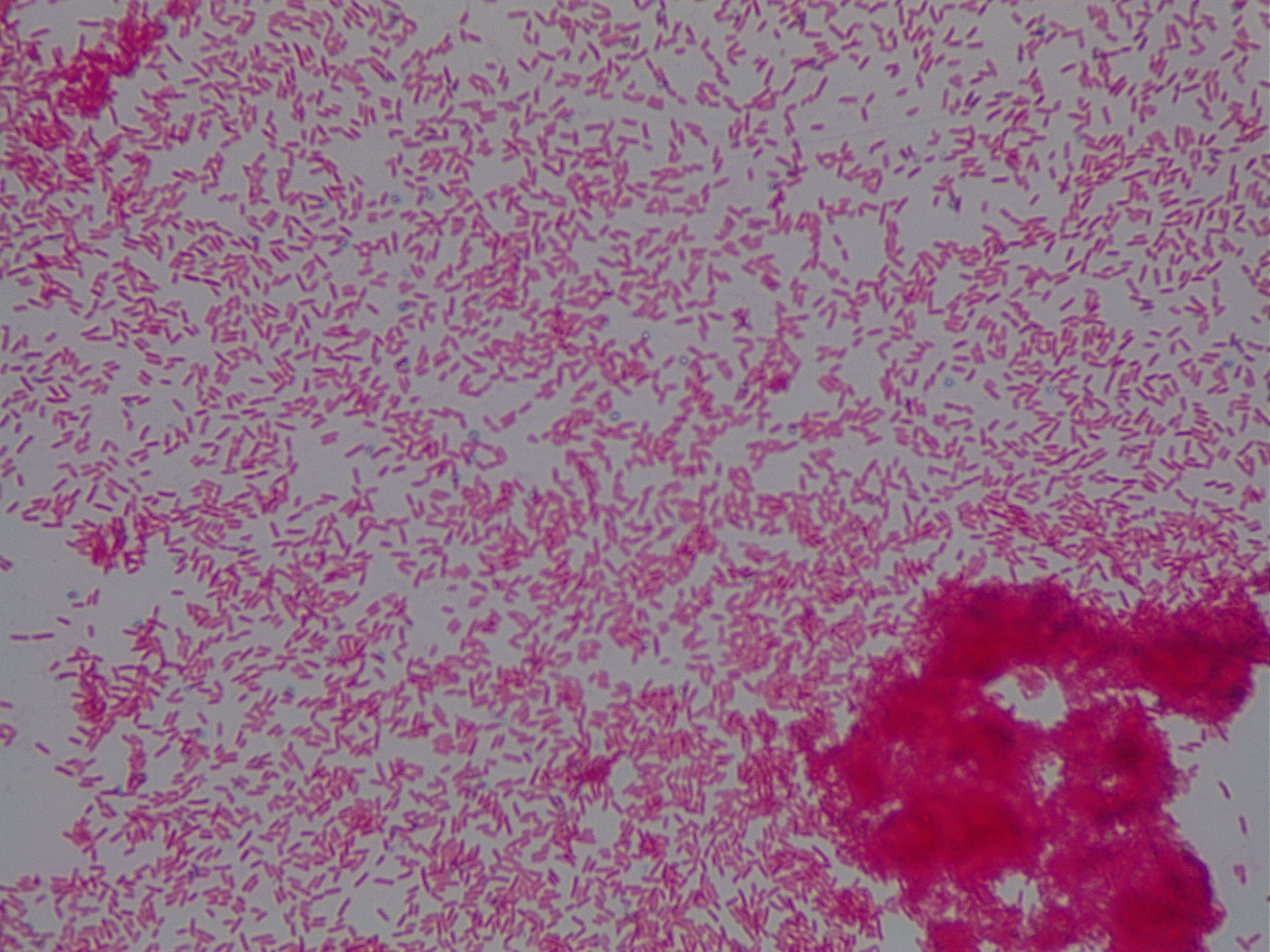
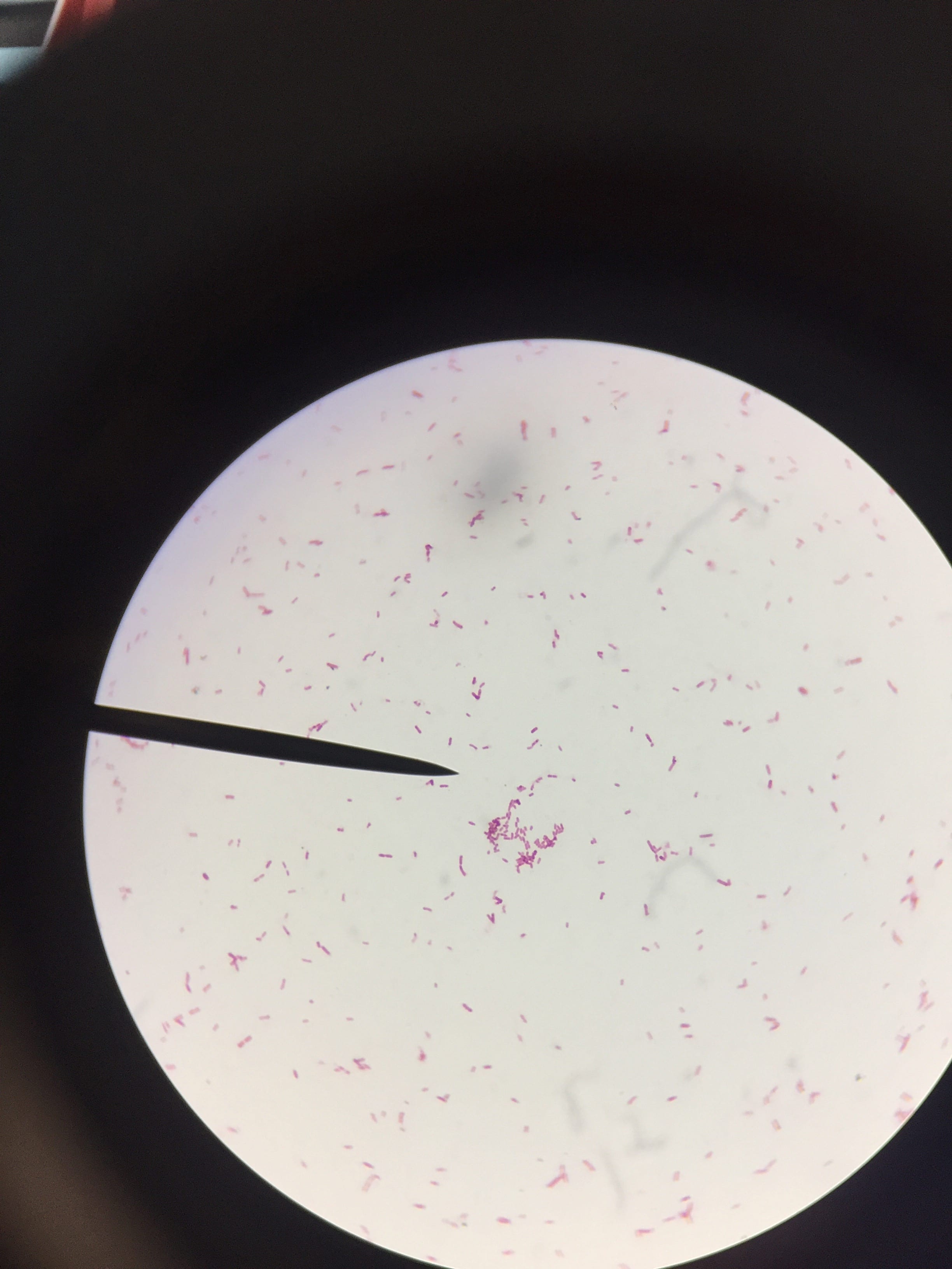
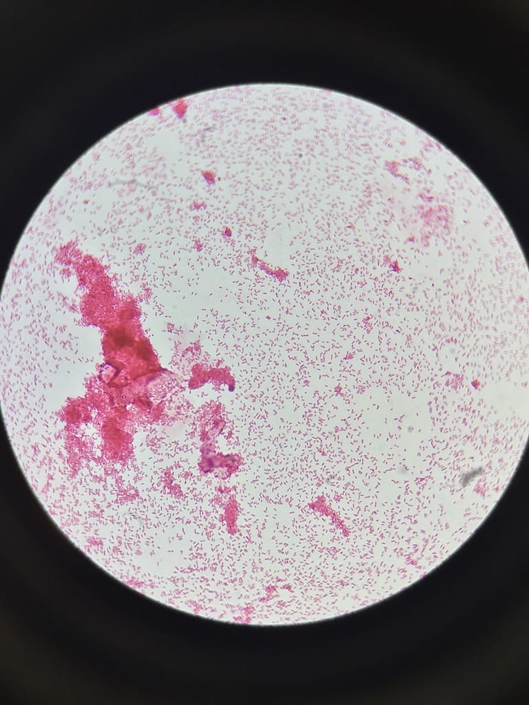
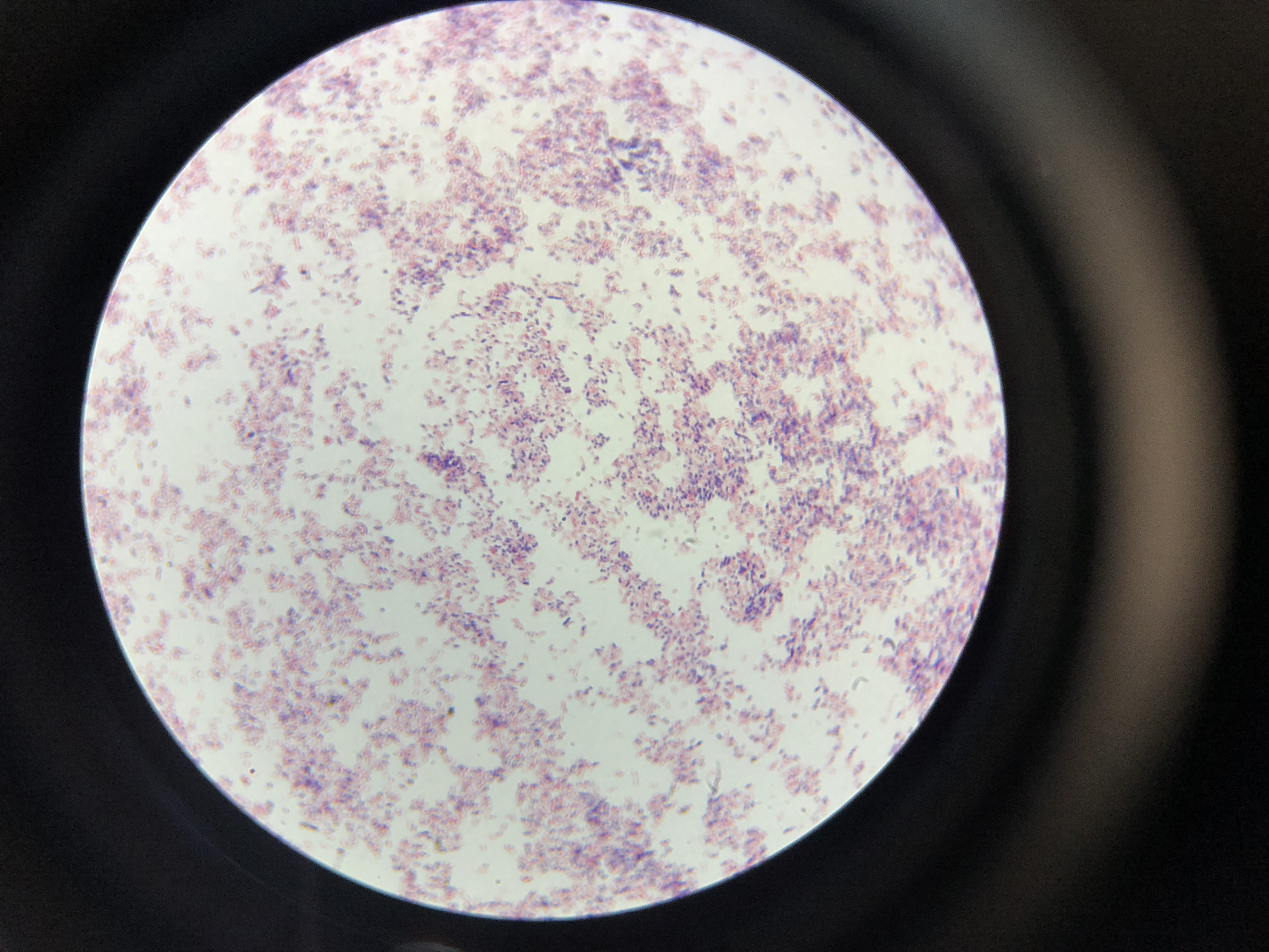
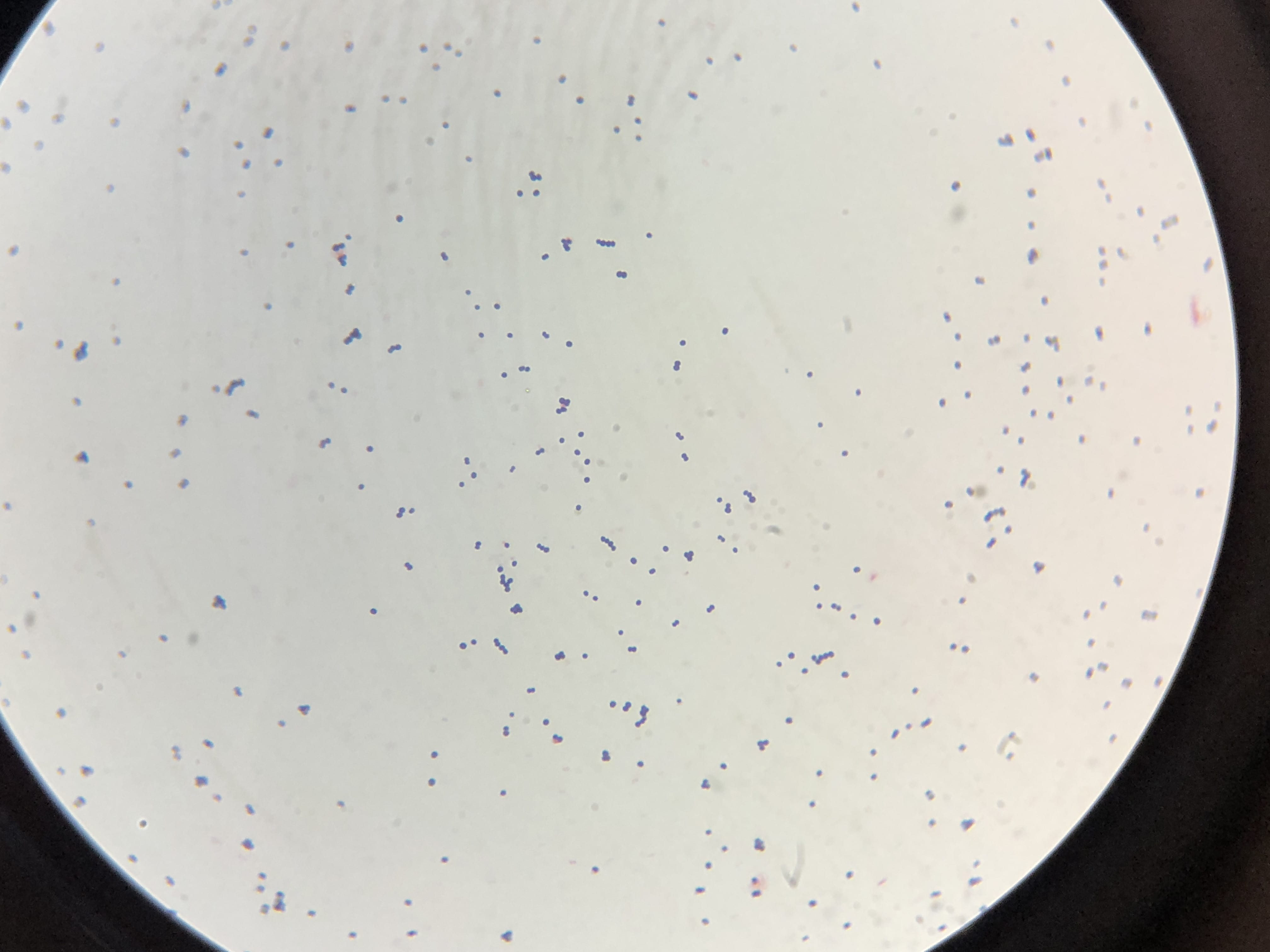
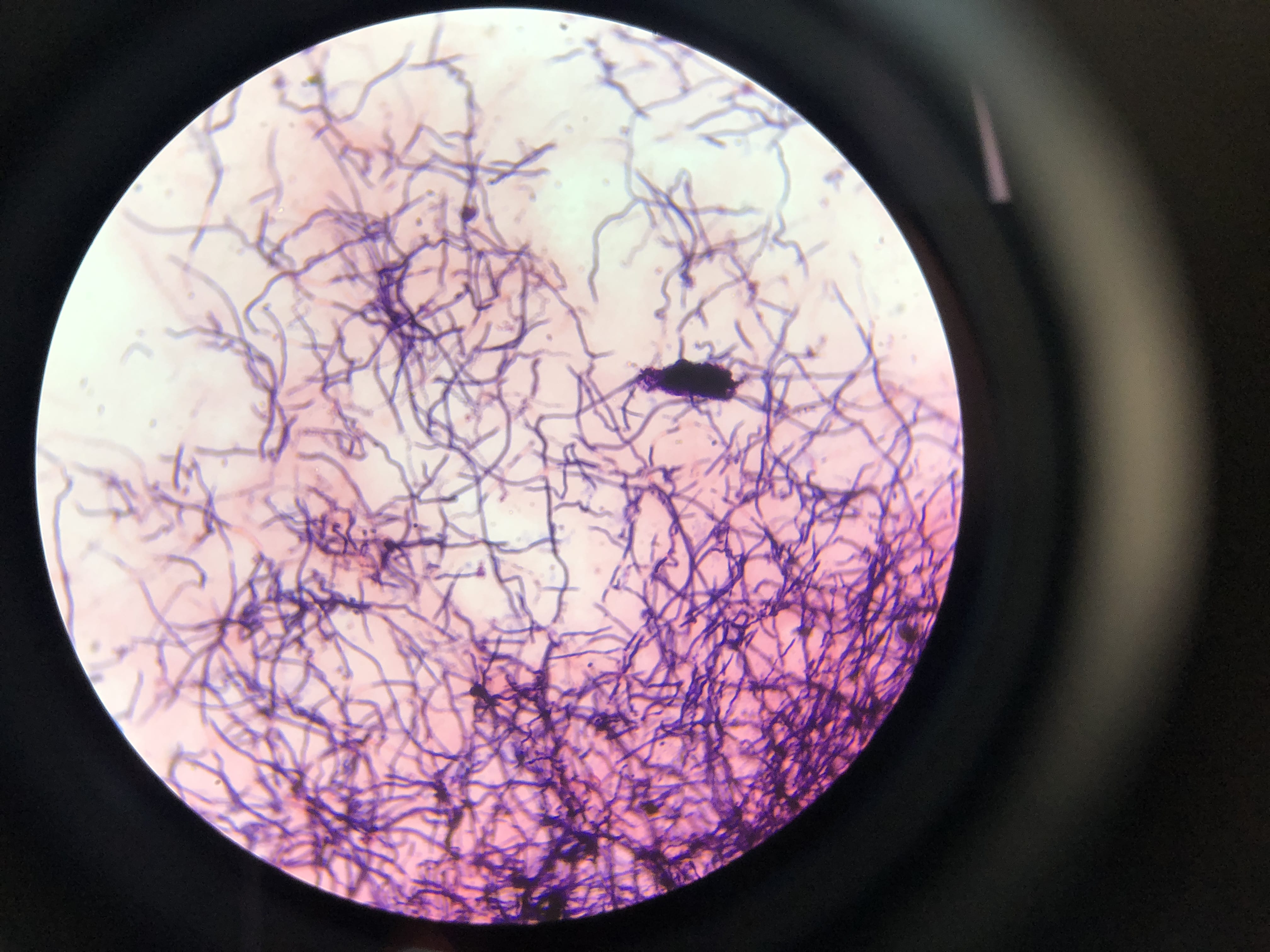
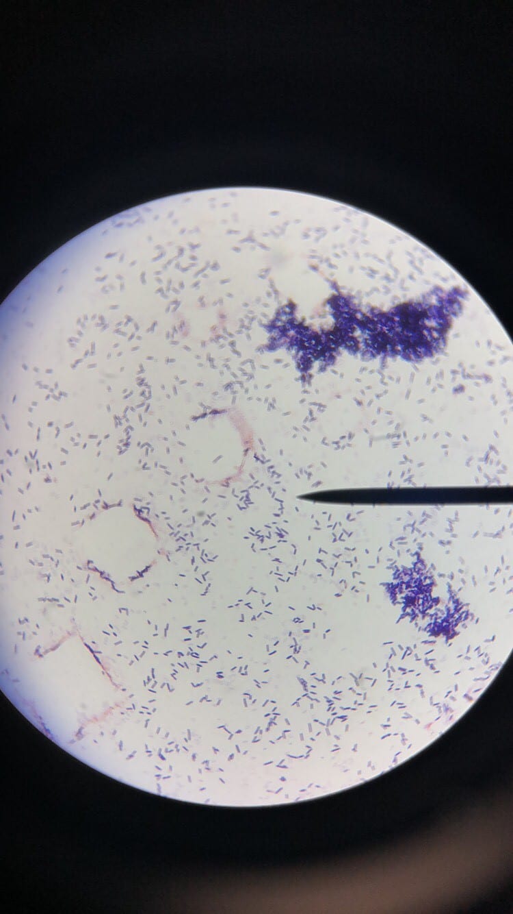
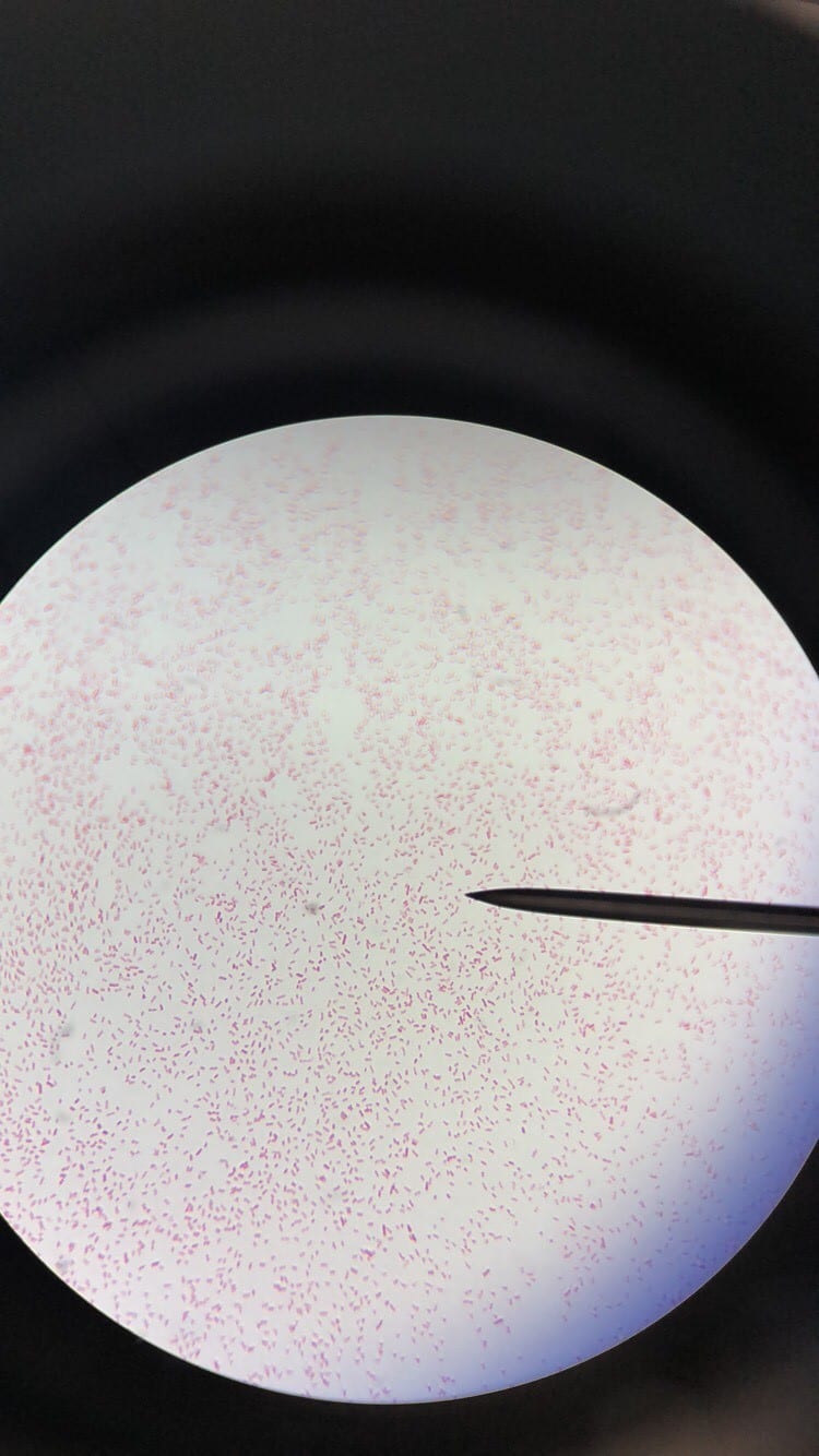
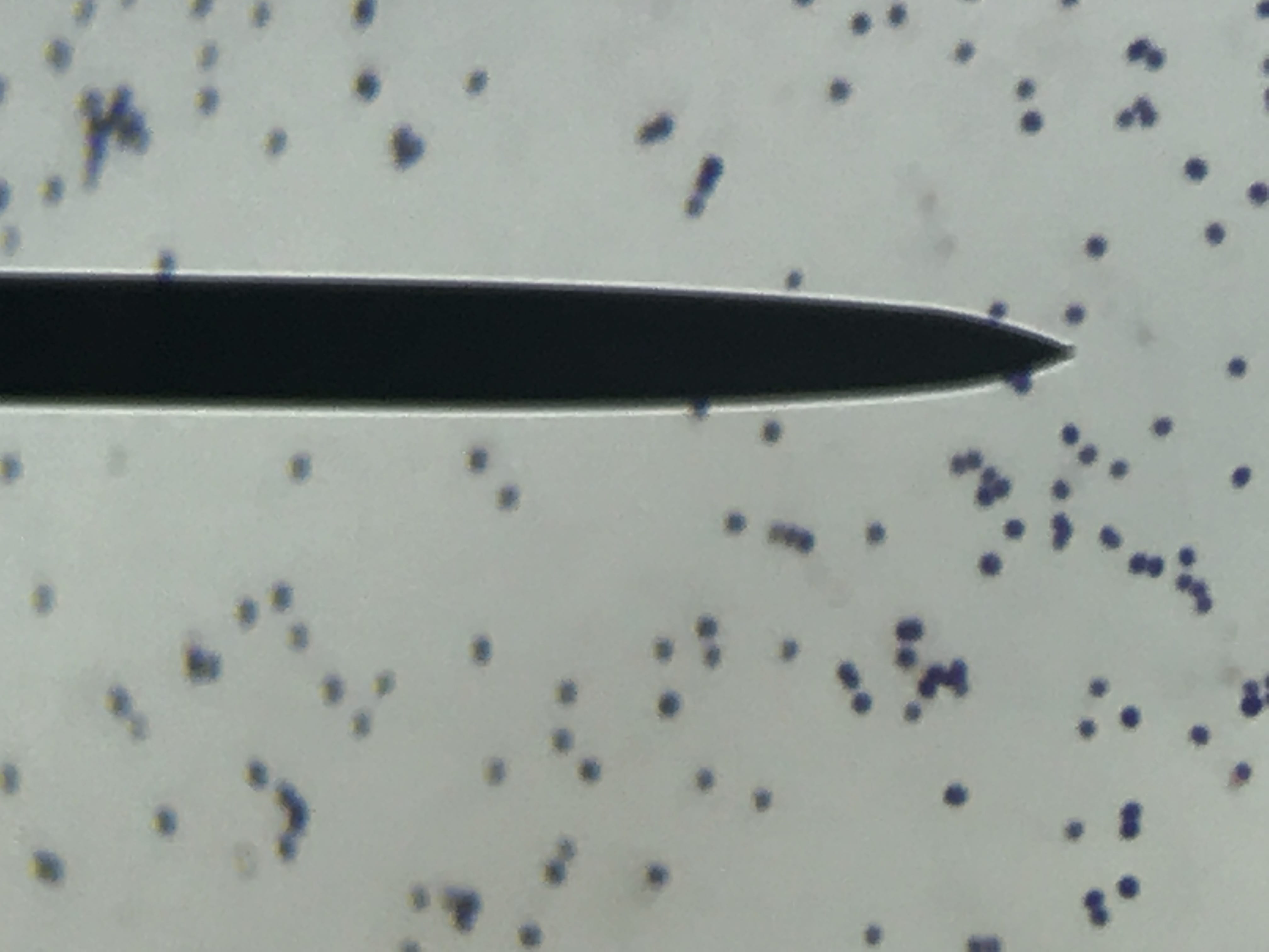
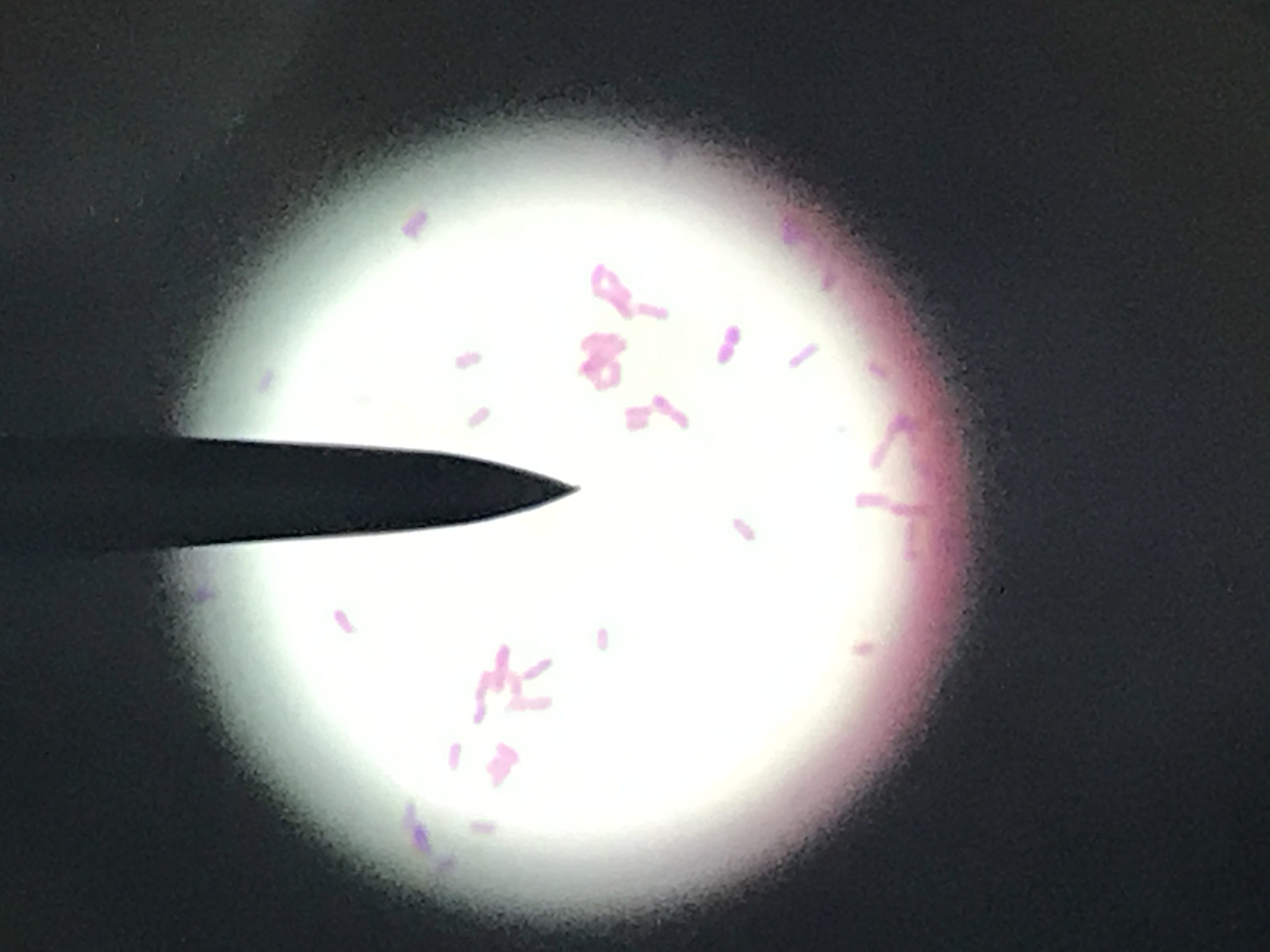
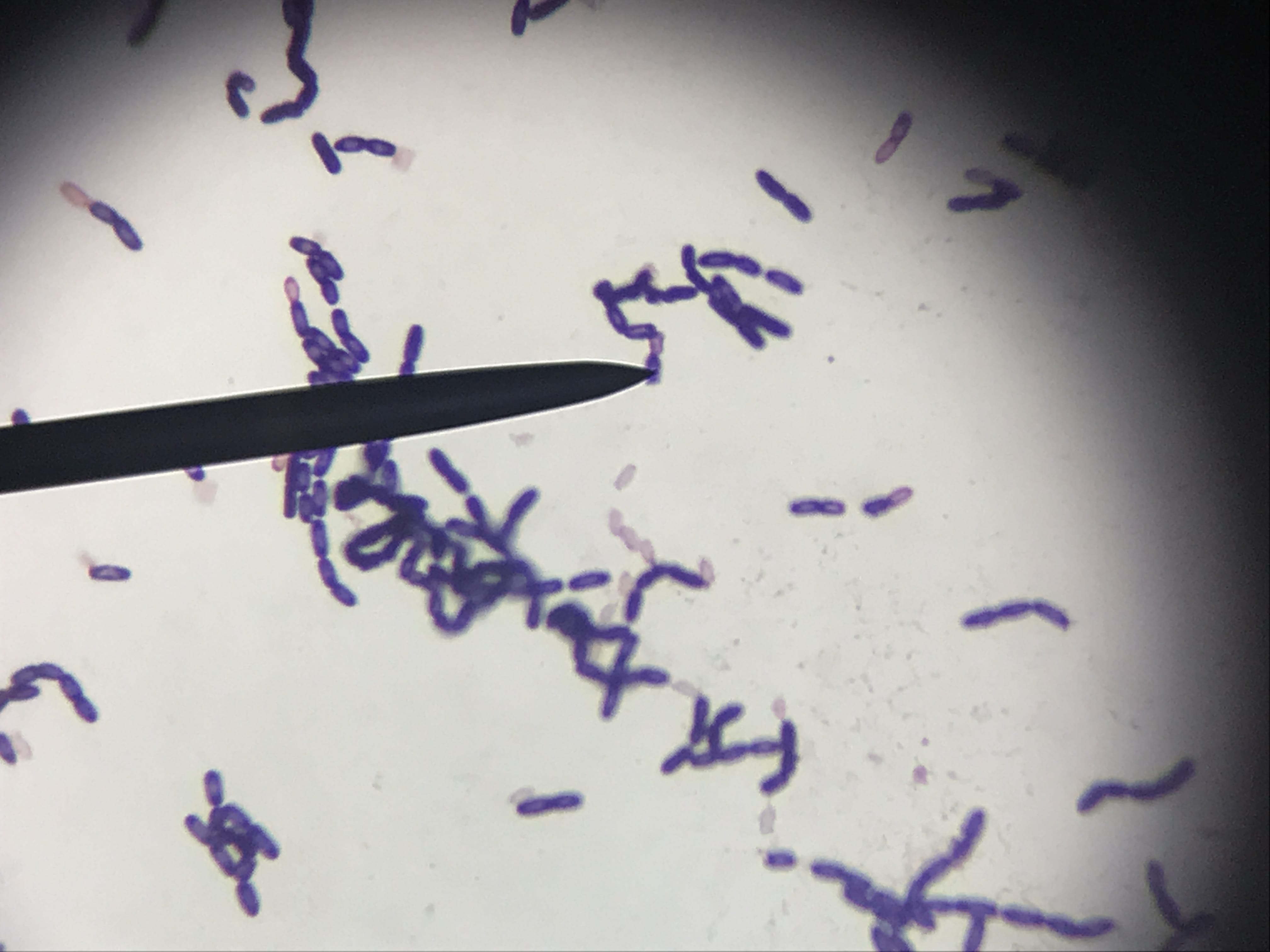
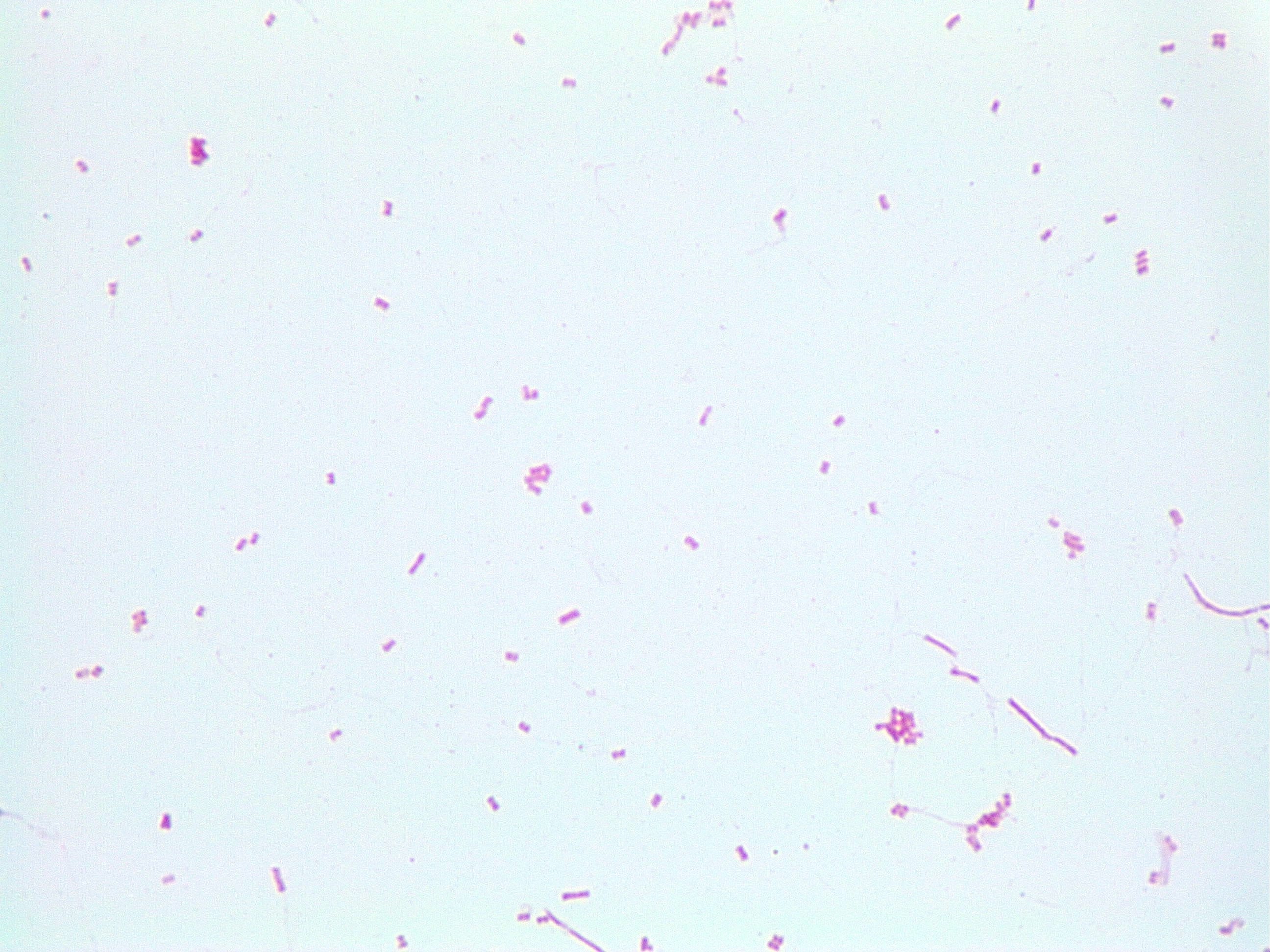
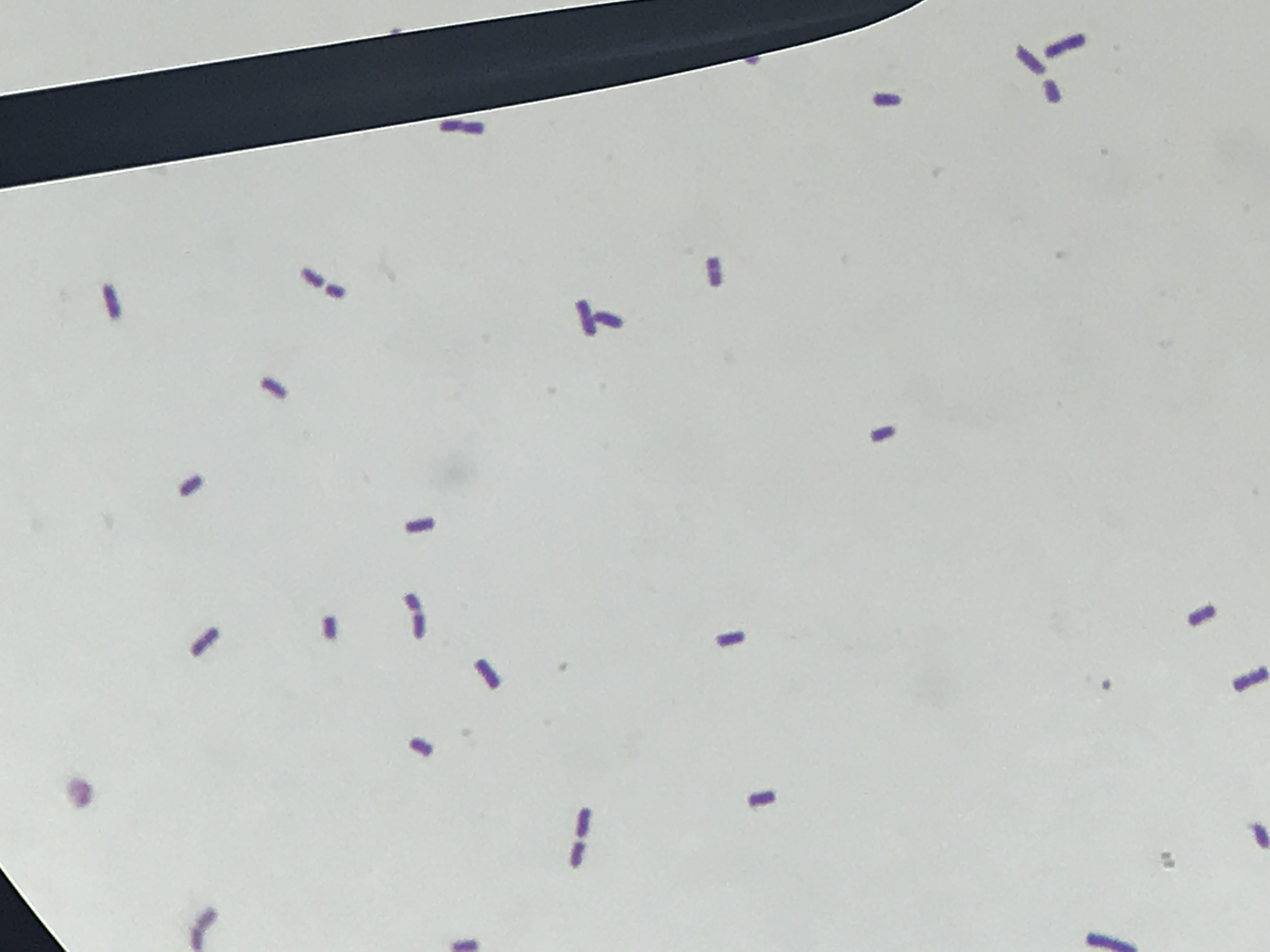
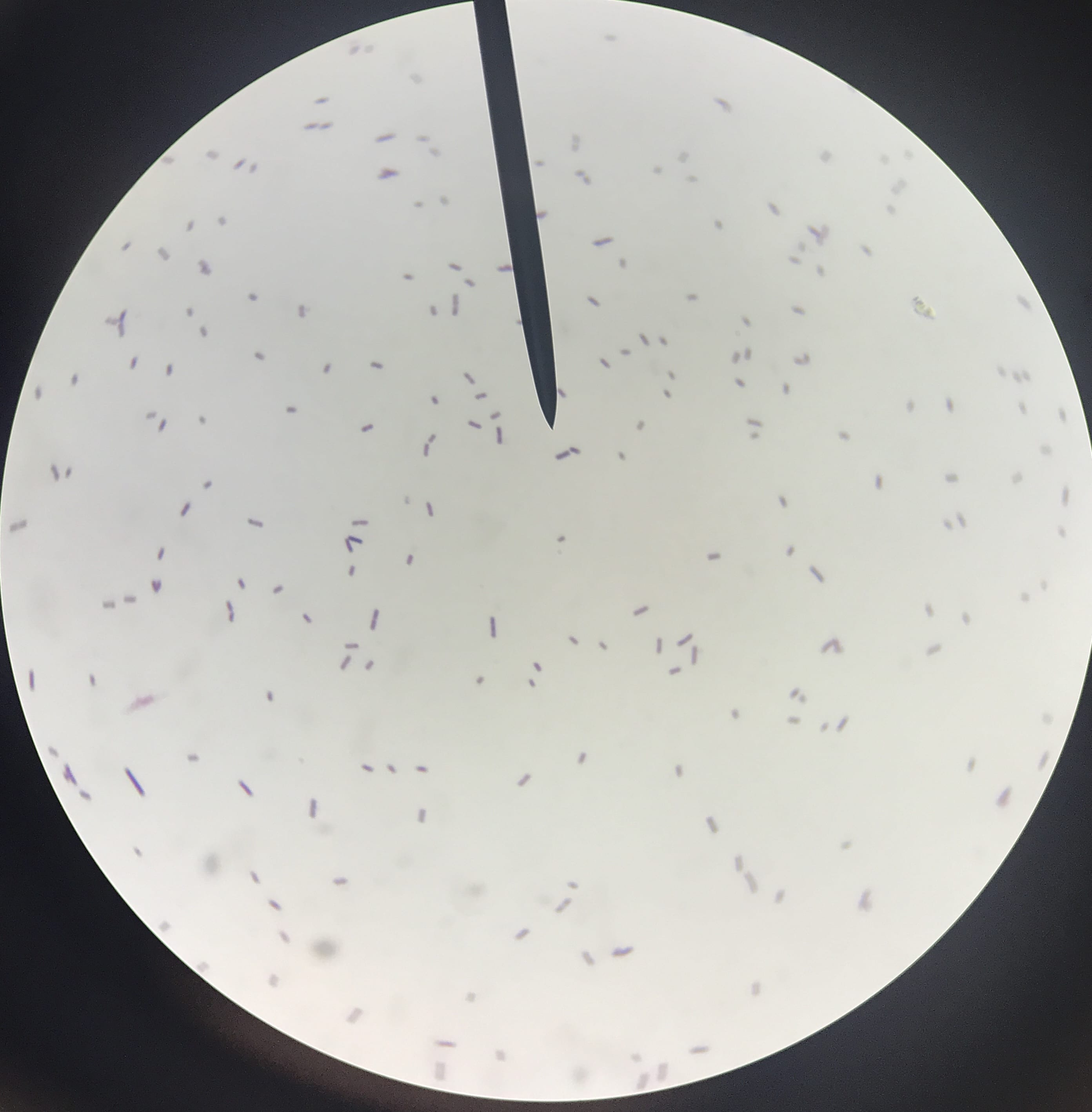
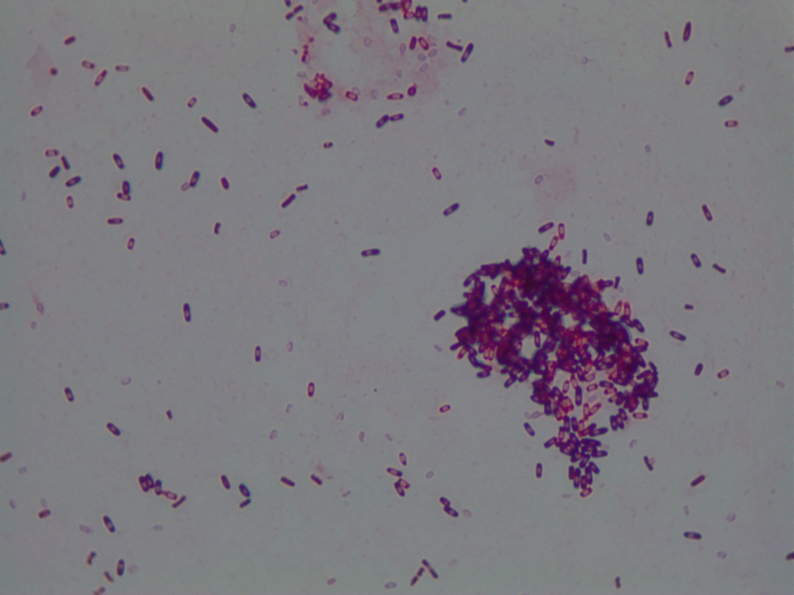
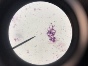
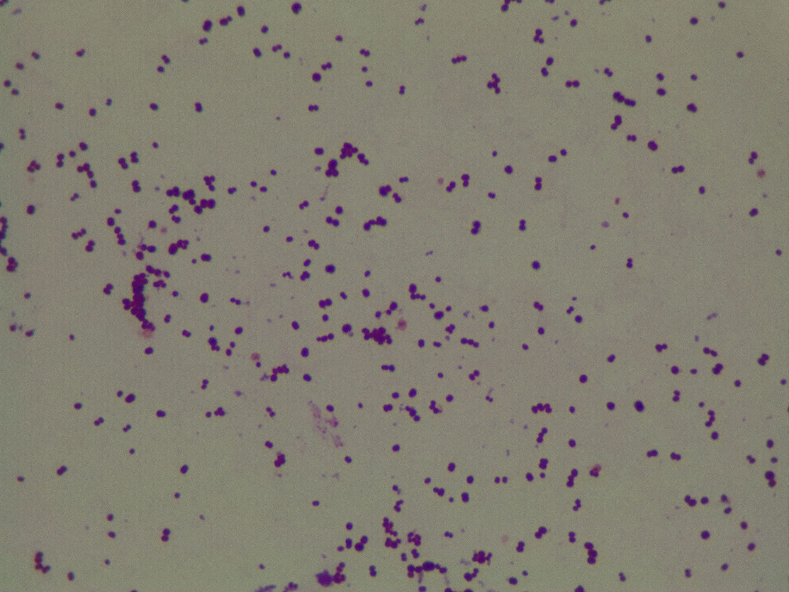
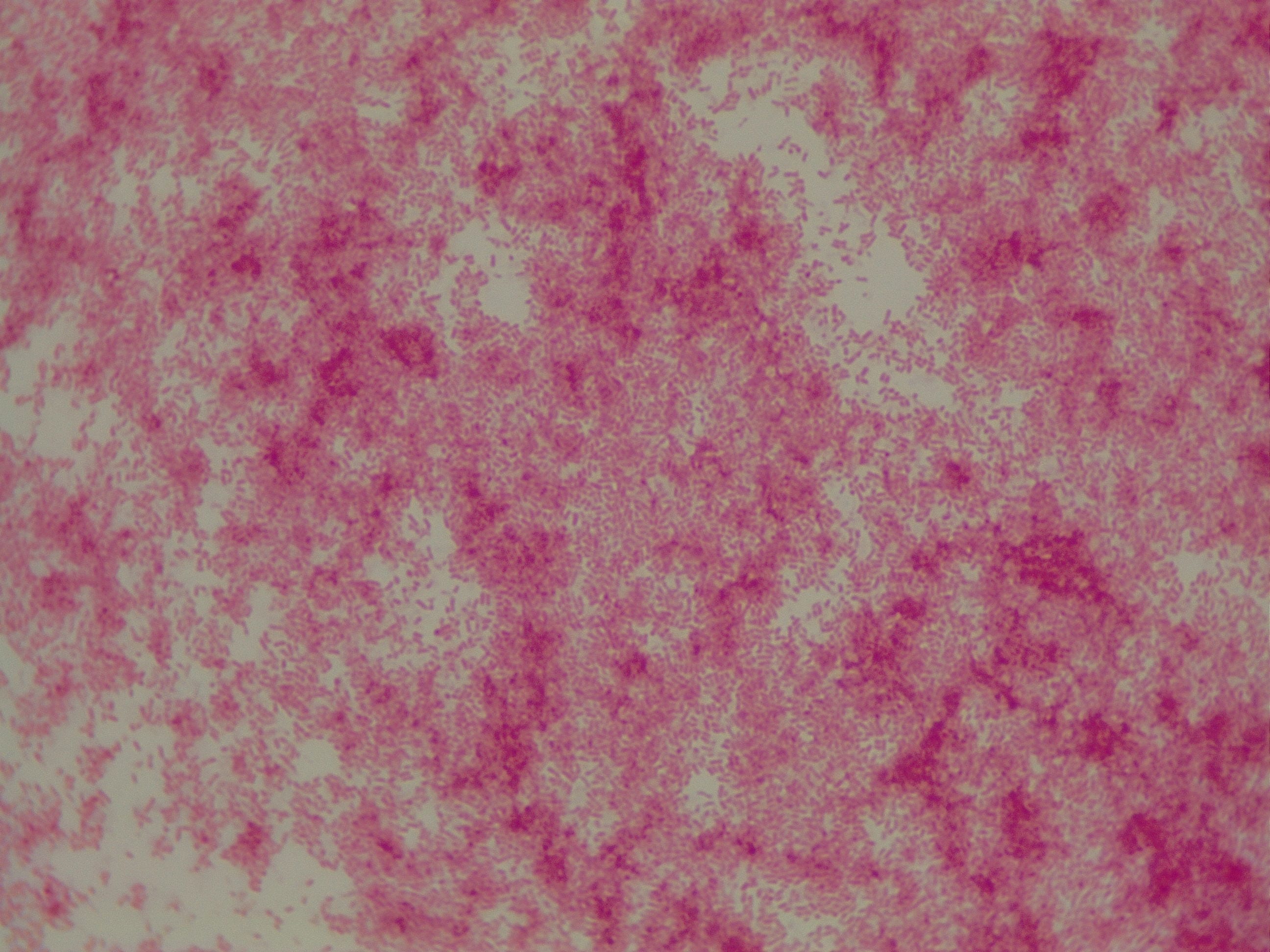
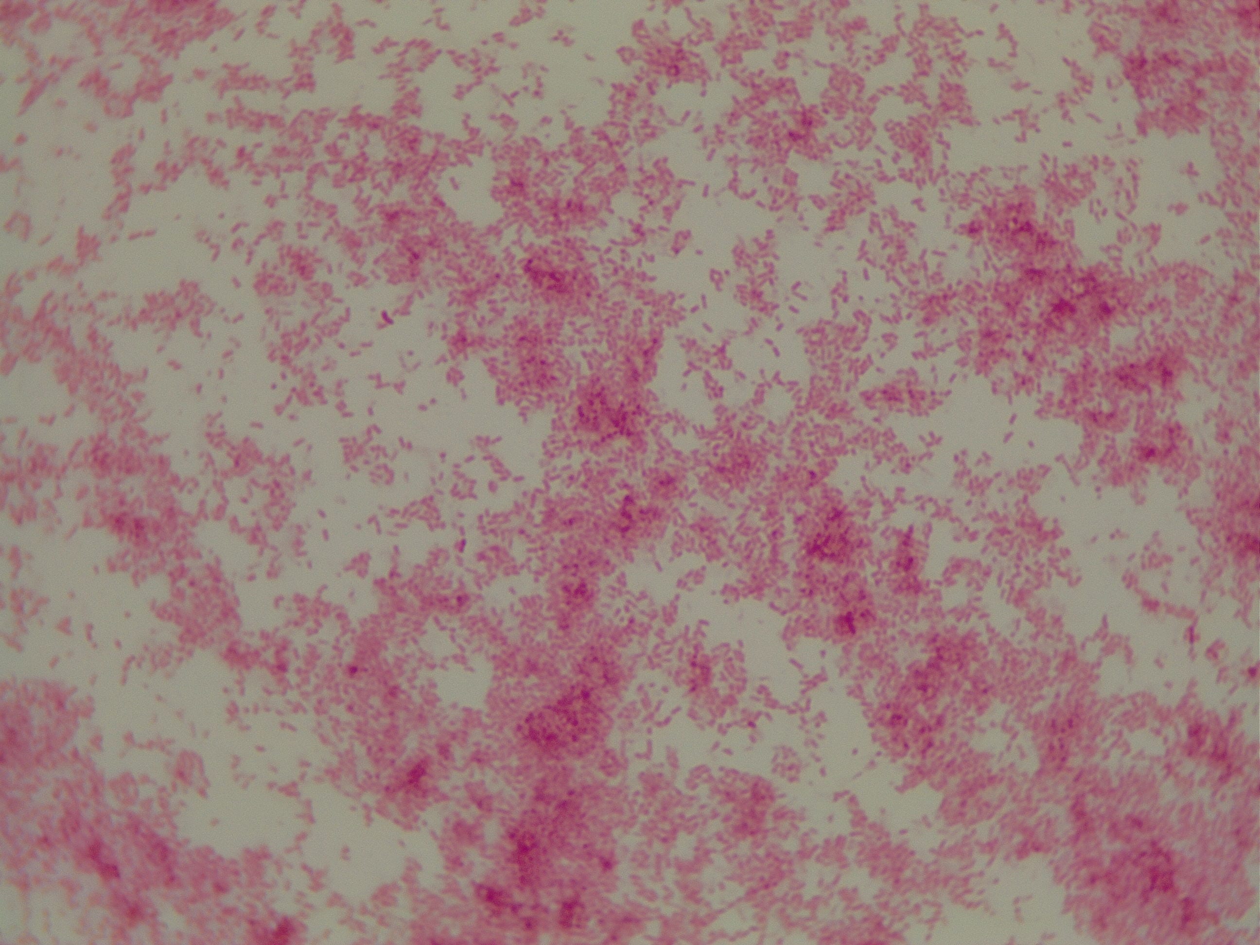
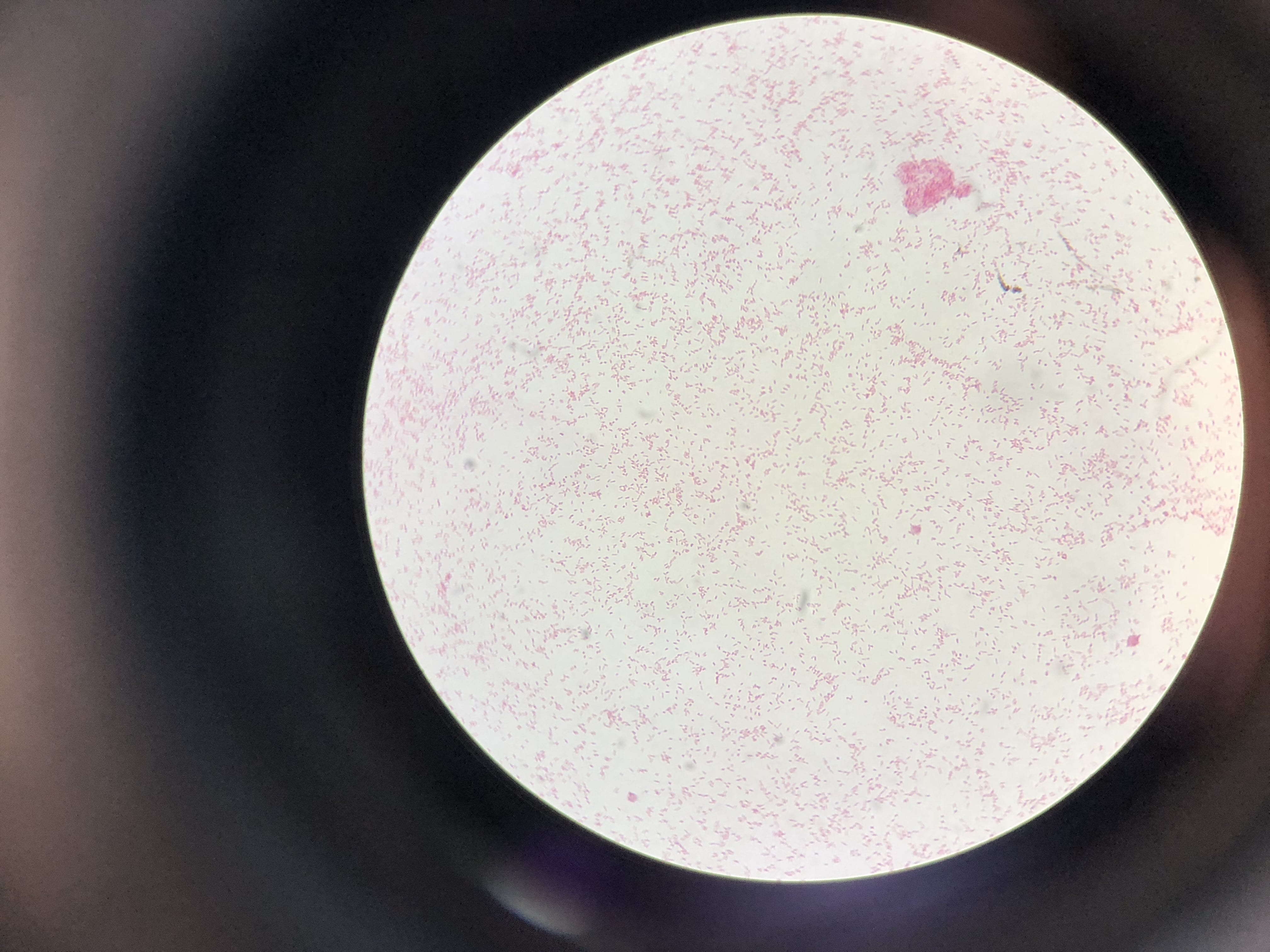
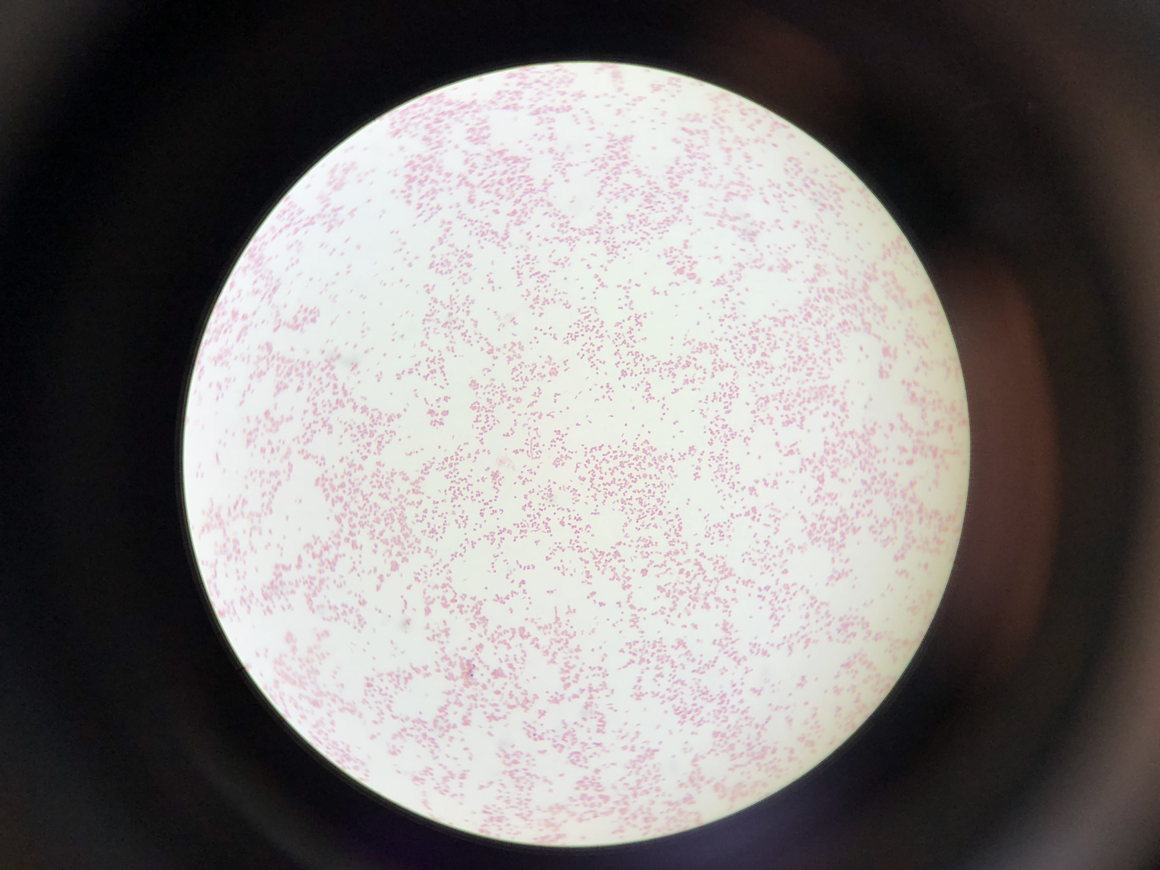
So you might be asking yourself, what kind of bacteria am I actually looking at here? Have no fear! After we isolated these strains we used a Gram staining method to determine if our bacteria were Gram positive of Gram negative.  If the bacteria were purple they were Gram positive, and if the bacteria were pink they were Gram negative.

Gram staining from bacteria colony sample 1A showing Gram negative cocci


Gram staining from bacteria colony sample 3A showing Gram negative rods


Gram staining from bacteria colony sample 2B showing  Gram negative cocci

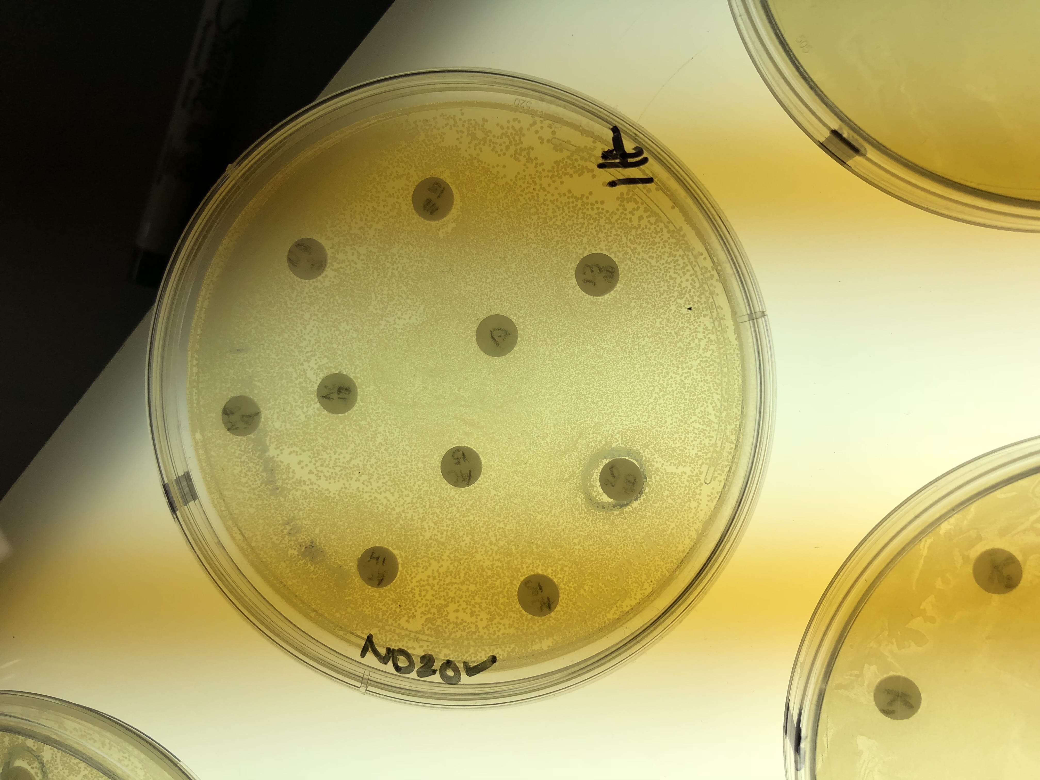
So you might be asking yourself, “Did these bacteria actually produce any antibiotics?”  And your answer is YES! They did!  After testing all of the antibiotics against four different strains of bacteria it was revealed that they each inhibited at least one strain!

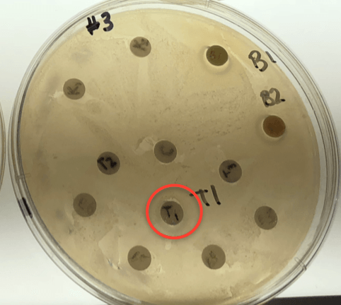
Antibiotic success in stain 1A, seen on plate T1

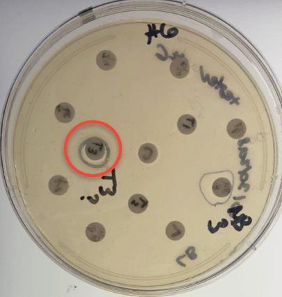
Antibiotic success in stain 3A, seen on plate T3

Antibiotic success in stain 2B, seen on plate T2

If you look at the individual plates (in the red circles) you’ll see that there are little halos around them!  These halos are the proof that the antibiotics worked, as they successfully killed off the bacteria!  While these rings are small it is still super cool that the antibiotics actually worked.  Hopefully in the future someone will be able to determine the mechanism by which it works, and enhance the function of the antibiotic or further the search for antibiotics in these types of bacteria! Who knew a little soil sample from outside the construction zone would be so interesting…

Meet my Microbes! -Alexa

This is a picture of my first set of spread plates. From left to right, the dilutions are 10^-2, 10^-3, and 10^-4. From top to bottom, the media used is PDA, LB, and AC.

This is a picture of my three patch plates. From top to bottom, the media is LB, AC, and PDA.

This is my patch plate on PDA. I just love the colors of the bacteria on this!

These are two of my patch plates against tester strain #6. The top plate is on AC, and colony #15 showed inhibition. The bottom plate is on LB, and #14 showed inhibition.

These are two of my patch plates against tester strain #3. The plate on the left is on AC, and colony #23 showed slight inhibition. The plate on the right is on LB, and #13 and #14 showed inhibition.

The plate on the left is the same as the LB plate against tester strain #6 as seen above. The plate on the right is my patch plate against tester strain #6 and colony #17 is showing inhibition.

The plate on the left is the same as the LB plate against tester strain #3 as seen above. The plate on the right is my patch plate on PDA against tester strain #3 and #13 showed inhibition.

These are all of my patch  plates on AC, LB, and PDA (from left to right).

This is a streak plate on PDA of two of my potential antibiotic producing bacteria. I love the pink color of colony 13!

These are streak plates of the rest of my interesting colonies. From top to bottom, there’s my colony #15 and Nikki’s colonoy #7 on AC, then my colonies #3 and #23 on PDA, then my #13 and #23 on PDA, and lastly my #13 and #14 on LB.

I patched four of my isolates against tester strains on PDA. Shown above is a patch plate against tester strain #9 and my colony #17 is showing great inhibition!

Gram stain of colony #13. It’s gram positive and rod shaped.

Gram stain of colony #14. It’s gram negative and rod shaped.

Gram stain of colony #15. It’s gram positive and coccus shaped.

Gram stain of colonoy #17. It’s gram negative and rod shaped.

Meet my microbes – NIcole


I sort of wish I had better photos.

My first 10% TSA patch plate. Look at the variety of colors. If you look closely, since I don’t know how to edit this picture, you can see that the microbes in spaces 3, 9, 11, and 21 have a check mark by them, meaning that they had zones of inhibition when tested on the safe ESKAPE strains.

The antibiotic producers that I chose to try to put on streak plates. I don’t know why this picture is sideways.

A Gram stained image of the bacteria I called v13.

A Gram stained image of the bacteria I called v19. I don’t know why it is a bluer purple than the other image.

Meet my microbes! – Nikki

Location of the soil sample collection: Octopus Community Garden at Union College in Schenectady, NY.                                                                                       GPS coordinates- Latitude: 42.8 degrees Longitude: -73.9 degrees

Example of 10% TSA original plate at a 10^-4 dilution (top), patch plate (middle) and streak plate of #9, 15, 20, and 23 isolates (bottom).

Gm+/Gm- Inhibition

Inhibition of both Gm+ B. subtilus (top) and Gm- E. coli (bottom) are seen for isolates 15, 20, and 23 on 10% TSA.


Gram – Control

Gram + Control

Isolate #15 Gram Stain

Isolate #20 Gram Stain

Isolate #23 Gram Stain

Gram Stain Results: Isolates 15, 20 & 23 all appear to be G+ and streptococcus, bacillus/filamentous, and coccus in shape, respectively.

Inhibition against all tester strains: Isolate #15 is inhibiting strains 1. Staphylococcus epidermidis (+)  and 2. E. coli (-), isolate #20 is inhibiting strain 6. B. subtilis (+), and isolate #23 is inhibiting strain 1. Staphylococcus epidermidis (+), all on 10% TSA.

Extraction Test Results:

Extractions were obtained for isolates #15, 20, &23. Only extract #20 showed inhibition on tester strain 1, S. epidermidis.

Results from 16S Analysis and Blast/Ribosome Database Project (RDP) Search.  I was only able to obtain results for isolate #20 (677 bp). The results show that the closest relative is Streptomyces of the vinaceus or cirratus strains with 99% identity and query coverage (Accession numbers: NR_041131.1 and NR_112388.1).

Meet my microbes! – Tommy

Here is a photo of my PDA patch plate. PDA plates gave me a lot of mold over the course of the term, but also displayed some really interesting and unique microbe morphologies. Unfortunately, none of these inhibited the ESKAPE tester strains – neither Gram positive nor Gram negative. But they look awesome!


Gram negative control.


Gram positive control. Cocci.


Streak plate of isolate 18. This is the isolate whose extraction strongly inhibited a Gram positive tester strain and a Gram negative tester strain. It was my best producer!


Unknown 18. Gram positive. Appear to be diplococci that form some longer chains as well and like to aggregate. This is what my best antibiotic producer looks like up close!


Streak plate of isolate 11


Isolate number 11. Gram positive rods. Looks like they might be spore formers.

Meet My Microbes! – Brianna

Here are (some of ) my original plates from the soil sample extracts.

The first is my 10-3 dilution plated onto R2A , the second is a 10-2 dilution on PDA, and the third is 10-3 dilution on LB media. All are photographed on day three of incubation. For the most part, PDA showed the most diverse colonies with a variety of colors and shapes. From these and other dilution plates, 24 colonies (per plate type) were picked and selected to be further investigated.

Here are my master patch plates after four days of incubation, in the same order as before. A few interesting things occurred at this stage. The diversity of the colonies was much easier to identify. They went from being generic-off-white-round colonies to having slightly different pigments, shapes, and morphologies. Additionally, some of the picked colonies did not grow on the new plate (RIP), so who knows if I lost the answer to the antibiotic crisis.


Then, all of these colonies were tested for antibiotic producers. Below is my champion of my antibiotic producers, PDA #5. As you can see, a vary large halo formed around colony #5, clearing the tester ESKAPE #6, B. Subtillis.  I also had 3 other producers from other plates and colonies, but they produced only a small halo against the background.

From all the ESKAPE testing, I narrowed down my colonies to a total of 4 producers. These are the streak plates for each of these microbes. In order, they are: PDA #2, PDA #5, LB #2, and R2A #18.

These four microbes were then stained to determine if they were gram positive or negative. All images are taken under a 100X oil emersion microscope lens. For reference, here is the positive (Staphylococcus epidermidis) and negative (E. Coli) controls.


The following are of my isolates. The first image is of R2A #18. This microbe is gram positive, and appears in diploid rods. The second is LB#2, which is gram negative and appears to be a shortened more ovular rod, and tends to be in diploid clusters. The third, PDA #2,  is a gram negative cocci, and is generally diploid, but forms larger clusters as well. Lastly, PDA#5, is gram-negative, and the cells are shaped like a typical rod bacterium.


Meet My Microbes! – Le Minh Nguyen

This is of one of my very first plates from the first week of lab. I plated my soil sample on the LB + cycloheximide plate with a 10^-4 dilution. This plate was unique because it contained those brown circular colonies with a surrounding halo-fade that also caused pigmentation in the media. As I later found out, this isolate showed some antibiotic production as it inhibited the growth of some of our testers and I eventually chose it for my PCR reaction analysis.


These plates also come from the first week of lab. The plate on the left is also a plate of my soil sample, plated on PDA + cycloheximide plate with a 10^-3 dilution. This plate shows a great diversity of bacteria as presented by a variety of different colors. A lot of colonies were picked from this plate and were patched. Its master plate looks as interesting as shown by the diverse and colorful isolates that were able to grow. 

This is a PDA + cycloheximide plate from the first round of ESKAPE testing. The tester strain is a Gram-negative E. caratovora. As seen on the plate, the tester strain did not grow very well, so the ESKAPE testing was repeated with another Gram-negative E. coli. However, what is more interesting is that only a few colonies were able to grow (#1, 4, 13, 14) while the rest seemed to be contaminated as they appear as red smears. This most probably occurred due to using the same toothpick for ESKAPE testing. 

This is an LB plate, also from the first round of ESKAPE testing with a Gram-negative E. caratovora. This time the tester strain was able to grow and my isolates were tested. As shown on the plate, colonies 5, 6, and 11 showed promise to be antibiotic producers as indicated by the presence of a halo. 


These are pictures of one of my master plates obtained during the ESKAPE testing. These isolates are grown on the PDA plate and as mentioned above, the diversity of the obtained bacteria is great; they are significantly different in morphology (as indicated by the difference in color and shape) and look really cool. 

This is one of my streak plates of the bacteria that I streaked for PCR reaction. The bacteria is grown on the PDA plate and is the same one from the previous PDA plates, indicated as #1. The single colony has a circular egg-like shape and is of deep brown color. Furthermore, after a few days, the colony causes the media to absorb the bacteria’s pigment. 


These are my Gram-positive (Staph epi) and Gram-negative (P. putida) controls. They were visible on my slide with the expected coloration so I could identify the Gram identity of my bacterium sample. 

This is my stained colony #5 from LB plate that I picked to identify as it was the only sample that produced the desired PCR product. It is a Gram-negative rod-shaped bacterium. 

Meet my microbes – Kara

This is the PDA patch plate I made, and where I isolated my only producer. The other producers I have were generously given to me by Elizabeth Altman.

this is an image of my producer. It appears to have both a mix of both gram + and gram – bacilli.  I’m not sure how to characterize this, it could be a mixed culture.


Both isolates from Elizabeth looked very similar.  They show individual gram positive bacilli on the left, and filamentous “clusters” of cells on the right.

Meet My Microbes- Carly Burns

These are my master patch plates from 9/17 and 9/25.  The LB plates were labeled group “a”, the R2A plates were labeled group “b”, and the AC plates were labeled group “c”.

These are the patches from 9/25 from group a and b tested against the Gm+ tester strain E. raffinosus and the Gm- tester strain E. carotovora.  No patches showed possible antibiotic production against them.

One set of these plates are patches from group c made on 9/25 against the Gm+ tester strain E. raffinosus and the Gm- strain E. carotovora.  None of these patches showed possible antibiotic production so a new patch plate with different colonies from the original plate were made and tested against E. raffinosus and E. carotovora on 9/27.  These patches also showed no possible antibiotic production.

Since no patches showed possible antibiotic production, all of those plates were tossed in the trash and I took two possible producers from Elizabeth and made this streak plate on 9/27 for further tests.  After testing these two bacteria against all the tester strains I found that only #10 was a possible producer so I took this bacteria and two bacteria from Tommy and performed a PCR reaction with them.  These PCR reactions were then run via gel electrophoresis and this showed that only the two cultures I got from Tommy contained the band we were looking for so I sent these two out to be sequenced.

I performed the Gram stain test on the two cultures and one of them ended up being Gm+ and the cells had a deep purple color and were rod shaped (above).  This is a picture of one culture under 100x magnification.

The Gram stain test for the other culture showed that the cells were Gm- and the cells had a pink color and were oval shaped (above).  This is also a picture under 100x magnification.


Meet My Microbes – Elizabeth Altman

These are Professor Salvo’s initial patch plates of interesting organisms from her soil sample (Jackson’s Gardens). The first two were on LB and the last three were on TSA. I took interesting organisms from each media to get 1 set of 24 organisms on LB plates and 2 sets of 24 organisms each on TSA plates.


These were the LB plates from the first round of ESKAPE testing. Organisms 2 and 7 were identified as producers on the Gm- plate. These two were patched onto all the ESKAPE safe relatives by someone else.


These plates are the first round of ESKAPE testing on the first set of organisms on TSA. Organisms 1, 2, 4, 8, 13, 15, 16, 20, 22, 23, and 24 were identified as producers on the Gm+ plate. I patched all 11 of these onto all ESKAPE safe relatives myself.


These plates are the first round of ESKAPE testing on the second set of organisms on TSA. Organisms 3 and 10 were identified as producers on the Gm+ plate. These two were patched onto all of the ESKAPE safe relatives by someone else.


This is one of the patch plates from the second round of ESKAPE testing. Only organisms 8, 16, and 20 from the original TSA first set were found to be producers at all during this round of testing. These three were put through colony PCR.


The PCR reactions with the degenerate primer worked for all three organisms, so I moved forward with all three.


These are the positive and negative control organisms on each slide. They were visible on all slides with the expected coloration, so the stains for all unknowns were trusted. These two, along with all microscope images, were taken at 100x.


This is unknown organisms 8. It is a gram positive bacillus that seems to connect in diplobacilli chains.


Organism 16 was a very small gram negative cocci that existed in groups from two up to about twenty cells.


These are pictures of unknown organism 20. It is gram positive, but has a pretty unique appearance, looking more like rectangles than normal bacilli or cocci.

Meet My Microbes- Kelsey


This is a picture of where I got my soil sample! There were lots of decomposing elements to this soil including rusty beer cans, paper, organic matter, etc. The soil was most likely in the A Horizon.


These are two of my streak plates with the four producers that I found in lab. (slightly blurry due to the plastic bag).


These are my R2A plates, where I found most of my producers. You can see the mycoides growing on the 10^-2 and 10^-3 dilution plates. The second photo is a close up of the mycoides on a light box at a dilution factor of 10^-2.


A close up of my R2A plate with a 10^-4 dilution. This photo was taken on the light box.


These are three examples of patch plates. These were the first set of patch plates that we did. These photos were all taken on the light box. In both the LB and AC media plates, you can see an overgrowth of mycoides. The mycoides overtook the patches in this section and rendered them unusable. I found minimum mycoides growth in the R2A plates throughout my experiment.


These are two pictures of my Gm+ bacteria. I know it is Gm+ due to the purple color. My bacteria are most likely bacillus because they are rods and spore forming. I know that they are spore formers due to the central pink spore between the purple ends of the rods. These photos were taken at 100x.


This is another photo of my bacteria. You can see a large aggregation of colonies on the right side of the slide. This photo was taken at 1000x.


These are pictures at 1ooox of my Gram positive control (S. epi).


These are pictures at 1000x of my Gram negative control (P. putida).