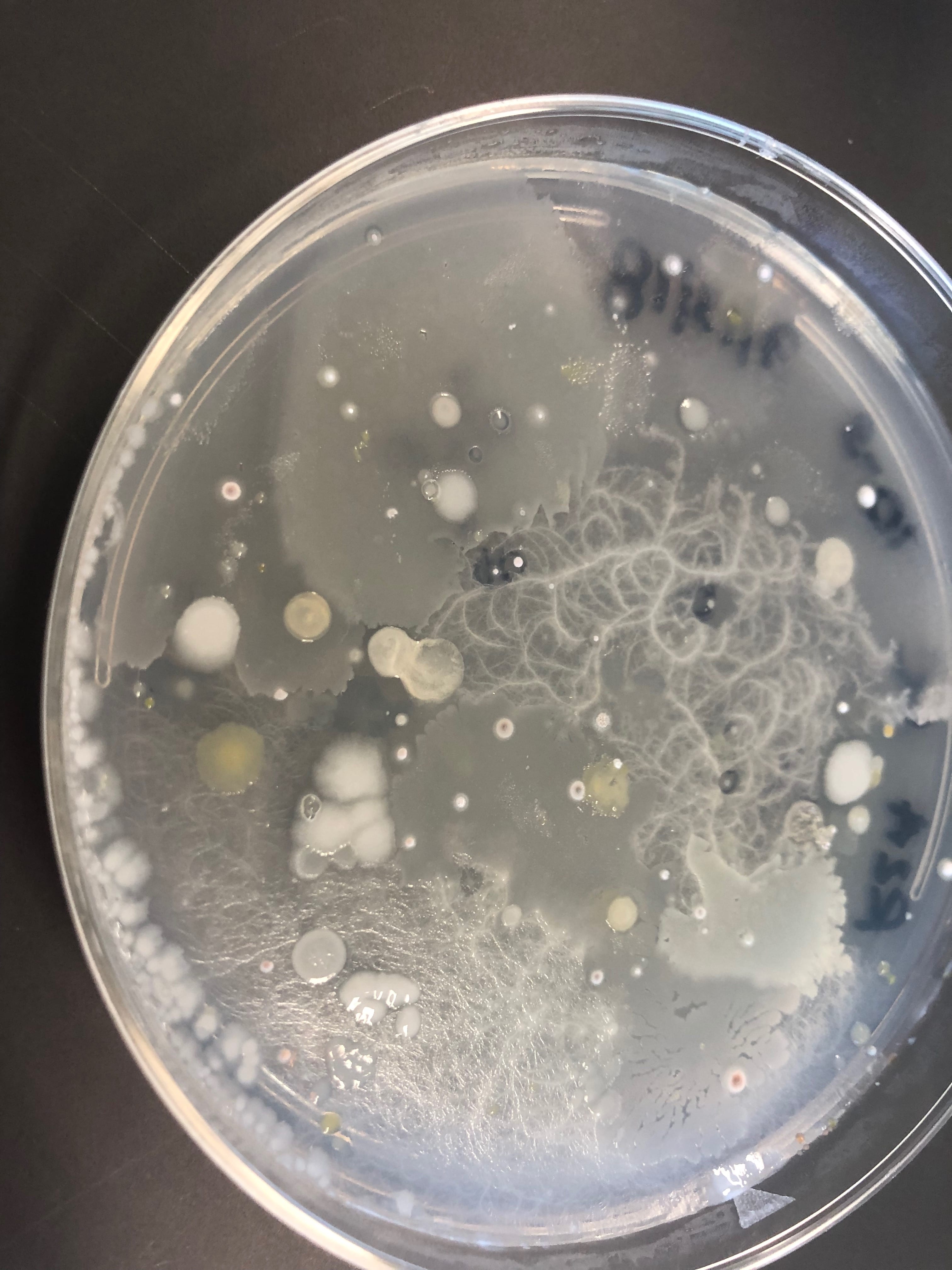
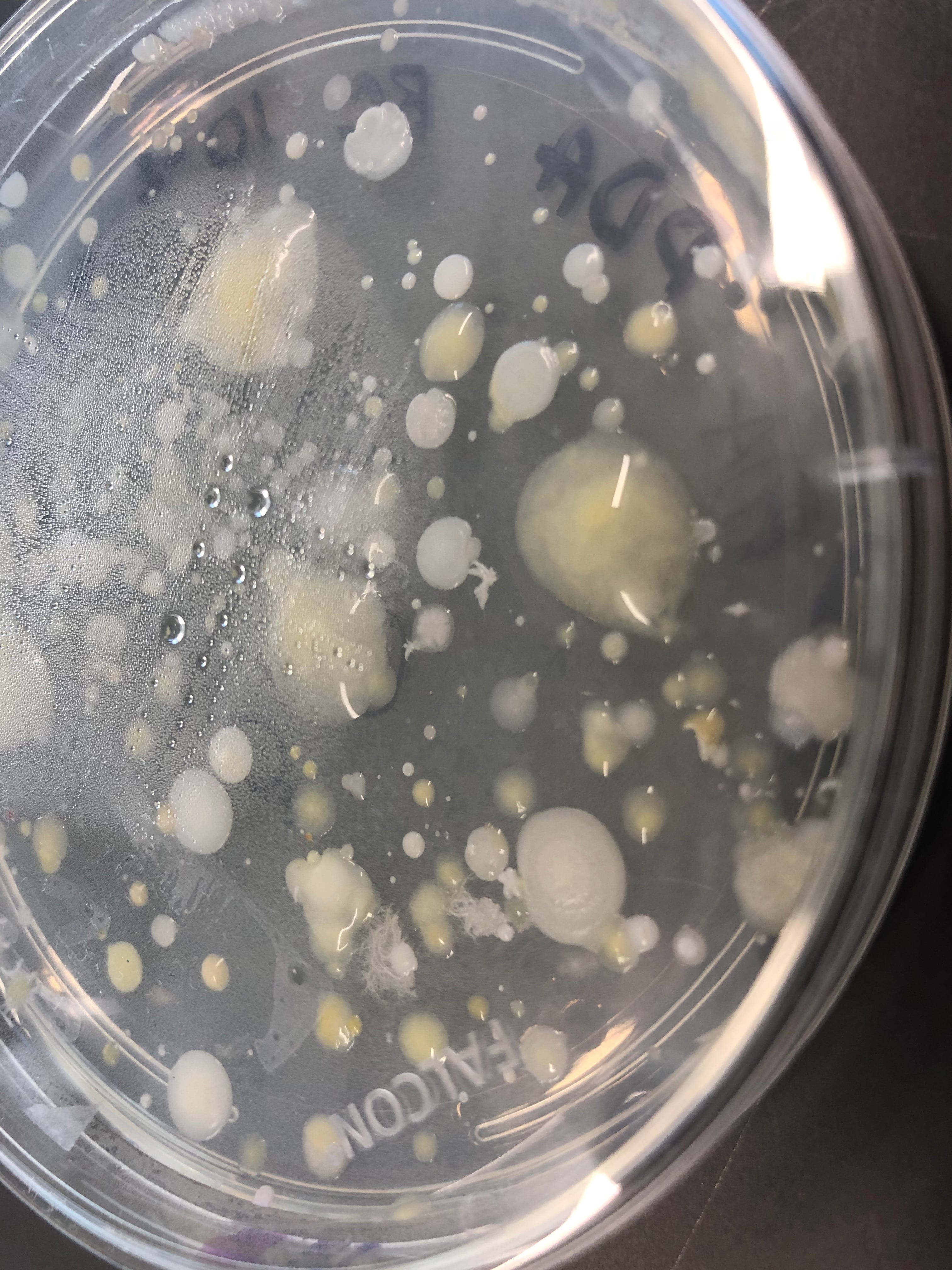
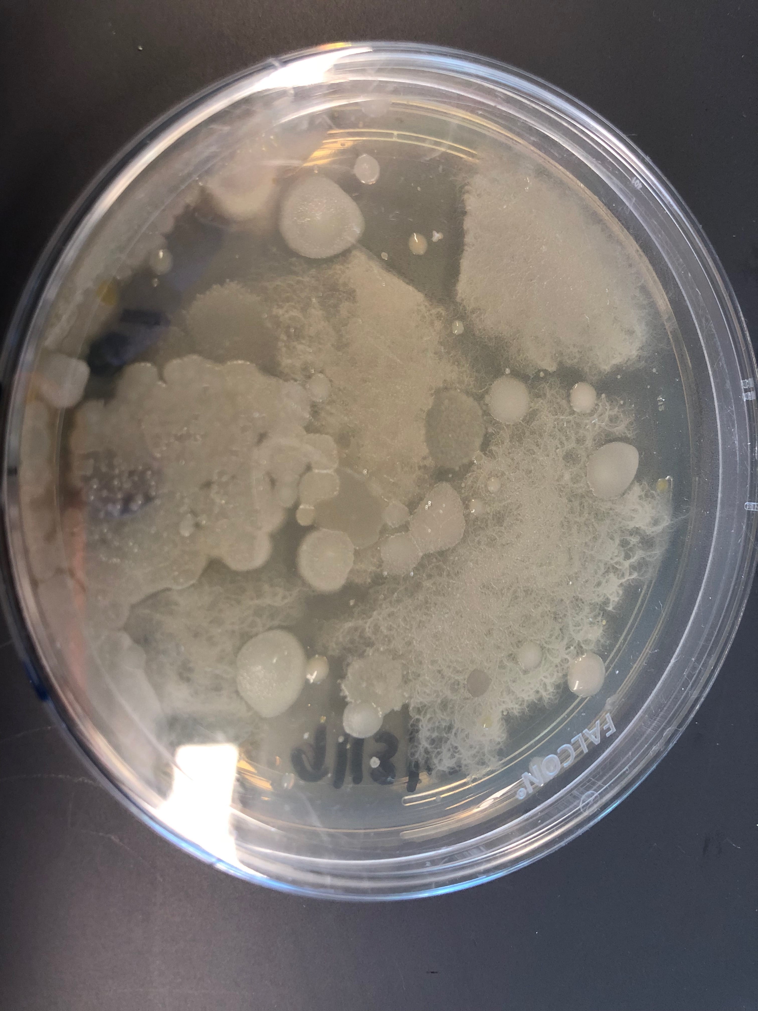
Here are (some of ) my original plates from the soil sample extracts.
The first is my 10-3 dilution plated onto R2A , the second is a 10-2 dilution on PDA, and the third is 10-3 dilution on LB media. All are photographed on day three of incubation. For the most part, PDA showed the most diverse colonies with a variety of colors and shapes. From these and other dilution plates, 24 colonies (per plate type) were picked and selected to be further investigated.
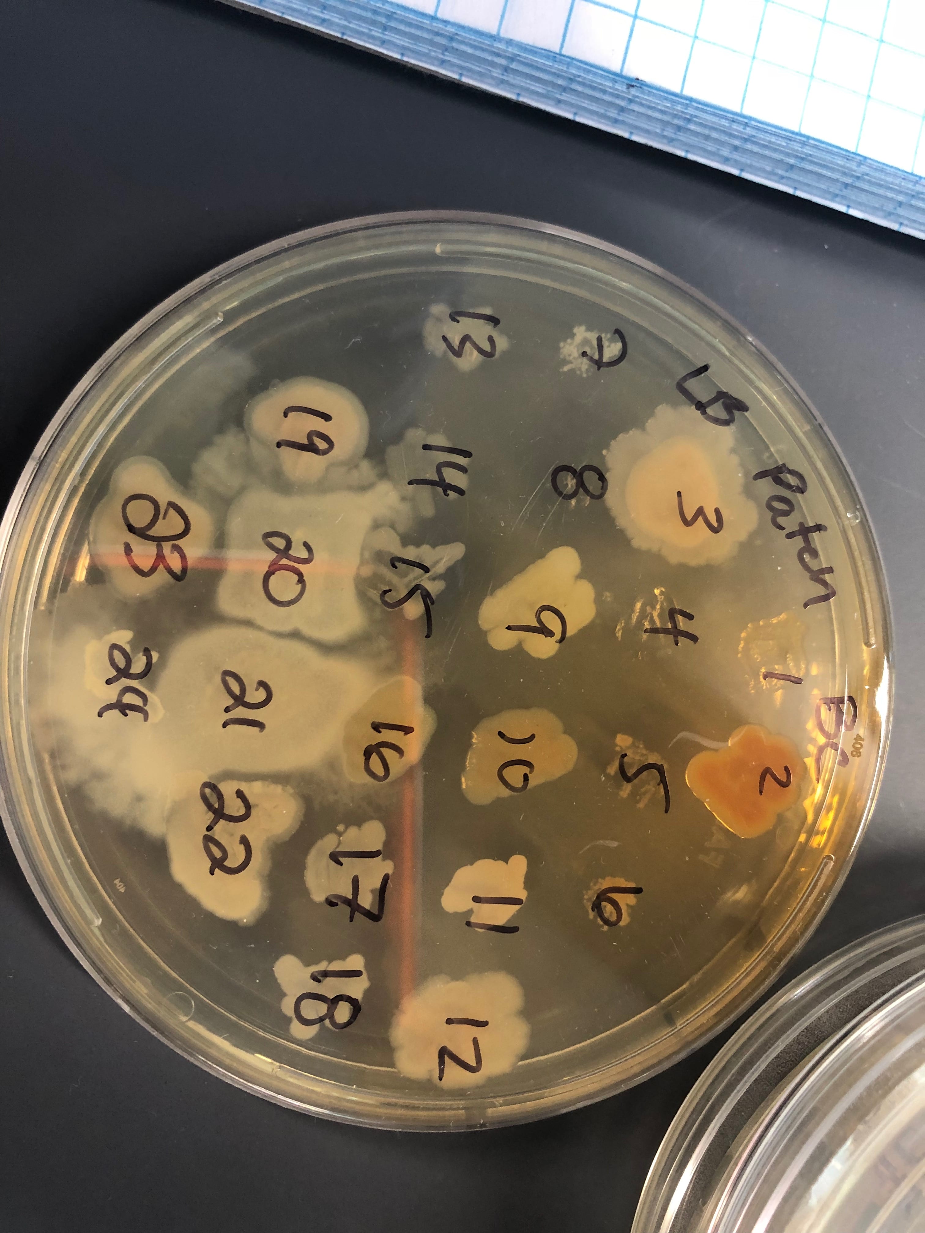
Here are my master patch plates after four days of incubation, in the same order as before. A few interesting things occurred at this stage. The diversity of the colonies was much easier to identify. They went from being generic-off-white-round colonies to having slightly different pigments, shapes, and morphologies. Additionally, some of the picked colonies did not grow on the new plate (RIP), so who knows if I lost the answer to the antibiotic crisis.


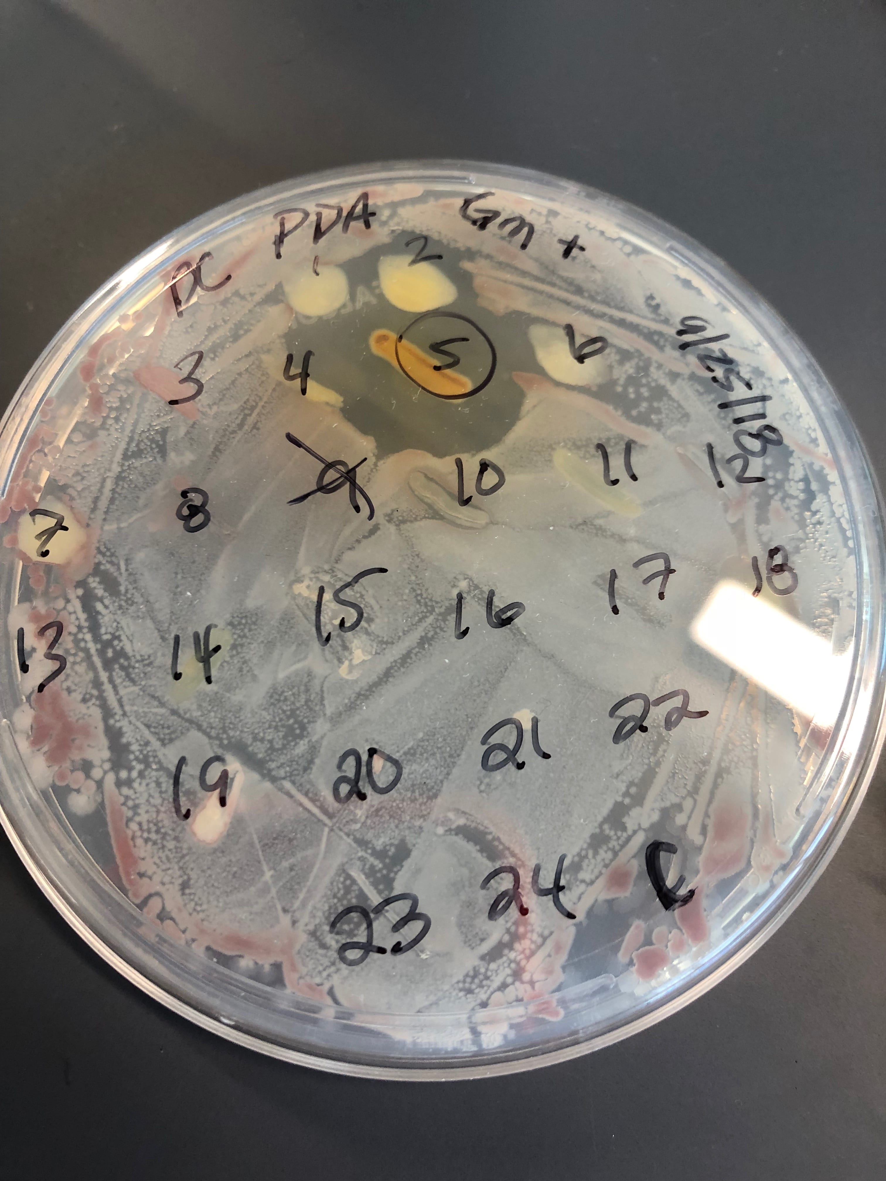
Then, all of these colonies were tested for antibiotic producers. Below is my champion of my antibiotic producers, PDA #5. As you can see, a vary large halo formed around colony #5, clearing the tester ESKAPE #6, B. Subtillis. I also had 3 other producers from other plates and colonies, but they produced only a small halo against the background.
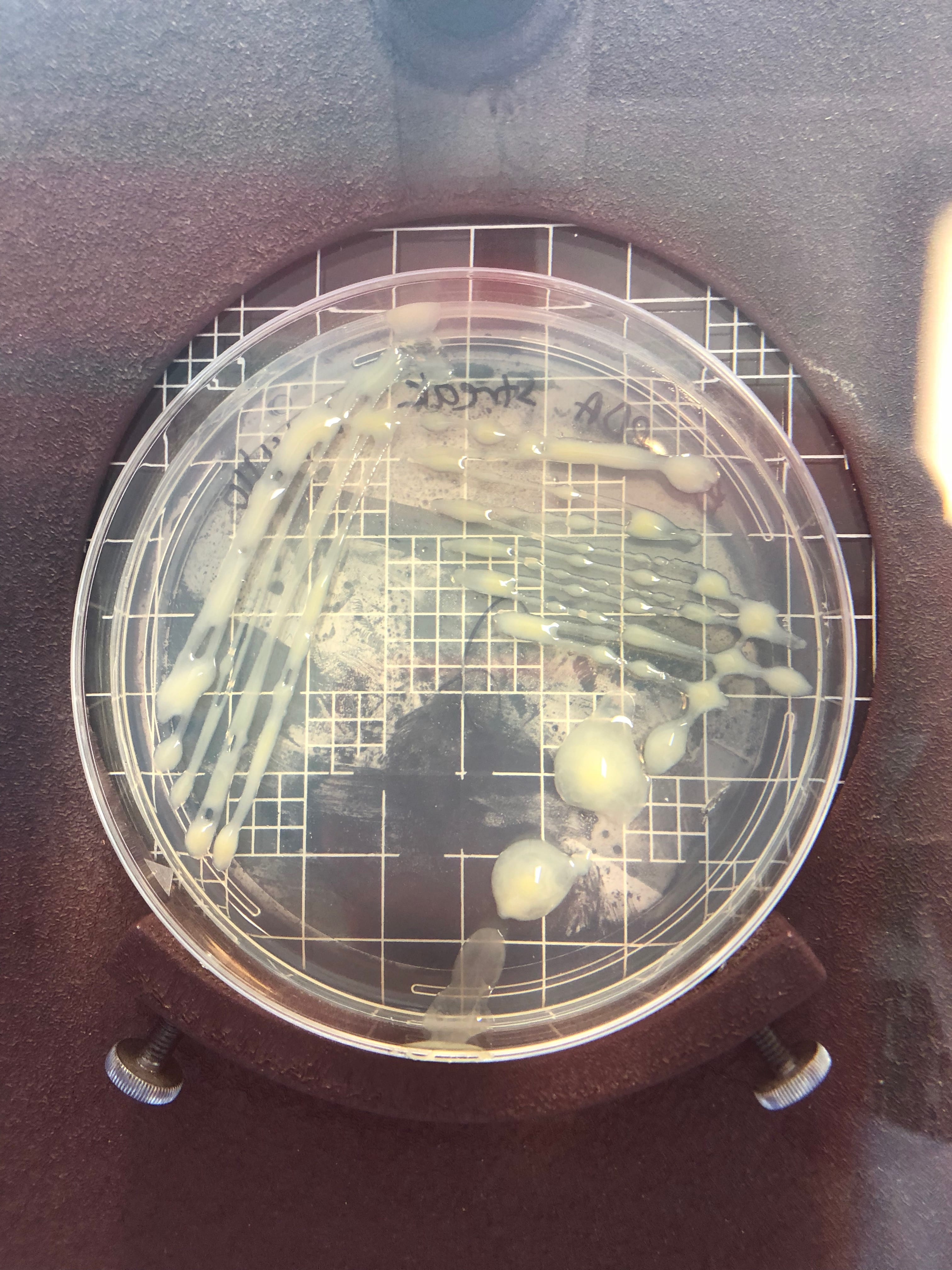
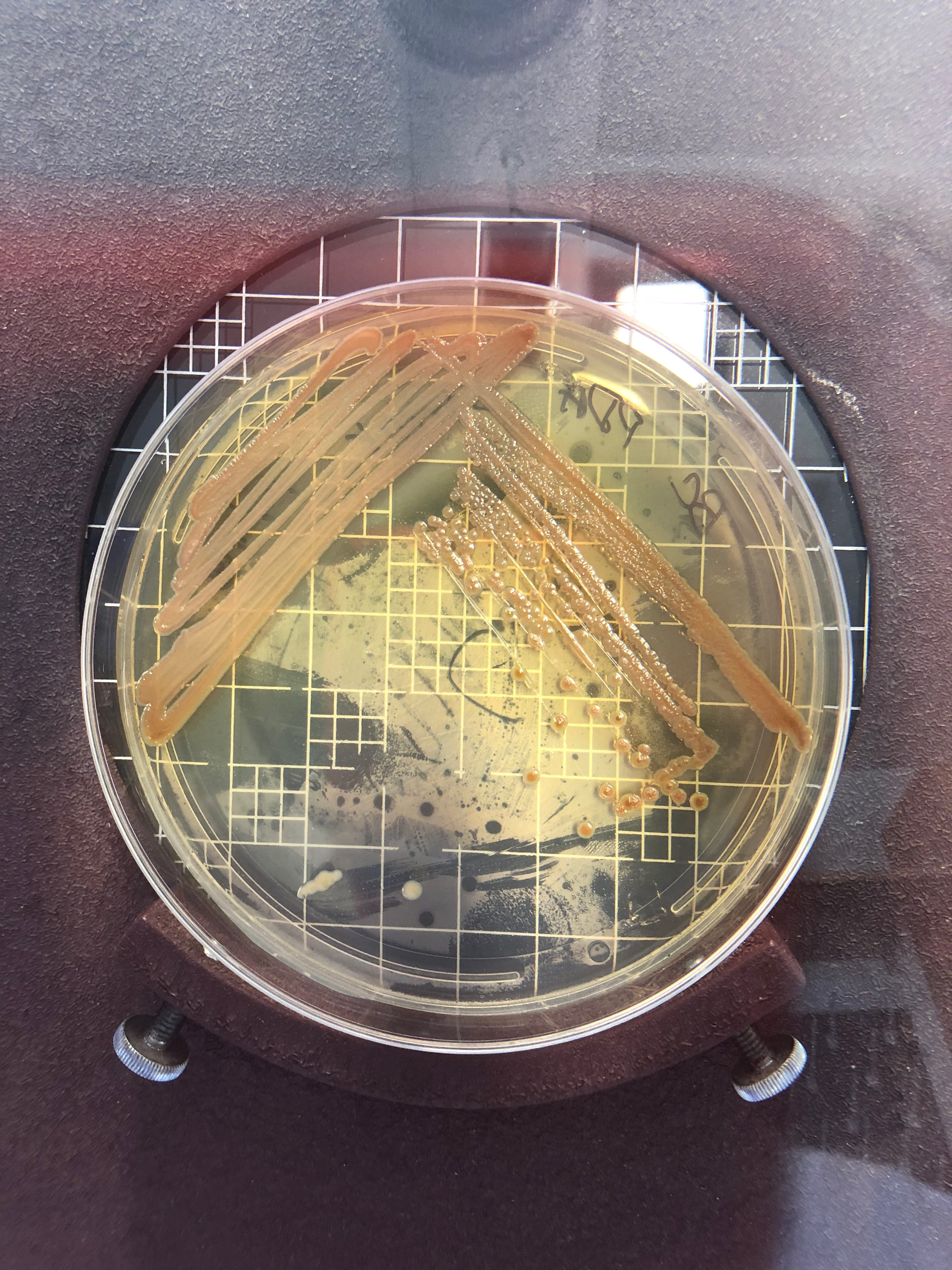
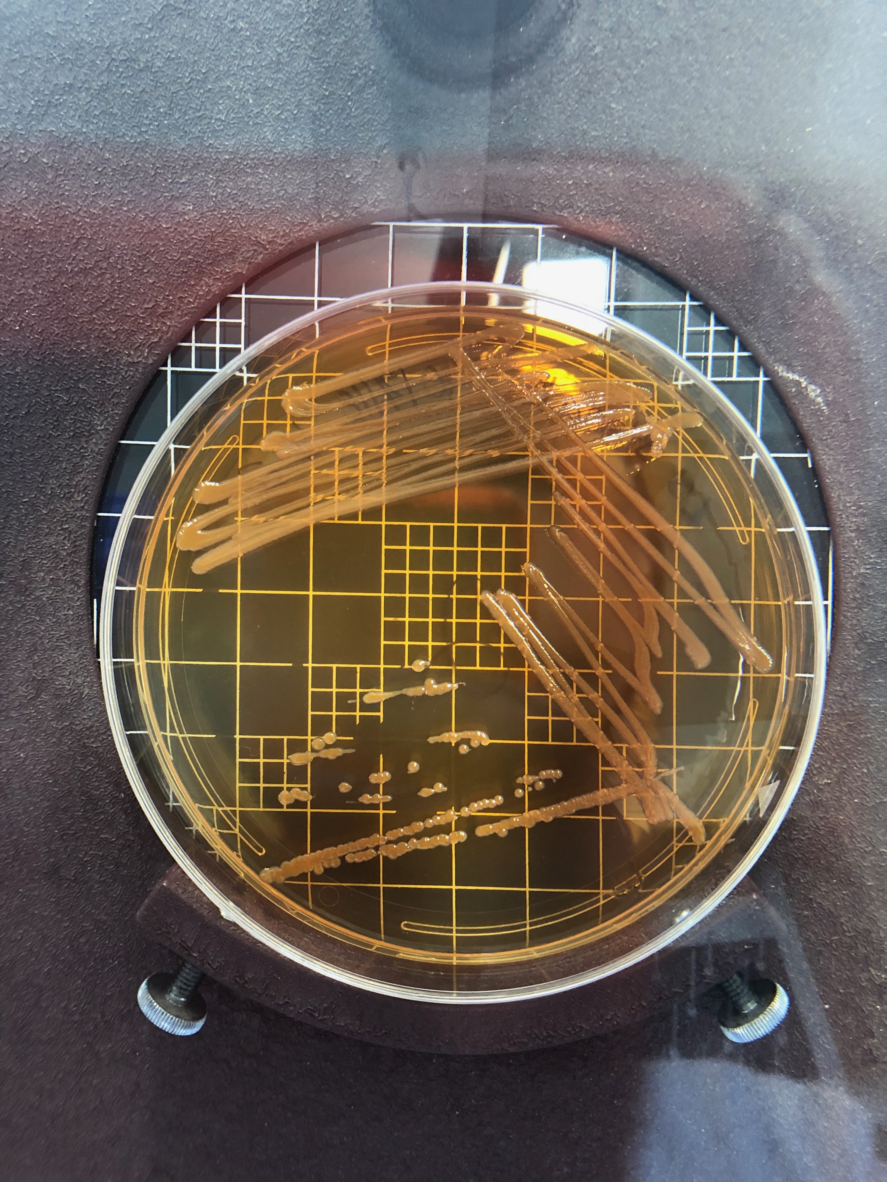
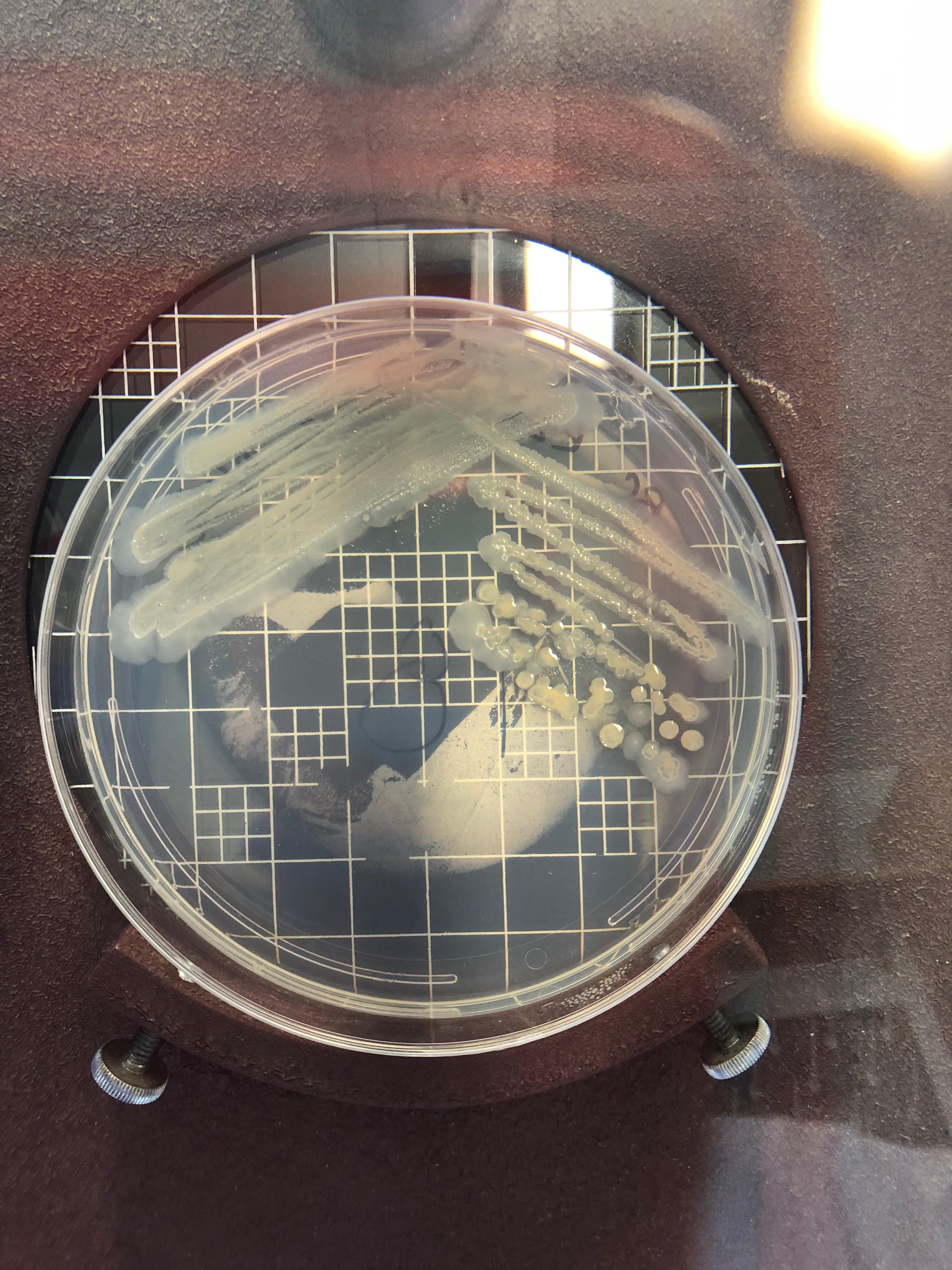
From all the ESKAPE testing, I narrowed down my colonies to a total of 4 producers. These are the streak plates for each of these microbes. In order, they are: PDA #2, PDA #5, LB #2, and R2A #18.
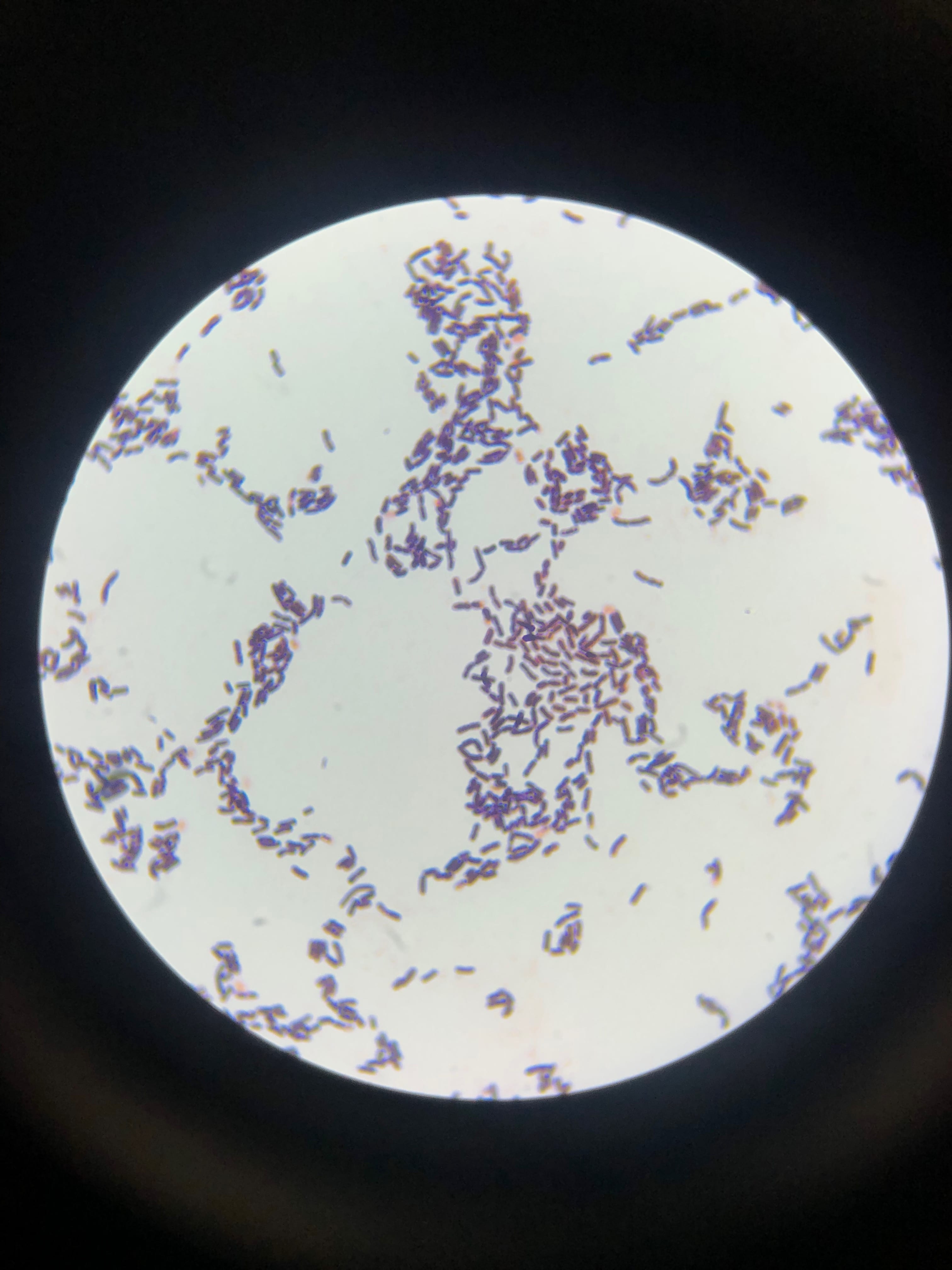
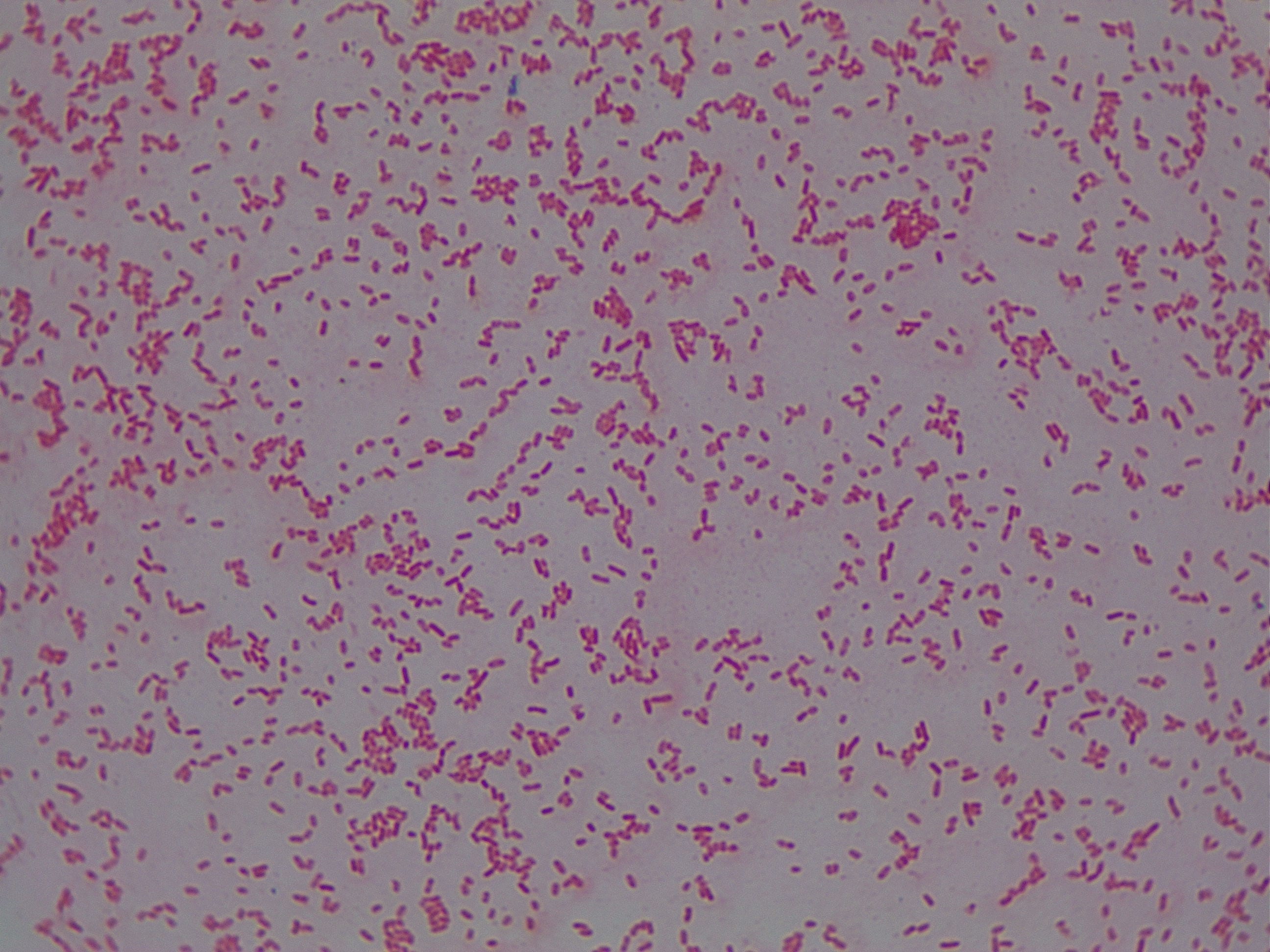
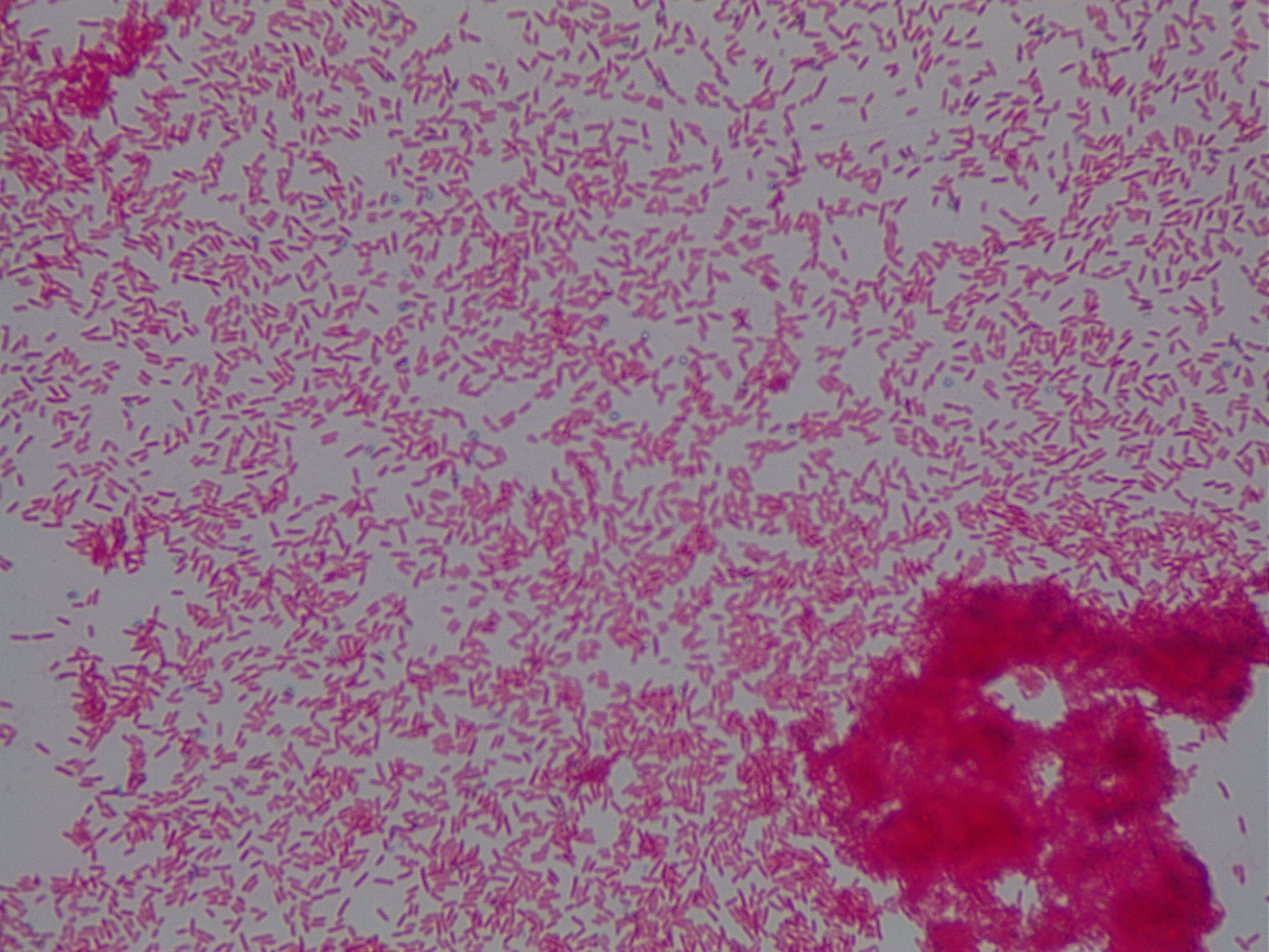
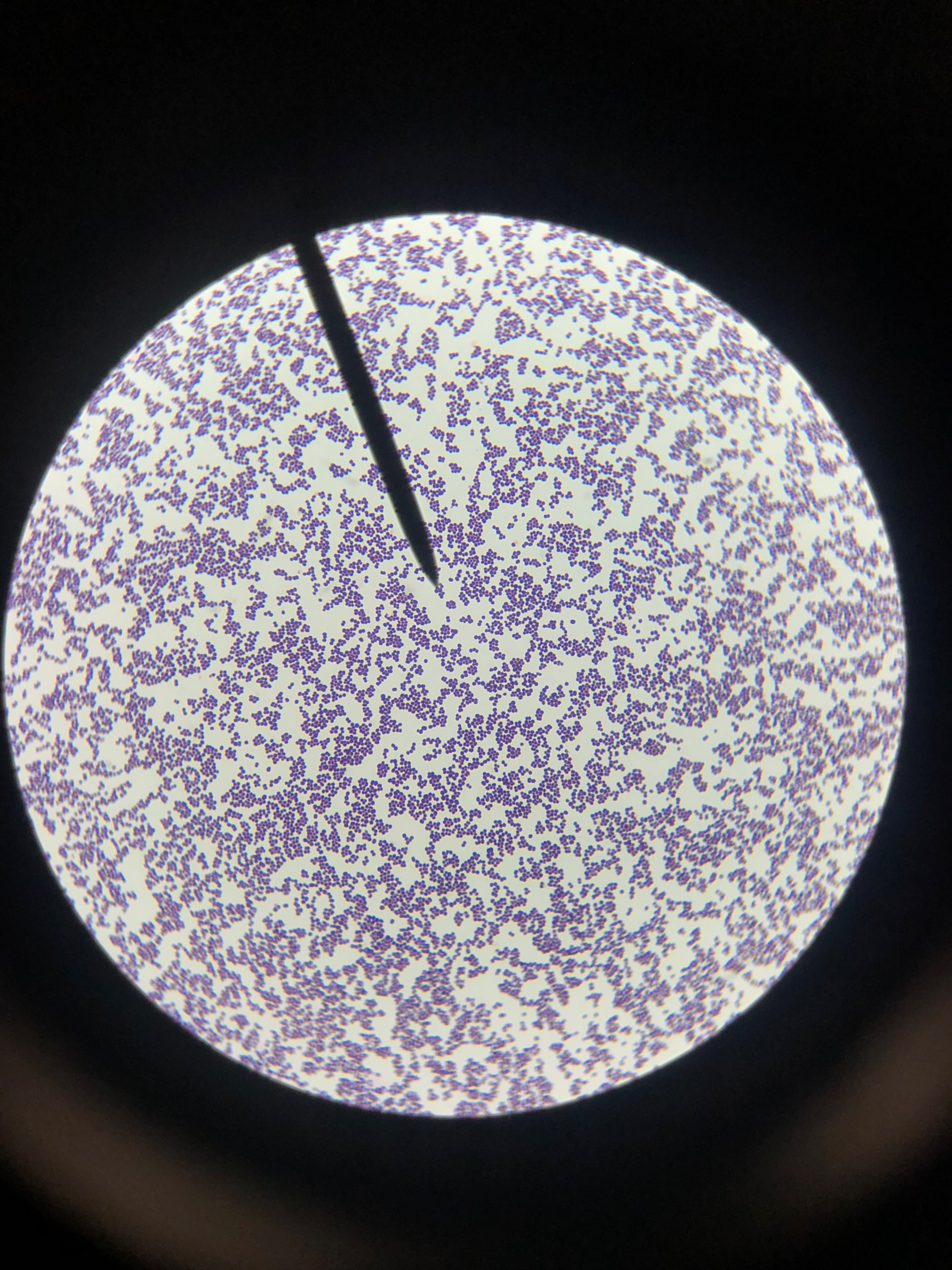
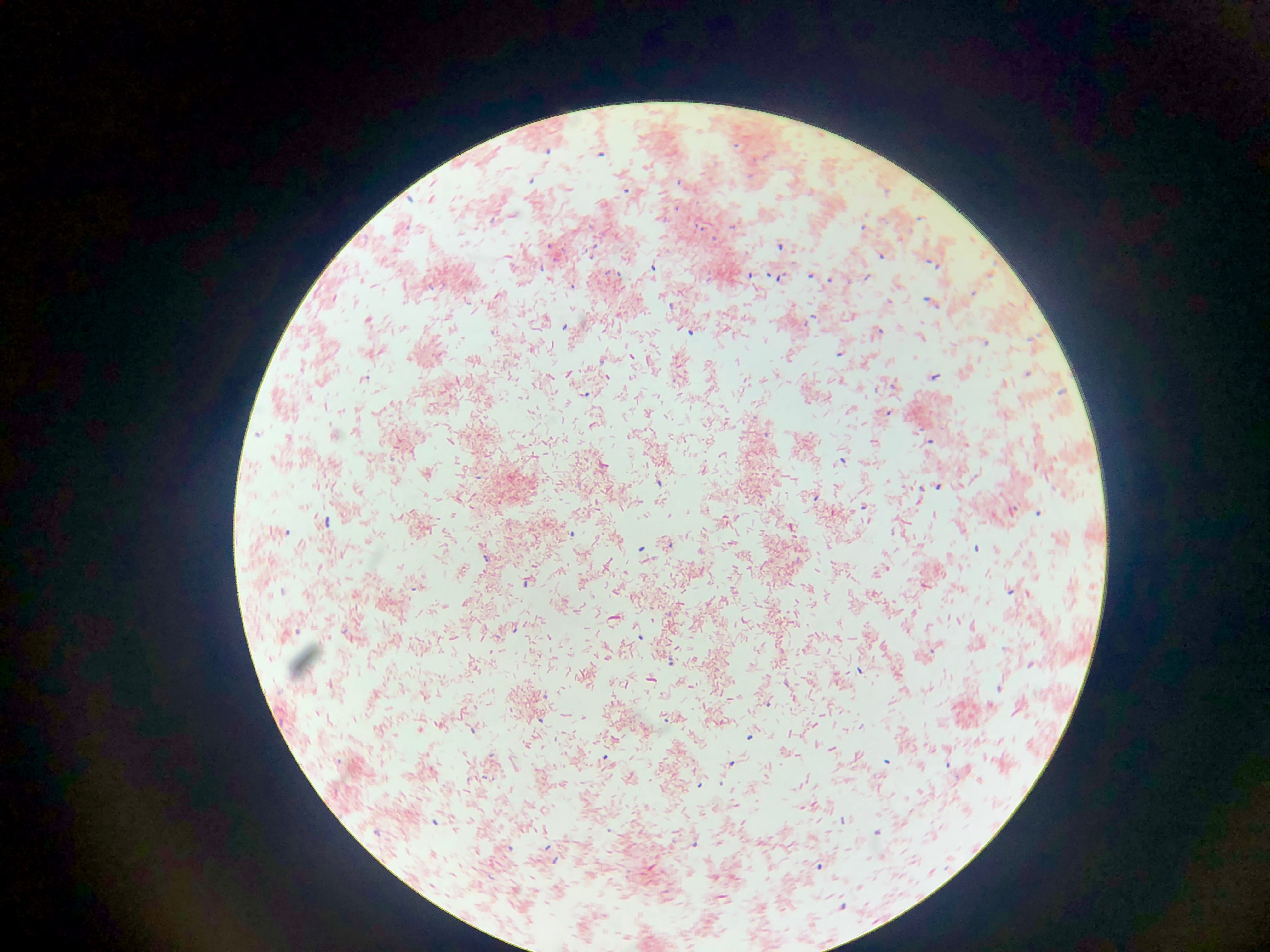
These four microbes were then stained to determine if they were gram positive or negative. All images are taken under a 100X oil emersion microscope lens. For reference, here is the positive (Staphylococcus epidermidis) and negative (E. Coli) controls.
The following are of my isolates. The first image is of R2A #18. This microbe is gram positive, and appears in diploid rods. The second is LB#2, which is gram negative and appears to be a shortened more ovular rod, and tends to be in diploid clusters. The third, PDA #2, is a gram negative cocci, and is generally diploid, but forms larger clusters as well. Lastly, PDA#5, is gram-negative, and the cells are shaped like a typical rod bacterium.