Meet my microbes – NIcole


I sort of wish I had better photos.

My first 10% TSA patch plate. Look at the variety of colors. If you look closely, since I don’t know how to edit this picture, you can see that the microbes in spaces 3, 9, 11, and 21 have a check mark by them, meaning that they had zones of inhibition when tested on the safe ESKAPE strains.

The antibiotic producers that I chose to try to put on streak plates. I don’t know why this picture is sideways.

A Gram stained image of the bacteria I called v13.

A Gram stained image of the bacteria I called v19. I don’t know why it is a bluer purple than the other image.

And the name of my microbe is… Nicole

Neither of my bacteria had a successful 16S rRNA analysis. While mine look sort of like Streptomyces under the microscope, I’m going to call v13 Sheila, and v1Manuel. Those are good names.

The two DNA sequences you gave me are either Burkholderia or the really closely related Paraburkholderia (the only difference between these two geneses is that Paraburkholderia is never pathogenic and just lives quietly in dirt. In fact, people realized that they are distinct clades only very recently).

When looking at the data for the 08E isolate, the most related species was Paraburkholderia phenazinium, though of course there are other really similar species. I can’t find morphology through the links provided by BLAST, but a brief Google search says that bacteria in the Paraburkholderia genus are Gm-, slightly curved rods. They are soil bacteria, but a group grew it on what looks to be a rich media because there are 3 grams of meat. The bacteria can create Iodinin, which induces apoptosis by possibly breaking the DNA. While that is technically an antibiotic, I wouldn’t recommend using it.

Extract News -Nicole

I tried to extract organic molecules from the plate containing the colonies I called v1 3 and v1 9. I tested it against the Gm+ Staph epi and B. subtilis, and the Gm- E. caratavora and P. putida, since one or both of my isolates inhibited each one. There was only a faint zone of inhibition in the B. subtilis from the v1 3 extract. Since the isolates were only grown for one day, it could be that there wasn’t much of any production of secondary metabolites yet.

Meet the ESKAPE pathogens: Staphylococcus aureus (Nicole B)

About 30% of people have staph bacteria, usually living commensally on the skin and in the nostrils. Occasionally, staph can break the skin and cause infections ranging in seriousness from small boils to sepsis and pneumonia.

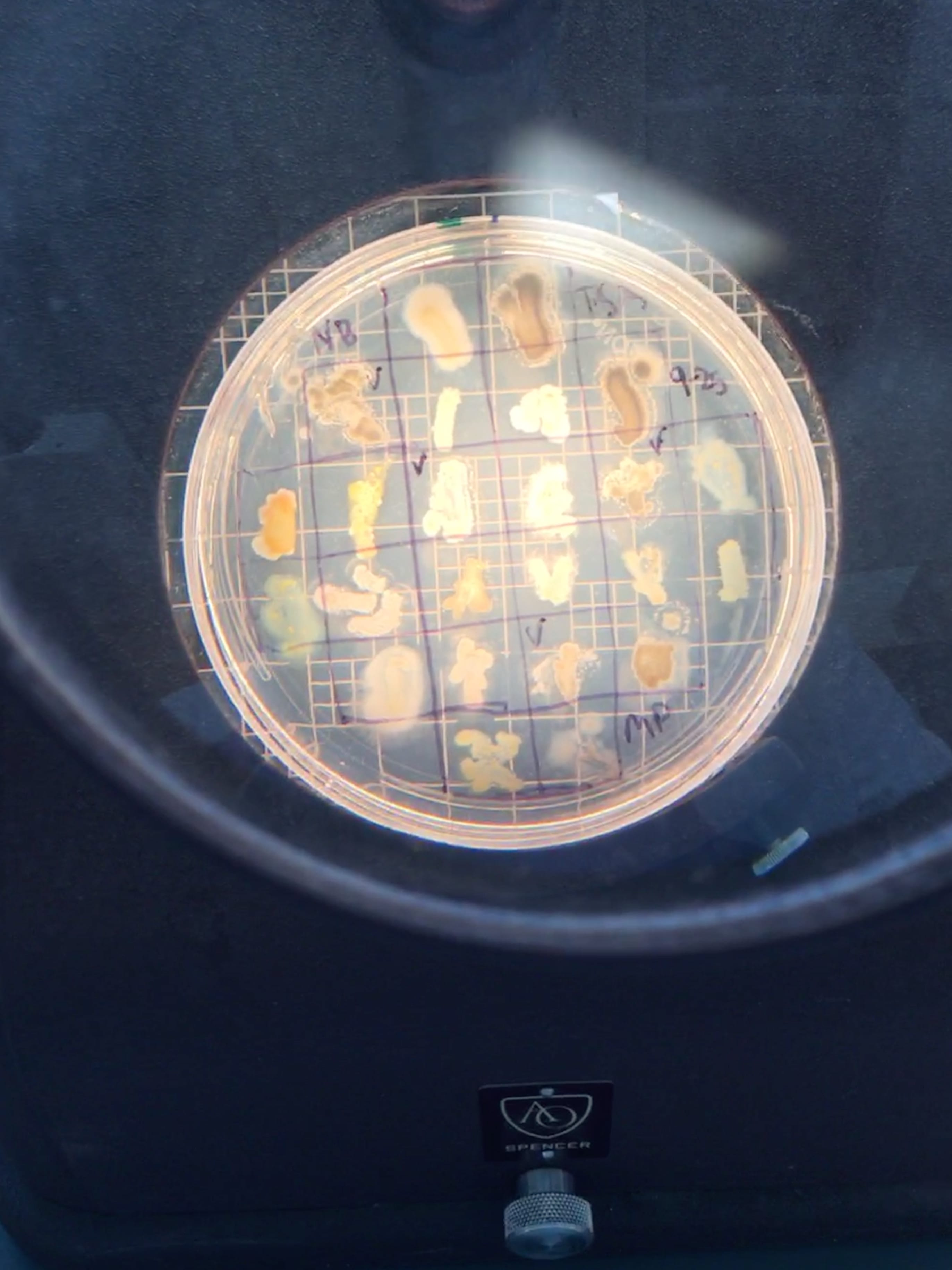
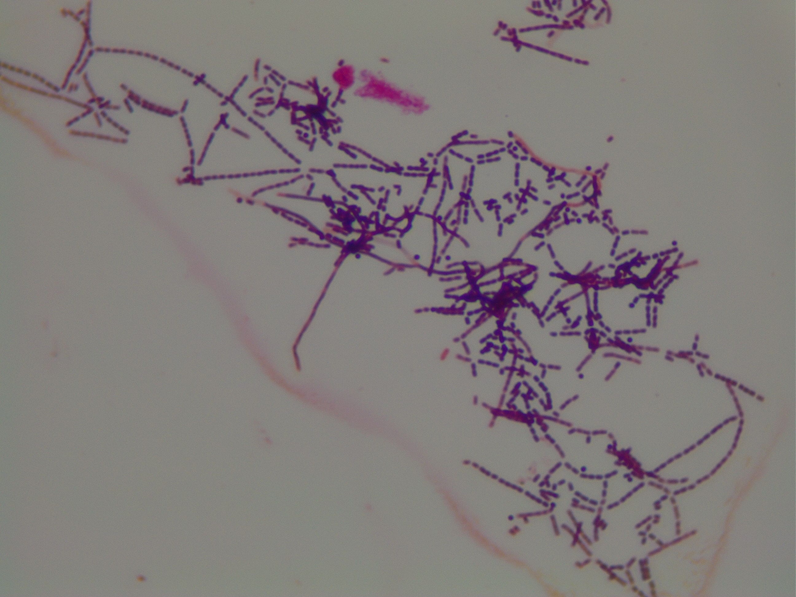
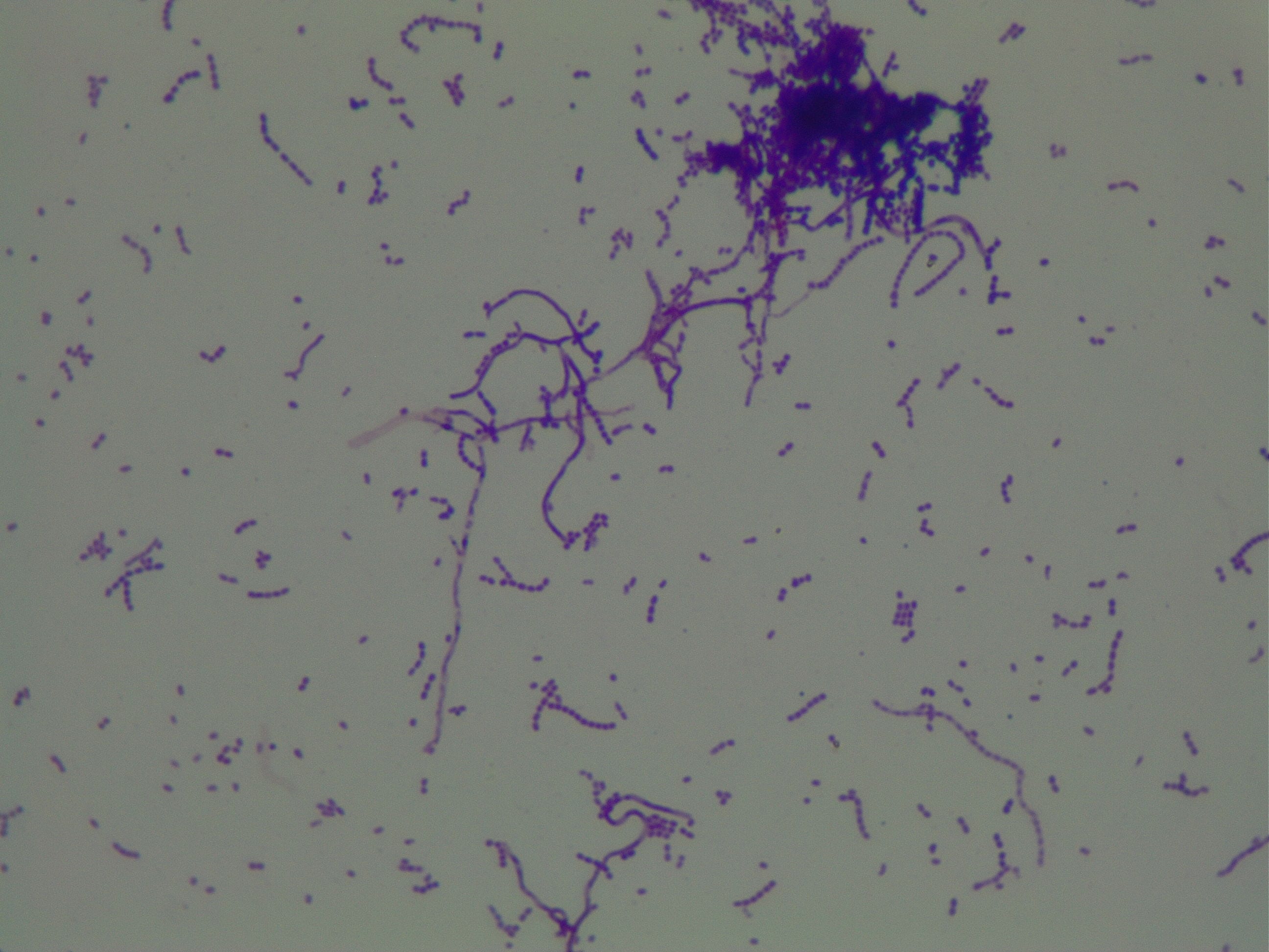
Staphylococcus bacteria are named so because they are round, or coccus shaped, and they are arranged in grape shaped clusters (the Greek word for grapes is transliterated to staphule). On plates, S. aureus usually create yellow or golden colonies. They grow readily between 18 and 40 degrees Celsius, and are faculitative anaerobes, meaning they will use oxygen if available but can live without it.

Staphylococcus aureus is believed to be living peacefully on the skin or in the nose of 1/3 of the world’s population. In 2006, it was found that for every 10,000 visits to the hospital, 410 of those visits were for a staph skin infection such as abscesses and cellulitis; the number of skin infections in general might be much higher due to the relative lack of seriousness of some skin problems like impetigo and boils. Staph bacteremia, or presence of staph bacteria in the blood, is the cause of an estimated 23% of all cases of sepsis, and can lead to infection of the heart and infection of the bones. Staph is also a common complication of pneumonia, and S. aureus is implicated in more than 40% of healthcare acquired pneumonias. As you can see, staph infections can occur from a variety of ways; the best prevention is simply commonsense risk mitigation, such as cleaning open wounds and having good hygiene.

The antibiotic-resistant form of staph that everyone is afraid of is MRSA (methicillin-resistant Staphylococcus aureus). MRSA infections are commonly acquired in health care settings, but up to 12% of MRSA infections now are from the broader community. Oddly, MRSA refers to staph resistant against other antibiotics, though most antibiotics it is commonly resistant to is in the beta-lactam class, which includes methicillin and penicillin. Resistance happens when staph creates the enzyme beta-lactamase to cleave an important bond in the antibiotic. It recently became resistant to vancomycin, the antibiotic doctors usually go for when they treat MRSA. The antibiotics Bactrim, clindamycin, minocycline, and doxycycline are still effective and are still widely prescribed to patients.

Instead of playing with Stapholococcus aureus in the lab, we are using Staphylococcus epidermidis.


https://www.ncbi.nlm.nih.gov/books/NBK441868/

https://www.cdc.gov/mrsa/community/index.html

https://cmr.asm.org/content/10/3/505.long

https://www.ncbi.nlm.nih.gov/pmc/articles/PMC4451395/

https://www.ncbi.nlm.nih.gov/pmc/articles/PMC3367609/

https://en.oxforddictionaries.com/definition/staphylococcus

https://en.oxforddictionaries.com/definition/aureus

https://www.ncbi.nlm.nih.gov/pmc/articles/PMC3372331/

https://www.the-hospitalist.org/hospitalist/article/125986/what-best-treatment-adult-patient-staphylococcus-aureus-bacteremia

https://academic.oup.com/femsre/article/41/3/430/3608758

https://www.uptodate.com/contents/methicillin-resistant-staphylococcus-aureus-mrsa-beyond-the-basics

Fun with soil- Nicole B

Where did you obtain your soil sample?
I got my soil sample in the manure pile at the barn my aunt rides at, Graystone Stables. I tried to go to the older and farther back part of the pile, and I took my sample from a layer about two inches below the surface of the pile.

Why did you choose this location?
I figured that not many other people have searched through a manure pile, and going to an actively decomposing mix of hay, wood shavings, and horse poop would reveal more than taking a sample from already spread dirt. I went to the back to try to get at a point where the feces was already decomposed to avoid fresh ones. I noticed three distinct layers in the manure pile: a top layer of dry but still presumably nutrient rich stall products, a middle layer of moist, dark, and slightly warm stall products that I assume was actively decomposing, and a bottom layer of dry and crumbly hay and soil. I took my sample from the middle layer.

Do you expect a lot of isolates? Why or why not? Have your initial observations supported this?
Yes, I did. Not only did I know that the results of a manure pile can support a lot of growth, the soil was warm to the touch due to the decomposers (I washed my hands afterwards). My observations have definitely supported this because I have an uncountable number of colonies even on my 10^-3 dilution plate.

What media did you choose? What dilutions?
I selected LB and AC media because they were nutrient rich and I figured the bacteria in my soil sample would like that, and I chose 10% TSA because it would make a good representation of bacteria that didn’t thrive in rich environments. I also didn’t have many others to choose from. Due to a misunderstanding of dilution, I made 10^-1, 10^-2, and 10^-3. I also made a 10^0 dilution just to see what that would look like, and threw it away after realizing it wouldn’t give me much data.

Will you need to redo any?
Over the weekend, I noticed that my 10^-3 dilution plates had a ton of colonies, making it uncountable (or really difficult to count). On Tuesday, I made 2 spread plates at a 10^-4 dilution for each media I used. The plates are in the incubator right now and are growing.

How did your sample differ on the different media?
So far, I’ve noticed that there are more green and blue tinted bacteria colonies on the 10% TSA plates than on the other plates, but the colors could simply be more noticeable due to the whiter color of the media. The 10% TSA plate also had white/grey colonies with rhizoid arms on the border. There were a couple of colonies on the AC plate that appeared to tower over the rest of the bacteria, rising about 0.5 cm above the plate. The LB plate had more fuzzy colonies on it, and I noticed more brighter yellow colonies on the plate compared to AC.