
What were the results of your 16S analysis?
The PCR results were successful in amplifying the DNA in all three strains that were tested, and their sequences were used to run individual BLAST procedures. Strain 1A (seen in figure 1) yielded BLAST results (figure 2)that revealed that it was a Gm- bacteria belonging to the Klebsiella genus.

Figure 1: Strain 1A isolated streak plate

Figure 2: BLAST sequencing results for unknown strains show relatedness of different strains of bacteria to the proposed DNA sequence via the 16S analysis tool.
Successful results were also observed with strain 3A and 2B, while the BLAST sequences for these two retrieved 99% similarity between the tester strain’s DNA sequences and the BLAST 16S database [1]. It is important to note that not 100% similarity was observed with any of the BLAST results – which may be a cause of the PCR DNA sequences were not completely accurate. Strain 3A’s 16S analysis was in line with strain 1A’s, as they were both of the Klebsiella genus. 2B’s 16S analysis, however, indicated a different genus than strains 1 and 3A. 2B’s results suggested that it was in the genus Massilia, another common soil bacteria [2].
Does your gram stain agree?
My gram staining agreed for the Gm+/-, but I found a more difficult time in identifying the shape of the individual bacterial cells. The gram staining that I produced suggested that samples 1A and 2B were coccus-shaped Gm- bacteria, while sample 3A was a rod-shaped Gm- bacteria (Figures 3, 4, and 5). 1A and 2B analysis contradicted the 16S analysis, however, after analysis of the strains with BLAST [1]. It was observed that all three bacteria strains were rod-shaped Gm- bacteria. A reason for this mislabeling may have been that the bacteria cells were simply too small to accurately identify their shapes.

Figure 3: Gram staining from bacteria colony sample 1A showing Gm- rod-shaped Klebsiella.
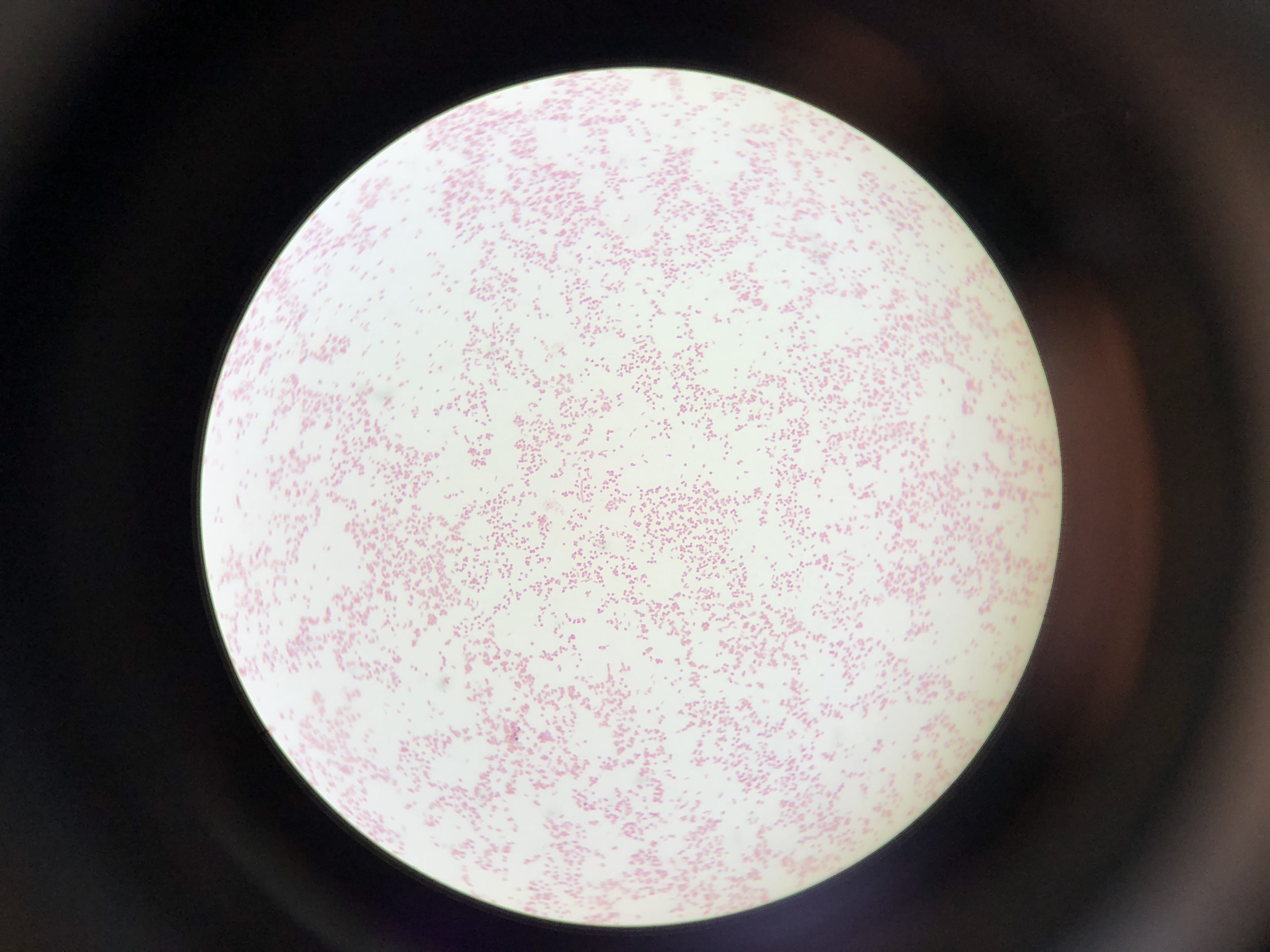
Figure 4: Gram staining from bacteria colony sample 2B showing Gm- rod-shaped Massilia.
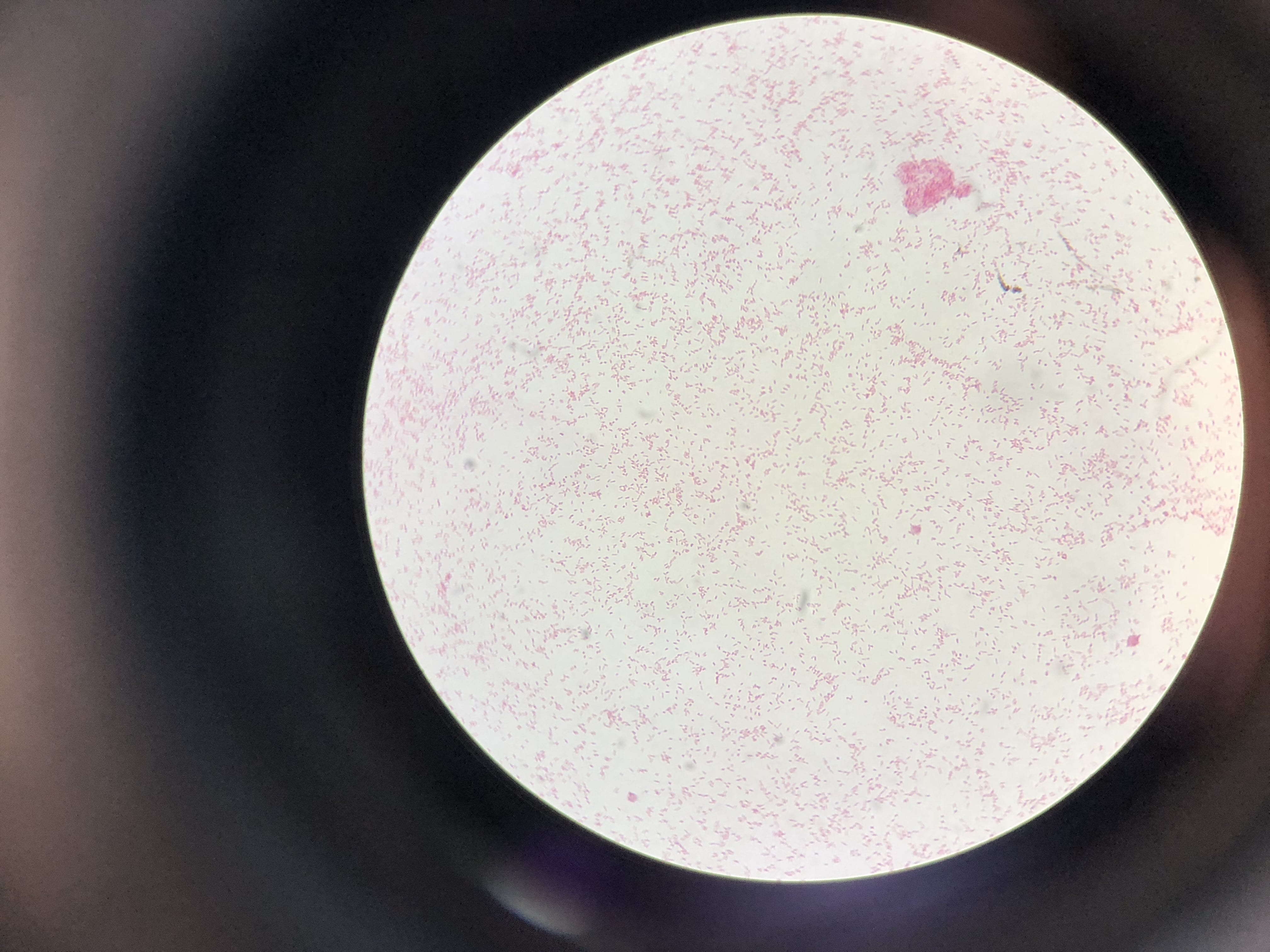
Figure 5: Gram staining from bacteria colony sample 3A showing Gm- rod-shaped Klebsiella.
Pick one of your isolates and find out more about the genus (it is unlikely you will be able to determine the species).
a) General cellular and morphological characteristics of the genus (taxonomic classification, nutrition, cell shape, habitat).
Klebsiella bacteria are an aerobic bacteria that are part of the phylum proteobacteria. They are of the class gamma proteobacteria, order Enterobacteriales, family Enterobacteriaceae, and genus Klebsiella [3]. They are gram negative rod-shaped bacteria that are often found in soil. They do not require any special nutrients to grow, as they can survive off of citrate, glucose, and ammonia; this makes them effective at growing in low-nutrient environments. They are normally found in the nose, mouth, and gastrointestinal tract in humans, but they can also be spread to other parts of the body to cause diseases like pneumonia and urinary tract infections.
b) Information regarding antibiotic production in this genus.
Not much information is known about the production of antibiotics in the genus Klebsiella, but they are the subject of antibiotic research as they are growing more and more resistant to normal antibiotics. Their ability to survive in low-nutrient environments allows them to stay alive in dire environments, and they have recently evolved an ability to cleave carbapenem antibiotics with an enzyme carbapenamase. This enzyme has rendered the entire line of carbapenem antibiotics useless against Klebsiella bacteria[4]. It is mutations like this that has researchers working toward discovering new antibiotics, because bacteria are constantly evolving to survive – posing a greater threat to humanity every day.
Works Cited
- “BLAST: Basic Local Alignment Search Tool.” National Center for Biotechnology Information, U.S. National Library of Medicine.
- Shen, Liang, et al. “Massilia Eurypsychrophila Sp. Nov. a Facultatively Psychrophilic Bacteria Isolated from Ice Core.” International Journal of Systematic and Evolutionary Microbiology, Microbiology Society, 1 July 2015.
- Thomas, Gavin. “Klebsiella Pneumoniae.” What Is Electron Microscopy?.
- Daikos, George L., et al. “Carbapenemase-Producing Klebsiella Pneumoniae Bloodstream Infections: Lowering Mortality by Antibiotic Combination Schemes and the Role of Carbapenems.” Antimicrobial Agents and Chemotherapy, American Society for Microbiology Journals, 10 Feb. 2014.