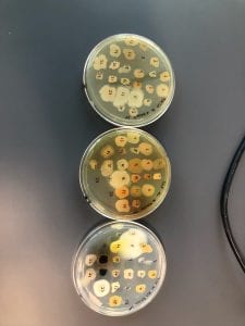
This is a picture of my first set of spread plates. From left to right, the dilutions are 10^-2, 10^-3, and 10^-4. From top to bottom, the media used is PDA, LB, and AC.
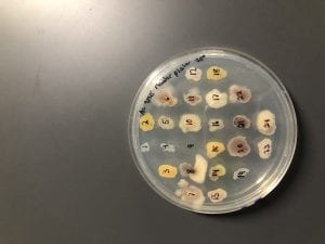
This is a picture of my three patch plates. From top to bottom, the media is LB, AC, and PDA.
This is my patch plate on PDA. I just love the colors of the bacteria on this!
These are two of my patch plates against tester strain #6. The top plate is on AC, and colony #15 showed inhibition. The bottom plate is on LB, and #14 showed inhibition.
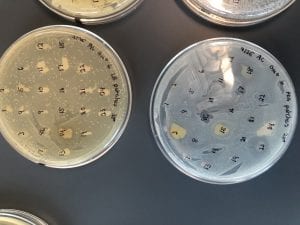
These are two of my patch plates against tester strain #3. The plate on the left is on AC, and colony #23 showed slight inhibition. The plate on the right is on LB, and #13 and #14 showed inhibition.
The plate on the left is the same as the LB plate against tester strain #6 as seen above. The plate on the right is my patch plate against tester strain #6 and colony #17 is showing inhibition.
The plate on the left is the same as the LB plate against tester strain #3 as seen above. The plate on the right is my patch plate on PDA against tester strain #3 and #13 showed inhibition.
These are all of my patch plates on AC, LB, and PDA (from left to right).
This is a streak plate on PDA of two of my potential antibiotic producing bacteria. I love the pink color of colony 13!
These are streak plates of the rest of my interesting colonies. From top to bottom, there’s my colony #15 and Nikki’s colonoy #7 on AC, then my colonies #3 and #23 on PDA, then my #13 and #23 on PDA, and lastly my #13 and #14 on LB.
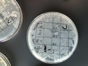
I patched four of my isolates against tester strains on PDA. Shown above is a patch plate against tester strain #9 and my colony #17 is showing great inhibition!
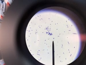
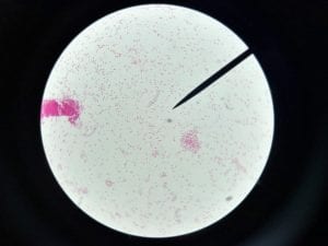
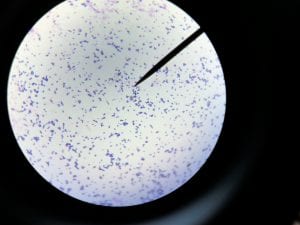
Gram stain of colony #13. It’s gram positive and rod shaped.
Gram stain of colony #14. It’s gram negative and rod shaped.
Gram stain of colony #15. It’s gram positive and coccus shaped.
Gram stain of colonoy #17. It’s gram negative and rod shaped.


