And the name of my microbe is -Carly B

What were the results of your 16S analysis?

I ended up getting two of my samples sequenced, #2 and #4.  After analyzing the sequences from BLAST I found that #2 was from the Klebsiella genus with 1062 bp and 98% query cover (4). Sample #4 was from the Acinetobacter genus with 790 bp and 99% query cover (4).

Does your Gram stain test agree?

The Gram stain test for #2 matched the results from BLAST of the Klebsiella genus (2). It was a Gram negative bacteria with oval shaped bacteria.

The Gram stain test for #4 did not match with the Acinetobacter genus because this genus is Gram negative (1). I got a Gram positive bacteria with a rod shape from the Gram stain test.

General cellular and morphological characteristics of the genus

Klebsiella is a Gram negative bacteria that is rod shaped (3).   It is from the Enterobacterales order and the Enterobacteriaceae family (4).  It normally lives in the mouth and gut flora but can be pathogenic (3).  It can also be found in soil, water, and plants (3).  Almost all Klebsiella can be grown on minimal media with ammonium ions or nitrate (5).  Klebsiella grow as yellow, dome-shaped colonies that are also often mucoid (5).  They are an anaerobic, non-motile species (3).  The Klebsiella genus is resistant to many commonly used antibiotics and is responsible for some nosocomial infections (3).  The most common infection is pneumonia which is caused by the species Klebsiella pneumonia, a community acquired pulmonary infection (5).

Klebsiella pneumoniae Grown on Nutrient Agar

Picture from Medical-Labs: Medical Laboratories Portal.

Gram-negative Bacilli of Klebsiella pneumoniae

Information regarding antibiotic production in this genus

There is not a lot of information about the antibiotics produced by species of Klebsiella.  There is, however, a lot of information on the antibiotics they are resistant to, including aminopenicillins and carboxypenicillins (5). Many Klebsiella bacteria produce beta-lactamase which is an enzyme that gives them resistance to certain antibiotics (5).

References

  1. Nemec, A. et al. “Genotypic and phenotypic characterization of the Acinetobacter
    calcoaceticuse Acinetobacter baumannii complex with the proposal of
    Acinetobacter pittii sp. nov. (formerly Acinetobacter genomic species 3)
    and Acinetobacter nosocomialis sp. nov. (formerly Acinetobacter
    genomic species 13TU)”. Research in Microbiology, vol. 162, 2011, pp. 393-404.
  2. Diene, S. et al. “The Rhizome of the Multidrug-Resistant Enterobacter aerogenes Genome Reveals How New “Killer Bugs” Are Created because of a Sympatric Lifestyle”. Molecular Biology and Evolution, vol. 30, no. 2, 2013, pp. 369–383.
  3. Buckle, Jane. Clinical Aromatherapy. Elsevier, 2015.
  4. BLAST, NCBI, 2018. Accessed 11 November 2018.
  5. Brisse, S. Grimont, F., & P. Grimont. The Prokaryotes. Springer, 2006, pp. 159-196.

Extract news! -Carly B

I had two samples, 2 and 4, to perform the extract with.  Sample 2 did not evaporate so I could not add methanol to it but sample 4 did evaporate.  I performed the agar disc diffusion test with my two extracts against tester strains #3, #4, #6, and #7.  Strain #4 was E. raffinosis and #6 was B. subtilis and they were Gm+.  Tester strain #3 was E. carotovora and #7 was E. aerogenes and they were Gm-.  I chose E. raffinosis and B. subtilis because my samples previously showed inhibition against these two strains and then I randomly chose two Gm- tester strains because my samples did not show previous inhibition of any Gm- tester strains.  Extract 2 did not inhibit any of the tester strains.  Extract 4 inhibited E. raffinosis and B. subtilis, both Gm+ bacteria, so this sample seems to be an antibiotic producer against Gm+ bacteria.


Meet My Microbes- Carly Burns

These are my master patch plates from 9/17 and 9/25.  The LB plates were labeled group “a”, the R2A plates were labeled group “b”, and the AC plates were labeled group “c”.

These are the patches from 9/25 from group a and b tested against the Gm+ tester strain E. raffinosus and the Gm- tester strain E. carotovora.  No patches showed possible antibiotic production against them.

One set of these plates are patches from group c made on 9/25 against the Gm+ tester strain E. raffinosus and the Gm- strain E. carotovora.  None of these patches showed possible antibiotic production so a new patch plate with different colonies from the original plate were made and tested against E. raffinosus and E. carotovora on 9/27.  These patches also showed no possible antibiotic production.

Since no patches showed possible antibiotic production, all of those plates were tossed in the trash and I took two possible producers from Elizabeth and made this streak plate on 9/27 for further tests.  After testing these two bacteria against all the tester strains I found that only #10 was a possible producer so I took this bacteria and two bacteria from Tommy and performed a PCR reaction with them.  These PCR reactions were then run via gel electrophoresis and this showed that only the two cultures I got from Tommy contained the band we were looking for so I sent these two out to be sequenced.

I performed the Gram stain test on the two cultures and one of them ended up being Gm+ and the cells had a deep purple color and were rod shaped (above).  This is a picture of one culture under 100x magnification.

The Gram stain test for the other culture showed that the cells were Gm- and the cells had a pink color and were oval shaped (above).  This is also a picture under 100x magnification.


Meet the ESKAPE pathogens: Carly Burns and Sofia Boswell

Assigned ESKAPE Pathogen: Klebsiella pneumoniae

Why is this ESKAPE Pathogen of interest (in brief)
Klebsiella pneumoniae is a common nosocomial (originating in a hospital) pathogen that accounts for about 10% of all infections acquired within a hospital setting (1). It is also a common pathogen responsible for community-acquired infections, as well as a number of other diseases such as meningitis, septicemia, purulent abscesses, and pneumonia (1). Additionally, as some strains are nitrogen-fixing, K. pneumoniae is of agricultural interest because it has been observed to increase crop yields in certain agricultural conditions.

General Cellular and Morphological Characteristics of the Organism (taxonomic classification, nutrition, cell shape, habitat)
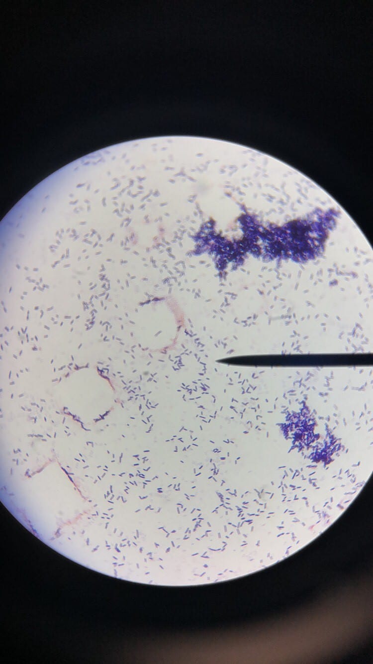
K. pneumoniae is a Gram negative, non-motile, lactose-fermenting, facultative anaerobic, rod-shaped bacterium. It is a part of the Enterobacteriacae family (6). K. pneumoniae is commonly found in the normal flora of the mouth, skin, and intestines, but can become destructive and cause damage if inhaled, specifically to the alveoli. K. pneumoniae occurs naturally in the soil and about one third of strains are nitrogen-fixing in anaerobic conditions.

https://chrislima90.weebly.com/blog/top-advice-on-klebsiella-pneumoniae

http://faculty.ccbcmd.edu/courses/bio141/labmanua/lab12/Mac_kpneumoniae.html
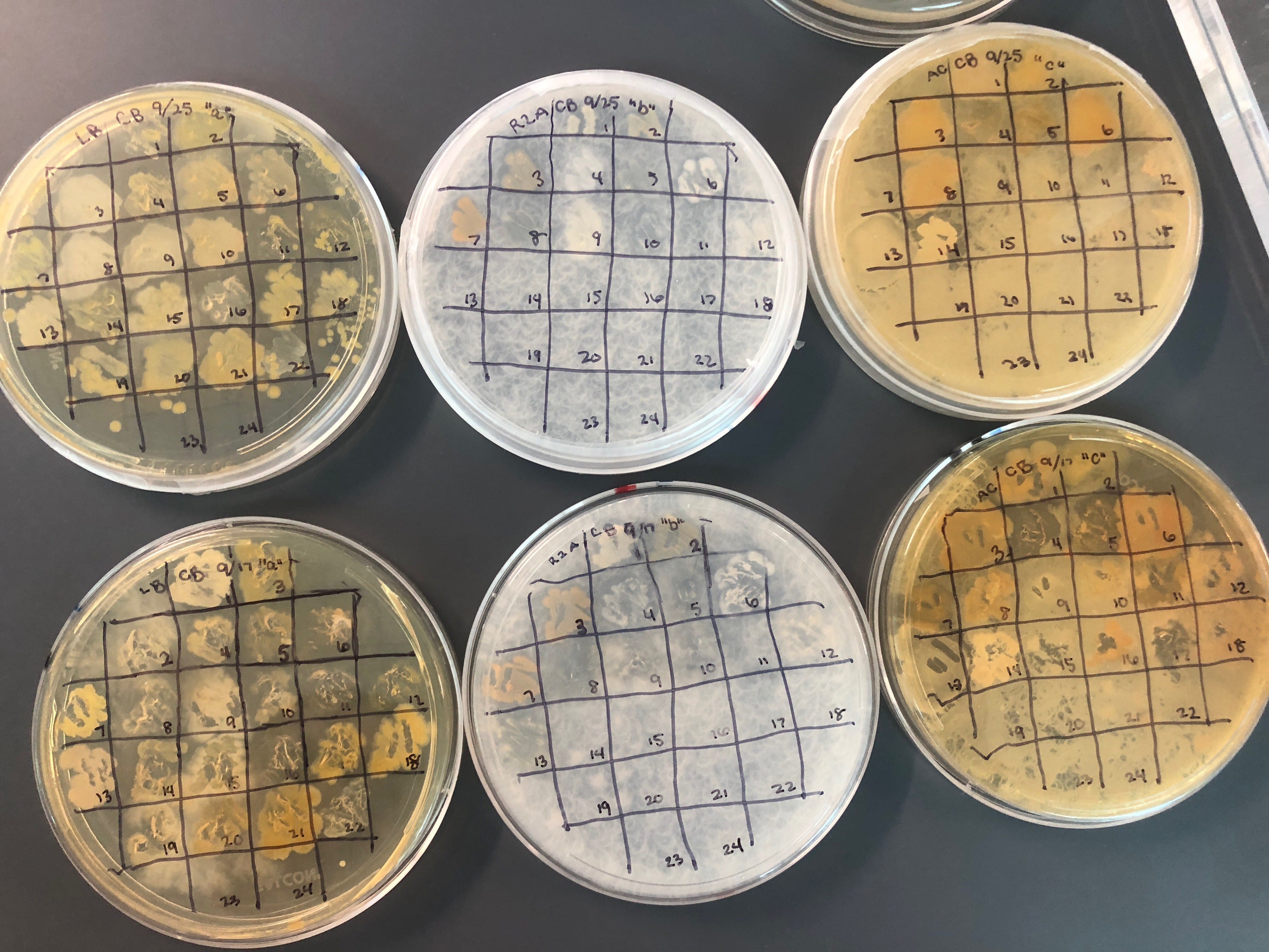
(Grows very well on MacConkey agar)

Clinical Importance and Prevalence
K. pneumoniae originated in a hospital setting and is now globally prevalent in hospital and community settings (6). Infections are more common in the young, the old, and the immunocompromised (6). Enterobacteriacae are becoming more and more difficult to treat because of their increasing resistance to antibiotics through the production of different enzymes such as extended-spectrum beta-lactamases and carabapenemases (6).

Infection (How does the infection occur and where is it localized?)
K pneumoniae infections are typically seen in individuals with a compromised immune system, and most infections are seen in middle-aged or older men who already have debilitating diseases. Healthy people typically do not get Klebisella pneumonia infections (3). Humans serve as the primary reservoir for this pathogen, and the pathogen is typical transmitted between individuals through fecal matter and nasal secretions. K. pneumoniae is rarely carried on the skin (2). Carrier rates of the pathogen increase significantly in hospital patients (2). Klebisella pneumonia is spread through contact, either person-to-person contact (i.e. touching the hands of an infected individual) or environmental contact (i.e. touching a contaminated surface). The bacteria cannot be spread through the air (3). Certain medical tools such as ventilators and catheters may cause an individual to be exposed as well (3). K pneumoniae are capable of infection because of fimbrial adhesins and a thick capsule that is comprised of two layers of polysaccharide fibers (6).

Pathology (What disease is caused? What are the symptoms?)
The most common disease caused by Klebisella pneumoniae is pneumonia. This occurs when Klebisella bacteria enter the respiratory tract and setting in the air sacs of the lungs, causing infection and inflammation (3). The most common symptoms of pneumonia are fever, cough, chest pain, difficulty breathing, and abnormal mucus production (4). Without treatment, pneumonia can become a very serious infection, especially in the elderly and those who are immunocompromised (4). Other diseases that can be caused by Klebisella pneumonia are septicemia, meningitis, endocarditis, and cellulitis (4).

Ineffective Antibiotics (Antibiotics to which the organism has acquired resistance)
Some K. pneumonia have become very resistant to a class of antibiotics called carbapenems (3). Resistant bacteria produce an enzyme called carbapenemase which renders carbapenems ineffective (3). These resistant bacteria are also known as KPC-producing organisms and are very difficult to treat (3).

Effective Antibiotics (Antibiotics known to inhibit the organism)
There are many effective antibiotics to treat K. pneumoniae (5). Beta-lactams, aminoglycosides, and quinolones are effective antibiotics against Klebsiella infections (5). Cephalosporins have also been used alone and in conjunction with aminoglycosides but should not be used if ESBL strains are present (5).

Corresponding Safe Relative
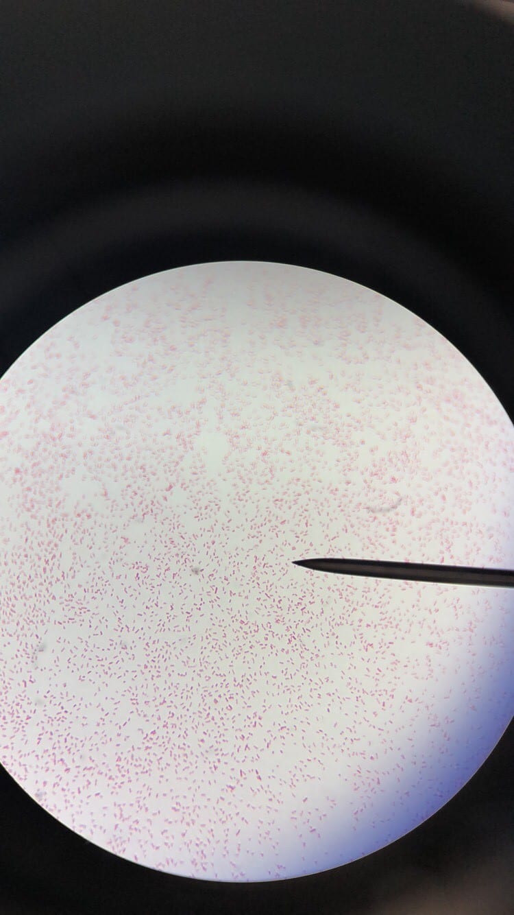
The corresponding safe relative is Escherichia coli (6). Most E. coli are commensal and make up about 0.1% of the of the normal intestinal flora, however some are pathogenic and has shown marked resistance to multiple antibiotics in the past decade (6).

Sources:
1. Guo, S., J. Xu, Y. Wei, Y. Li, and R. Xue. 2016. Clinical and molecular characteristics of Klebsiella pneumoniae venitllaor-associated pneumonia in mainland China. BMC Infec Dis. 16: 608
2. Yu, W.L., Y.C. Chuang. UpToDate. Clinical features, diagnosis, and treatment of Klebsiella pneumoniae infection. 2018. https://www.uptodate.com/contents/clinical-features-diagnosis-and-treatment-of-klebsiella-pneumoniae-infection
3. Centers for Disease Control and Prevention. Klebsiella pneumoniae in Healthcare Settings. 2012. https://www.cdc.gov/hai/organisms/klebsiella/klebsiella.html
4. WebMD. What is Klebsiella Pneumoniae Infection?. 2018. https://www.webmd.com/a-to-z-guides/klebsiella-pneumoniae-infection#1
5. S. Qureshi. Medscape. Klebsiella Infections Medication. 2017. https://emedicine.medscape.com/article/219907-medication#showall
6. Pendleton, J. N., Gorman, S. P., & B. F. Gilmore. Medscape. Clinical Relevance of the ESKAPE Pathogens. 2013.

2. Fun with Soil -Carly B

I collected my soil sample from the lawn in front of the Nott on campus. I dug down about an inch and then filled my test tube with soil and tried to avoid getting grass in it. The GPS coordinates were 42.8173 degrees N, 73.9300 degrees W.

I chose this location because I was interested in seeing what kind of bacteria was growing there since many students sit on the lawn in front of the Nott when it is nice out. I also wanted to see what was there because I know that lawn is constantly treated to keep up its appearance.
I do expect a lot of isolates because the lawn is treated so much that it makes it rich and good for sustaining the growth of the lawn and also bacteria.

So far this has been reflected on my culture plates. There has been a lot of growth and even a lot of overgrowth on most of my plates including the plates with less concentrated dilutions. I couldn’t count any of my plates from my first round of plating because they were so overgrown.

I chose to use LB, R2A, and AC for my media. I chose LB and AC because I figured the complex media would reflect their natural, rich environment of the lawn so I was confident I would get growth even with higher dilutions. I used R2A because I wanted to plate a minimal media since I figured the soil was so rich with bacteria that there might be almost too much bacteria to work with.

I plated a zero dilution, a 10^-1, and a 10^-2 dilution on R2A because this was the minimal media so I figured I should test out the more concentrated dilutions. I plated 10^-1, 10^-2, and 10^-3 dilutions for LB and Ac because they were the complex media so I figured the bacteria would grow more easily on this media and a less concentrated dilution would be the best to use.

Since pretty much all of my plates were overgrown and I couldn’t count cfu’s for any of them, I had to replate some dilutions. I replated a 10^-3 dilution on AC and R2A because I thought that if I left them in the incubator for a shorter amount of time before looking at them that I’d be able to count them and pick more isolated colonies and this was correct. I also replated a 10^-4 dilution on LB, AC, and R2A because there was overgrowth on all of my 10^-3 plates from the first round of plating so I thought doing a dilution with a lower concentration that it would be even better work with, however these plates barely had any growth after 2 days.

All three of my media had a lot of growth. I chose the fewest colonies from the R2A plates because those plates had some good colonies but there were a lot of extremely small colonies that were very close together which meant I couldn’t pick individual colonies up very easily. The LB and AC also had a lot of good colonies but they were very overgrown so it was hard to pick individual colonies. The colonies were overall larger on AC and LB and also more diverse.