And the name of my microbe is…Nikki

What were the results of your 16S analysis?

Isolate #/ Name

(Include # bp)

Closest Relative Identity

(%)

Query Coverage (%) Accession Numbers
#20

(677 bp)

Streptomyces

(vinaceus or cirratus strains)

99 99 NR_041131.1

NR_112388.1

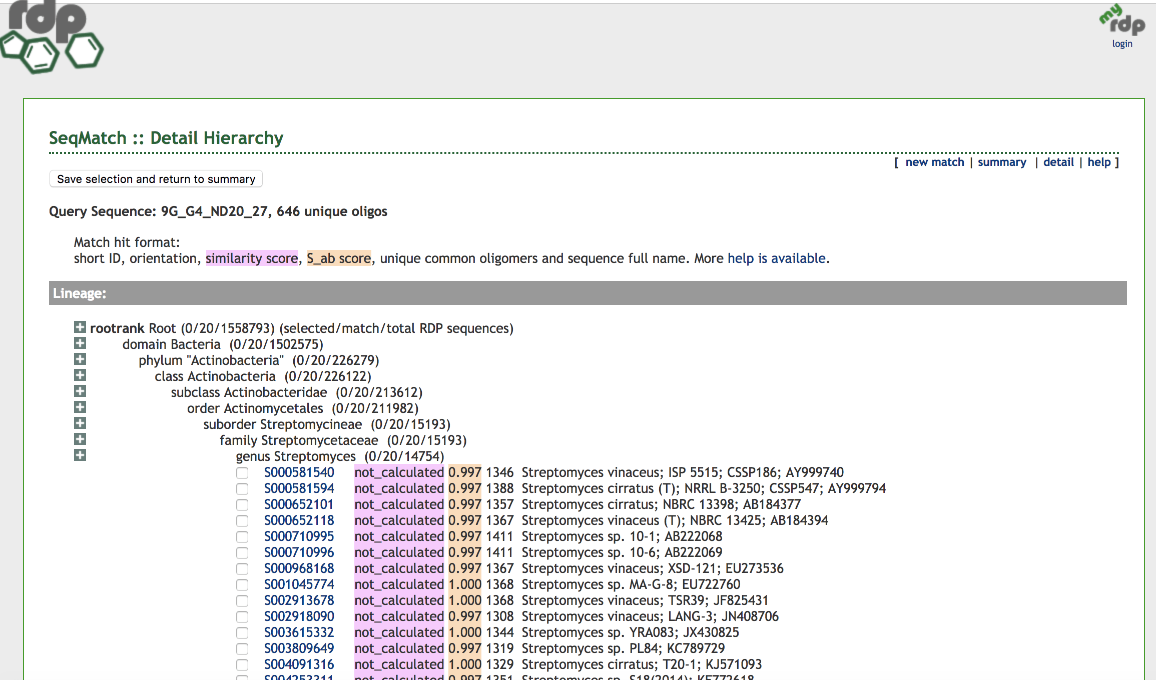
Unfortunately, I was only able to obtain 16S results for one of my isolates, #20, as the other two, #15 and #23 did not work. However, for #20, I was able to run 677 bp of reliable sequence in my Blast and Ribosome Database Project (RDP) searches. Both searches agreed on Streptomyces genus as the identify of my isolate with 99% identity match and 99% query coverage. Further, my searches determined the closest relative to be Streptomyces of the vinaceus or cirratus strains, but more informations and biochemical tests would be needed to distinguish any further.

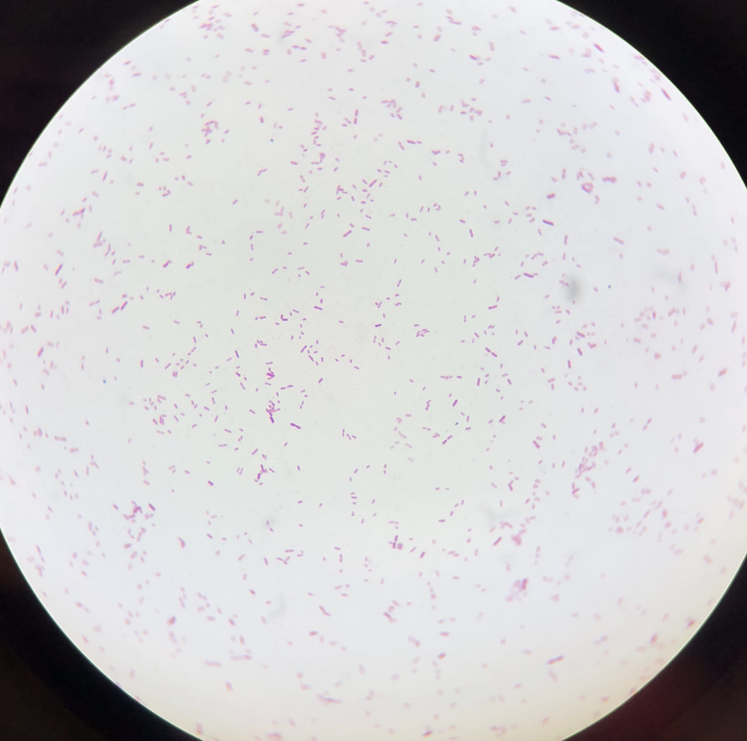
Does your gram stain agree?

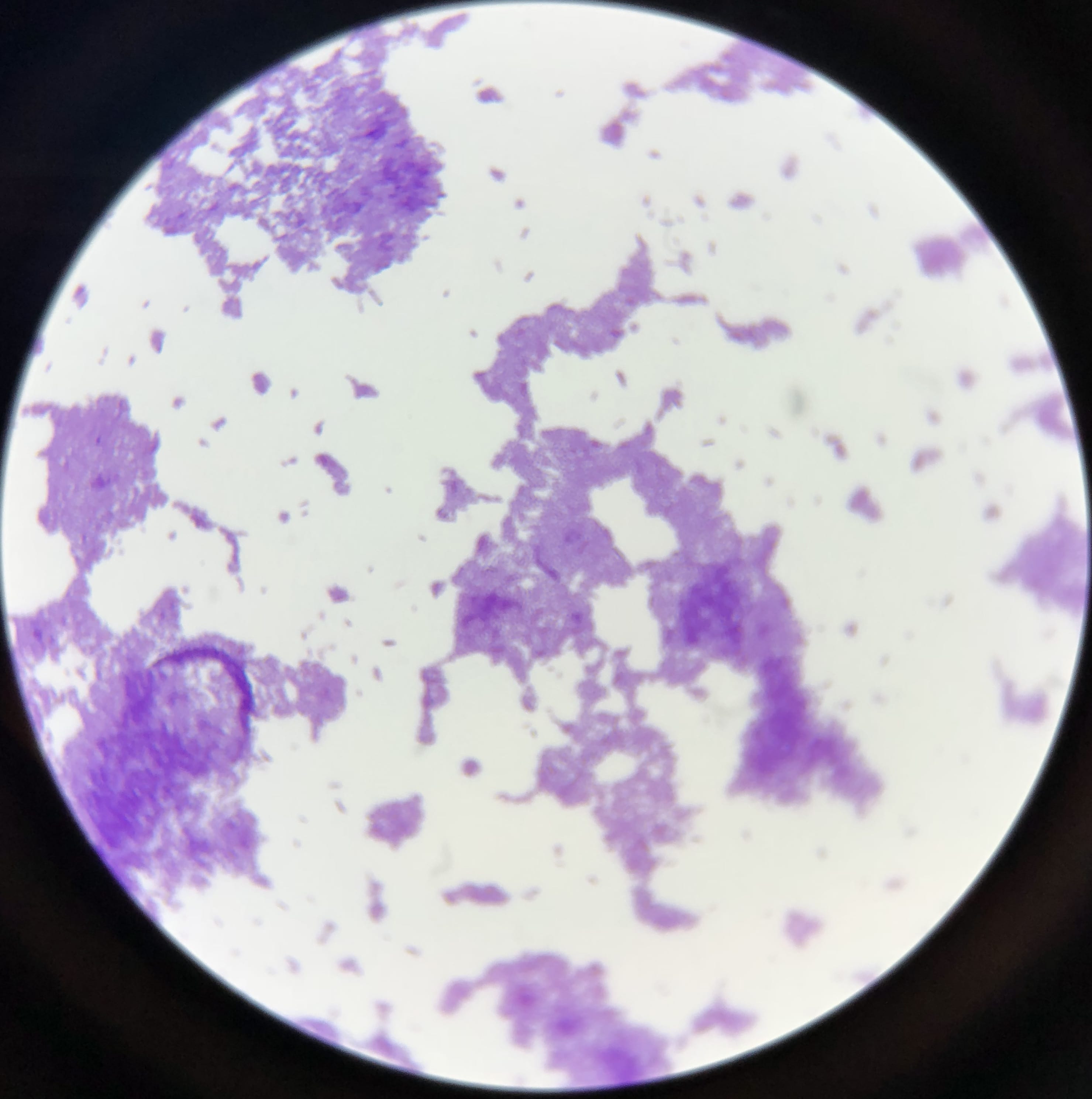
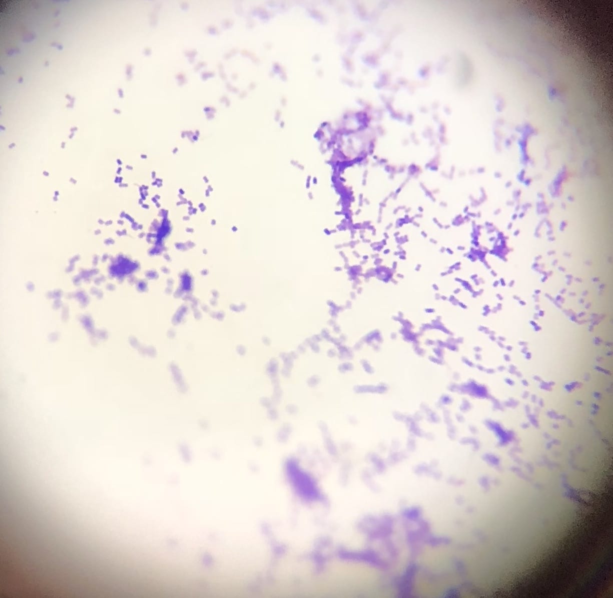
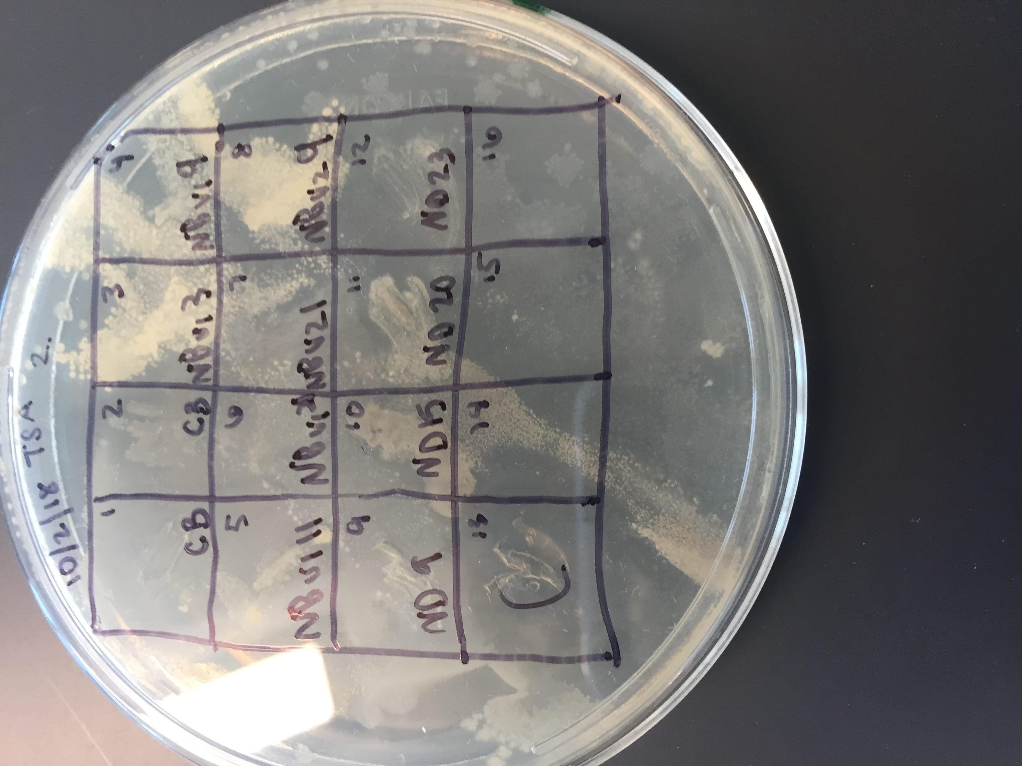
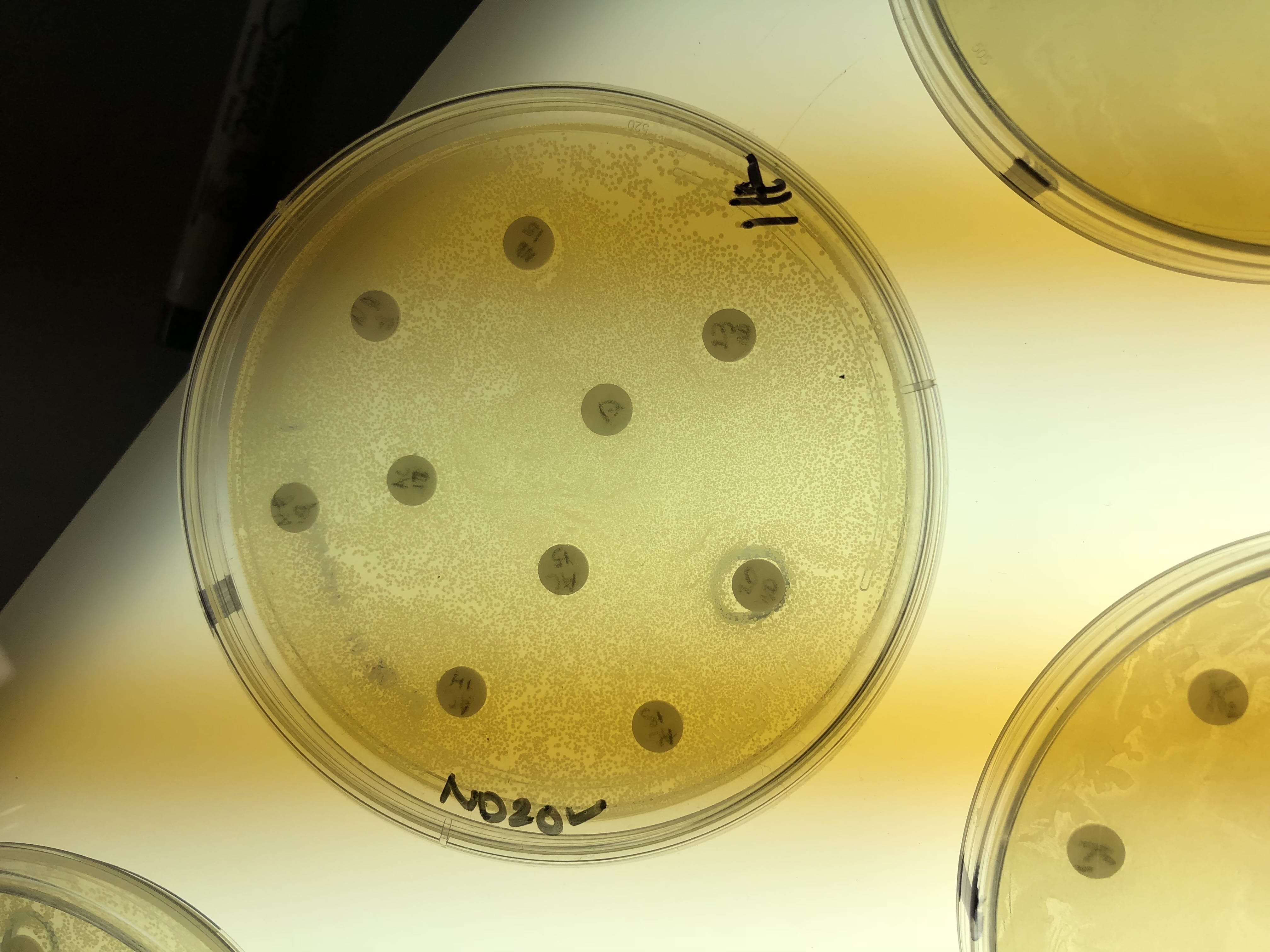
This is a photo of my gram stain for isolate #20 (photos of controls and other info in my “Meet My Microbes!” post). From my gram stain, I determined my isolate to be Gram + with a rod-shape/bacillus and filamentous microscopic morphology. Therefore, my gram stain agrees with my 16S data because Streptomyces is a genus of Gram positive bacteria with a filamentous shape [1].

Pick one of your isolates and find out more about the genus (it is unlikely you will be able to determine the species).

a) General cellular and morphological characteristics of the genus (taxonomic classification, nutrition, cell shape, habitat).

b) Information regarding antibiotic production in this genus.

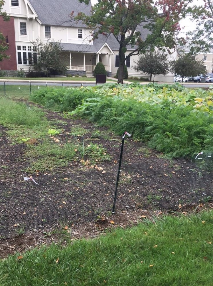
In terms of taxonomy, Streptomyces is the genus of the Streptomycetaceae family, which is further broken down into over 575 species, with more and more still being discovered. Additionally, Streptomyces belongs to the Bacteria Kingdom, Actinobacteria Phylum and Class, and Actinomycetales Order [2]. Streptomyces is known to be Gram positive bacteria that can grow in various environments, and has a filamentous morphology [1]. In their simplest form, Streptomyces are unicellular spheres and rods and are filamentous. However, in more complex forms, they are described as “mycelium of branching hyphal filaments, and reproduce in a mould-like manner by sending up aerial branches that turn into chains of spores” [3]. Streptomyces are primarily found in soil and decaying vegetation, which further confirms that this could be my isolate #20 because I isolated it from my soil sample from a garden [2, 4]. Interestingly, the first Streptomyces are thought to date back 400 millions years ago to when Earth last was first colonized by plants and as such their job was to solubilize the cell walls or other other components of plants, fungi, and insects. This function is further thought to be retained as certain species have genes for proteins that are involved in the binding and degradation of certain carbohydrates[4]. In terms of nutrition, the genus is primarily found in soil and so upon studies for optimum growth of Streptomyces in media, it was determined that various trace elements proved to be important including copper, manganese, and zinc, as well as using sodium nitrate, potassium phosphate, and magnesium sulphate [5].

In today’s society, Streptomyces are of utmost importance as a major source of antibiotics in medical, veterinary, and agricultural use [3]. Further, Streptomyces best known for their ability to produce bioactive secondary metabolites including antifungals, immunosuppressants, and perhaps most importantly in our case, antibiotics. The production of antibiotics is species specific and are important for them to compete with other microorganisms they meet [1]. Finally, the importance of this genus is expressed in the statistic that 80% of antibiotics commonly used today come from the Streptomyces genus [1].


[1] De Lima Procópio, Rudi Emerson, et al. “Antibiotics produced by Streptomyces.” Antibiotics produced by Streptomyces. https://www.sciencedirect.com/science/article/pii/S1413867012001341

[2] Streptomyces, Wikipedia. https://en.wikipedia.org/wiki/Streptomyces

[3] “Streptomyces inside-out: a new perspective on the bacteria that provide us with antibiotics” Philosophical transactions of the Royal Society of London. Series B, Biological sciences vol. 361,1469 (2006)

[4]. “Recent advances in understanding StreptomycesF1000Research vol. 5 2795. 30 Nov. 2016, doi:10.12688/f1000research.9534.1

[5] Spilsbury, J. F. “Observations on the nutritional requirements of Streptomyces griseus (Krainsky) Waksman & Schatz.” Transactions of the British Mycological Society.

Meet my microbes! – Nikki

Location of the soil sample collection: Octopus Community Garden at Union College in Schenectady, NY.                                                                                       GPS coordinates- Latitude: 42.8 degrees Longitude: -73.9 degrees

Example of 10% TSA original plate at a 10^-4 dilution (top), patch plate (middle) and streak plate of #9, 15, 20, and 23 isolates (bottom).

Gm+/Gm- Inhibition

Inhibition of both Gm+ B. subtilus (top) and Gm- E. coli (bottom) are seen for isolates 15, 20, and 23 on 10% TSA.


Gram – Control

Gram + Control

Isolate #15 Gram Stain

Isolate #20 Gram Stain

Isolate #23 Gram Stain

Gram Stain Results: Isolates 15, 20 & 23 all appear to be G+ and streptococcus, bacillus/filamentous, and coccus in shape, respectively.

Inhibition against all tester strains: Isolate #15 is inhibiting strains 1. Staphylococcus epidermidis (+)  and 2. E. coli (-), isolate #20 is inhibiting strain 6. B. subtilis (+), and isolate #23 is inhibiting strain 1. Staphylococcus epidermidis (+), all on 10% TSA.

Extraction Test Results:

Extractions were obtained for isolates #15, 20, &23. Only extract #20 showed inhibition on tester strain 1, S. epidermidis.

Results from 16S Analysis and Blast/Ribosome Database Project (RDP) Search.  I was only able to obtain results for isolate #20 (677 bp). The results show that the closest relative is Streptomyces of the vinaceus or cirratus strains with 99% identity and query coverage (Accession numbers: NR_041131.1 and NR_112388.1).

6. Extract News! – Nikki

Did you obtain an extract? Summarize the results of your testing and why you choose the tester strain(s) you did.

I was able to obtain an extract for each of my three isolates (#15, 20, and 23) following the extraction procedures we followed over the last few weeks in lab. I chose to use the following tester strains: #1 S. epidermidis (+), #2 E. coli (-), #6 B. subtilis (+), and #7 E. aerogenes (-). I chose these four tester strains because my isolates had shown inhibition previously of #1, #2, and #6 tester strains and then I chose #7 because it was a gram – strain to fulfill the 2 Gram + and 2 Gram – that we testing against in this stage. In my observations from the test extracts, I only saw a ring signaling potential inhibition around my #20 isolate/extract on the #1 S. epidermidis, a Gram + tester strain. There was no other antibiotic activity on the other plates/for the other extracts. I added a photo below of the #20 isolate on the #1 tester strain plate and it is the one circled and labeled “ND 20”. Note that this photo was taken on Thursday, November 1st, a day after Prof. Salvo took the plates out of the incubator and made the observations of antibiotic activity.


Fun with Soil – Nikki

Where did you obtain your soil sample?

I decided to collect my soil sample from Union’s Octopus Community Garden on campus. I took the sample an inch or two down, just below the surface, so that I did not get the hard topsoil/mulch and it was near bulbs that were growing (I assumed they were onion bulbs). GPS coordinates- Latitude: 42.8 degrees Longitude: -73.9 degrees.

Why did you choose this location?

I chose this location because I thought it would be interesting to look at something available right on our campus that many members of the community use or partake in on a regular basis. I also expected the soil to be “rich” due to its use in the garden to grow vegetables and various plants.

Do you expect a lot of isolates? Why or why not? 

Yes! I expected there to be a lot of isolates due to the rich nature of the soil being used for gardening purposes. I also thought that maybe because various plants and vegetables grow there that there may be more diversity depending on what has been grown in that location now and previously. In addition, there are a lot of people that work in the garden there that could contribute to what is in the dirt.

Have your initial observations supported this?

Yes, I have a lot of growth on my plates. It seems like I have a lot of growth and a decent variety because many of them look different in their coloring or morphologies. The only countable plates I had were on the 10^-4 dilution plates.

What media did you choose?

I chose 10% TSA, LB, and AC for my three. Prof. Salvo suggested that everyone in the class should use LB for one of the choices. Then I chose 10% TSA and AC because they were for cultivating a wide variety of microorganisms, according to their descriptions in the handout. I thought I would have a wide variety for my soil sample and so I chose these two.

What dilutions?

I plated 10^-2, 10^-3, and 10^-4 dilutions on my plates for each of the three media. (However, going by the way the book does it, these need to be changed to 10^-1, 10^-2, and 10^-3).

Will you need to redo any?

I had growth for all of my plates at the various dilutions, but I decided to re-plate a few. My plates with the AC medium still had too many colonies to count and lots of growth making it difficult to distinguish, so I went one dilution further, 10^-5 (which is actually 10^-4 according to the book) and plated two of these. I am still waiting for these to determine the CFU, but after two days there is no growth yet. I also did one more plate with LB and 10%TSA at a 10^-4 (actually 10^-3) dilution because these were countable, but there were a few large growths and I wanted to compare to get a more accurate CFU. I am still waiting a few more days to get a count because after 2 days there were 2 colonies on the 10% TSA plate and no growth on the LB plate.

How did your sample differ on the different media?

All three had a decent amount and variety on all the different media. AC had the least “pickable” colonies and I was only able to pick 14 (hopefully the new plates will provide more) because mycoides were present as well as really large growths. The LB and a few from the 10% TSA had a very dark brownish pigment sort of radiating from the circular shape and no other growths around it.