Meet My Microbes! Troy

First, bacteria were grown on three different culture media – LB, AC, and R2A – at three different dilution factors.  LB and AC were grown at 10-2, 10-3, and 10-4, while R2A was grown at 10-1, 10-2, and 10-3.  This was done, because they provided three different nutrient levels; Rich, somewhat rich, and somewhat sparse.

After the bacteria were grown they were selected for different traits that set them apart.  They were then picked and patched onto individual media corresponding to the media they were picked from.  12 cultures were picked from LB, 8 were picked from AC, and 16 were picked from R2A.

LB patch plate after selection for unique bacteria.

AC patch plate after selection for unique bacteria.

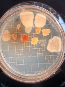
R2A patch plate after selection for unique bacteria.

I thought it was so cool how many different colors and textures were produced by these bacteria!  It’s safe to say that Professor Salvo was right – this soil is pretty diverse!

After testing all of these strains against different tester bacteria, the only colonies that actually exhibited any inhibition pattern were strains 1A, 3A, and 2B.  These colonies were isolated after they were tested, and even though I started off a little rough with streak plates I think it’s safe to say I got the hang of them by the end!

Strain 1A isolated streak plate

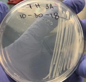
Strain 3A isolated streak plate

Strain 2B isolated streak plate

Just look at those beautiful streaks! Gotta bring these bacteria over to the Nott… (insert laughs here)

So you might be asking yourself, what kind of bacteria am I actually looking at here? Have no fear! After we isolated these strains we used a Gram staining method to determine if our bacteria were Gram positive of Gram negative.  If the bacteria were purple they were Gram positive, and if the bacteria were pink they were Gram negative.

Gram staining from bacteria colony sample 1A showing Gram negative cocci


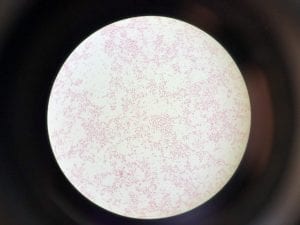
Gram staining from bacteria colony sample 3A showing Gram negative rods


Gram staining from bacteria colony sample 2B showing  Gram negative cocci

So you might be asking yourself, “Did these bacteria actually produce any antibiotics?”  And your answer is YES! They did!  After testing all of the antibiotics against four different strains of bacteria it was revealed that they each inhibited at least one strain!

Antibiotic success in stain 1A, seen on plate T1

Antibiotic success in stain 3A, seen on plate T3

Antibiotic success in stain 2B, seen on plate T2

If you look at the individual plates (in the red circles) you’ll see that there are little halos around them!  These halos are the proof that the antibiotics worked, as they successfully killed off the bacteria!  While these rings are small it is still super cool that the antibiotics actually worked.  Hopefully in the future someone will be able to determine the mechanism by which it works, and enhance the function of the antibiotic or further the search for antibiotics in these types of bacteria! Who knew a little soil sample from outside the construction zone would be so interesting…

And the Name of My Microbe Is… Troy

What were the results of your 16S analysis?

The PCR results were successful in amplifying the DNA in all three strains that were tested, and their sequences were used to run individual BLAST procedures.  Strain 1A (seen in figure 1) yielded BLAST results (figure 2)that revealed that it was a Gm- bacteria belonging to the Klebsiella genus.

Figure 1: Strain 1A isolated streak plate

Figure 2: BLAST sequencing results for unknown strains show relatedness of different strains of bacteria to the proposed DNA sequence via the 16S analysis tool.

Successful results were also observed with strain 3A and 2B, while the BLAST sequences for these two retrieved 99% similarity between the tester strain’s DNA sequences and the BLAST 16S database [1].  It is important to note that not 100% similarity was observed with any of the BLAST results – which may be a cause of the PCR DNA sequences were not completely accurate.  Strain 3A’s 16S analysis was in line with strain 1A’s, as they were both of the Klebsiella genus.  2B’s 16S analysis, however, indicated a different genus than strains 1 and 3A.  2B’s results suggested that it was in the genus Massilia, another common soil bacteria [2].

Does your gram stain agree?

My gram staining agreed for the Gm+/-, but I found a more difficult time in identifying the shape of the individual bacterial cells. The gram staining that I produced suggested that samples 1A and 2B were coccus-shaped Gm- bacteria, while sample 3A was a rod-shaped Gm- bacteria (Figures 3, 4, and 5).  1A and 2B analysis contradicted the 16S analysis, however, after analysis of the strains with BLAST [1].  It was observed that all three bacteria strains were rod-shaped Gm- bacteria.  A reason for this mislabeling may have been that the bacteria cells were simply too small to accurately identify their shapes.

Figure 3: Gram staining from bacteria colony sample 1A showing Gm- rod-shaped Klebsiella.

Figure 4: Gram staining from bacteria colony sample 2B showing Gm- rod-shaped Massilia.

Figure 5: Gram staining from bacteria colony sample 3A showing Gm- rod-shaped Klebsiella.

Pick one of your isolates and find out more about the genus (it is unlikely you will be able to determine the species).

a) General cellular and morphological characteristics of the genus (taxonomic classification, nutrition, cell shape, habitat).

Klebsiella bacteria are an aerobic bacteria that are part of the phylum proteobacteria.  They are of the class gamma proteobacteria, order Enterobacteriales, family Enterobacteriaceae, and genus Klebsiella [3].  They are gram negative rod-shaped bacteria that are often found in soil.  They do not require any special nutrients to grow, as they can survive off of citrate, glucose, and ammonia; this makes them effective at growing in low-nutrient environments.  They are normally found in the nose, mouth, and gastrointestinal tract in humans, but they can also be spread to other parts of the body to cause diseases like pneumonia and urinary tract infections.

b) Information regarding antibiotic production in this genus.

Not much information is known about the production of antibiotics in the genus Klebsiella, but they are the subject of antibiotic research as they are growing more and more resistant to normal antibiotics.  Their ability to survive in low-nutrient environments allows them to stay alive in dire environments, and they have recently evolved an ability to cleave carbapenem antibiotics with an enzyme carbapenamase.  This enzyme has rendered the entire line of carbapenem antibiotics useless against Klebsiella bacteria[4].  It is mutations like this that has researchers working toward discovering new antibiotics, because bacteria are constantly evolving to survive – posing a greater threat to humanity every day.


Works Cited

  1. “BLAST: Basic Local Alignment Search Tool.” National Center for Biotechnology Information, U.S. National Library of Medicine.
  2. Shen, Liang, et al. “Massilia Eurypsychrophila Sp. Nov. a Facultatively Psychrophilic Bacteria Isolated from Ice Core.” International Journal of Systematic and Evolutionary Microbiology, Microbiology Society, 1 July 2015.
  3. Thomas, Gavin. “Klebsiella Pneumoniae.” What Is Electron Microscopy?.
  4. Daikos, George L., et al. “Carbapenemase-Producing Klebsiella Pneumoniae Bloodstream Infections: Lowering Mortality by Antibiotic Combination Schemes and the Role of Carbapenems.” Antimicrobial Agents and Chemotherapy, American Society for Microbiology Journals, 10 Feb. 2014.

Extract News! Troy

Of all the fabulous bacteria that I had, there were three strains that were worthy of testing.  Coincidentally, all three were grown on R2A – a reason for which may be that there were fewer resources in this substrate.  The extracts were gathered on Thursday, October 25, by collecting the organic layer of our previously suspended colony plates.  I extracted three different ‘antibiotics’ from my three different strains, and prepared them to be dried.  On October 30, the extracts (which were not completely dried yet) were prepared to be tested against different bacteria strains.  They were suspended in methanol, and this solution was used to create antibiotic plates that were dropped onto the different tester bacteria plates.  I chose to test against 4 strains – 3, 4, 6, and 7 -, as these strains presented two GM(+) and two GM(-) bacteria strains.  On November 1, the results were observed, and of my three extracts I had antibiotic activity against both GM(+) and GM(-) bacteria.  Strain 1A – a GM(-) bacillus – produced antibiotics that inhibited growth on bacteria strain 3 – a GM(-) bacteria.  Strain 2B – another GM(-) bacillus – produced antibiotics that inhibited bacteria strain 7 – a GM(-) bacteria.  Strain 3A – a GM(-) rod-shaped bacteria – produced antibiotics that inhibited bacteria strain 4 – a GM(+) bacteria.

I was very happy to see that I had AB production from all three of my strains.  I was interested that the two bacilli strains produced AB’s that inhibited the GM(-) testers while the rod-shaped strains produced AB’s that inhibited the GM(+) tester strain (strain 4).  I believe that these strains could be further observed to be potential drugs against bacteria, as they target a wide range of GM(+) and GM(-) bacteria.


Meet the ESKAPE Pathogens: Troy Hansen

Assigned ESKAPE Pathogen

My assigned ESKAPE Pathogen was Pseudomonas Aeruginosa

Why is this ESKAPE Pathogen of interest (in brief)

Pseudomonas Aeruginosa is of interest, because it is a common infection in hospitals after surgery, burn victims, and in individuals with weakened immune systems.

General Cellular and Morphological Characteristics of the Organism (taxonomic classification, nutrition, cell shape, habitat)

Pseudomonas Aeruginosa is a gammaproteobacteria.  This means that it is a gram negative bacteria.  It is rod-shaped and does not produce spores, but it does have a flagellum that allows it to move efficiently. It thrives in temperatures between 25ºC and 37ºC, but it can also survive in temperatures up to 42ºC making them more deadly in hospitals and clinical environments. They produce pigments that are typically a greenish color, and it can grow aerobically or anaerobically under minimal nutrition – it has even been known to grow in distilled water!

Clinical Importance and Prevalence

Pseudomonas Aeruginosa is able to grow in a wide range of environments under minimal nutrition, so it becomes dangerous in hospitals as it can easily infect someone who is immunocompromised.  Typical patients who become infected are cancer patients and burn victims,

Infection (How does the infection occur and where is it localized?)

Pseudomonas Aeruginosa can occur in most areas in the body where there is some sort of a body cavity or mucus membrane.  It can also occur on the skin of burn victims, and most places excluding in the blood.

Pathology (What disease is caused? What are the symptoms?)

Bacterial infection is caused, and can lead to colony growth, tissue invasion, and eventually the spread of the bacteria into other parts of the body.  Pneumonia can also occur from the infection of Pseudomonas Aeruginosa.

Ineffective Antibiotics (Antibiotics to which the organism has acquired resistance)

The organism has acquired resistance to all antibiotics except fluoroquinolones, gentamicin, and imipenem.

Effective Antibiotics (Antibiotics known to inhibit the organism)

Same as above.

Corresponding Safe Relative

Pseudomonas Putida


Putty, Murali. “Pseudomonas Aeruginosa.” Testing Lab Analysis: Mold, Legionella, Asbestos, Environmental Microbiology, USP 797, Radon, Lead, March, 2007.

Prince, Alice S. “Pseudomonas Aeruginosa.” NeuroImage, Academic Press, 2012

Fun With Soil – Troy

Where did you obtain your soil sample?

I obtained my soil sample from outside the new S&E building next to the construction site.  Professor Salvo suggested this location, because she felt there would be interesting organisms there since the soil had not been turned over in many years.

Why did you choose this location?

Having stagnant soil allows for the growth of many different bacteria without disturbing any of them.  I chose this soil sample, because Professor Salvo and I knew there would be interesting organisms present that had not been disturbed in many years.

Did you expect a lot of isolates? Why or why not?

I expected some isolates, but I didn’t expect too many – the soil was sandy in texture and I thought that the organisms present would have a difficult time finding nutrients.  I did, however, think that this would make them more likely to produce antibiotics to fight for these nutrients.

Have your initial observations supported this?

My initial observations were that the bacteria grew out of control on my less dilute plates, but they grew beautifully on the more dilute plates.  I have also noticed a substantial amount of what appears to be AB production, but I will only know if this is the case after further analysis!  My observations that the less rich media (R2A) grew fewer bacteria was supported by the decrease in growth at the same dilution levels as the LB and AC plates.

What media did you choose?  How did your sample differ on the different media?

I chose the R2A (less rich), LB (rich), and AC (rich) medias.  I did this to support both bacteria that thrive in low nutrient environments as well as those that thrive in nutrient-rich environments.  I noticed that the bacteria grew at about the same rate in the R2A as the AC and LB when it was at a dilution 10 times less than the rich counterpart.  For example, when R2A was at 10^-1 and LB and AC were 10^-2 it seemed that they were closely related in growth.

What dilutions?

I ran my R2A at 10^0, 10^-1, and 10^-2 while I ran the LB and AC at dilutions of 10^-1, 10^-2, and 10^-3 dilution factors.

Will you need to redo any?

The dilutions worked beautifully.  The best example I can give of how well the dilutions worked was from the LB plates.   The 10^-1 plate had too many colonies to count, but the 10^-2 plate had 90 and the 10^-3 plate had 9 colonies.  This showed that the dilutions were accurate, and the plates were growing as expected.