BLAST results – Kara

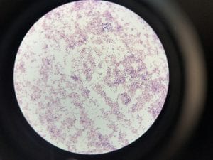
Two out of the three samples I sent out for sequencing came back with results.  My first unknown sample sample was a bit noisy, so I only blasted 687 nucleotides.  The closest genetic match was Acinectobacter baylyi strain B2 (Query 100%, Identity 96%). The majority of the bases that did not match were “n” so they could have been any nucleotide. Ironically, this is one of the tester strains used in lab. Colonies are described as “circular, convex, smooth and slightly opaque,” and the genus has general characteristics such as being aerobic, gram-negative bacilli (Carr et. al.).  This corresponds with my data. I put the picture of my gram stain below, because initially I thought that I had spore formation or a mix of 2 colonies, but with the BLAST data I can probably say this is not spore formation and is indeed gram-negative.

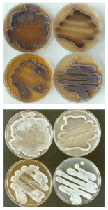
For my second sample, I was able to run 792 bases.  The BLAST results only had members of the genus streptomyces.  The closest match was Streptomyces spororaveus strain NBRC 15456 (query 99%, identity 99%).   Streptomyces spororaveus gram positive, forms dusty gray multicellular complexes, as well as spore chains, which I observed in lab (Podstawka). The spore chains made it hard to pick, spread, and gram stain this organism.  Below is an image of the spore chains from Streptomyces spororaveus, as well as spread plates on various media. My spread plates looked very similar.

 
Image citation: Joachim M. Wink, HZI – Helmholtz-Zentrum für Infektionsforschung GmbH, Inhoffenstr. 7, 38124 Braunschweig, Germany


Streptomyces is in the domain Bacteria, phylum Actinobacteria, class Actinobacteria, order Actinomycetales, and family Streptomycetaceae (Podstawka).  Optimal growth occurs at 28 degrees celsius, and streptomyces are commonly found in soil. (Podstawka). Streptomyces can survive in vastly diverse environmental / nutritional conditions due to their ability to form spores, and commonly form symbiotic relationships with plant roots (de Lima Procópio). “The most interesting property of Streptomyces is the ability to produce bioactive secondary metabolites, such as antifungals, antivirals, antitumorals, anti-hypertensives, immunosuppressants, and especially anitbiotics” (de Lima Procópio).  Over 60% of naturally derived antibiotics were isolated from members of the genus streptomyces.  The first, streptothricin, was discovered in 1942 (Wavte).  It is predicted that members of the genus streptomyces are capable of producing over 100,000 secondary metabolites that have antibiotic properties (Wavte). The antibiotic Streptomicin is derived from S. griseus, and Avermictin is derived from S. avermitilis (de Lima Procópio). Notice below how many antibiotics are from S. ___ indicating they are derived from streptomyces.

Image source: de Lima Procópio, Rudi Emerson. “Antibiotics produced by Streptomyces.” Braz journal of infect dis. 2012;16(5):466–471

Carr, E. L. “Seven Novel Species of Acinetobacter Isolated from Activated Sludge.” International Journal Of Systematic And Evolutionary Microbiology, vol. 53, no. 4, 2003, pp. 953–963., doi:10.1099/ijs.0.02486-0.

de Lima Procópio, Rudi Emerson. “Antibiotics produced by Streptomyces.” Braz journal of infect dis. 2012;16(5):466–471

Podstawka, Adam. “Streptomyces Spororaveus | Type Strain | DSM 41462, ATCC 43694, INMI 101, VKM Ac-318 | BacDiveID:16196.” BacDive | The Bacterial Diversity Metadatabase, bacdive.dsmz.de/strain/16196.

Watve, Milind, et al. “How Many Antibiotics Are Produced by the Genus Streptomyces ?” Archives of Microbiology, vol. 176, no. 5, 2001, pp. 386–390.,

Extract News! Kara

I tested 3 extracts from my 3 producers, one of which was isolated on PDA, while the other 2 were isolated on 10% TSA. I found that all three of my extracts inhibited strain #1 which was staph epi, but did not inhibit the other strains.  I picked the strains a bit arbitrarily, because one of my producers had inhibited all of the strains, so I chose 2 gram negative, and 2 gram positive.  I chose strains 1 (Staph epi), 2 (E. coli) , 5 (A. baylyi) and 6 (B. subtillis).  I made sure A. baylyi was one of the strains I looked at because my ESKAPE pathogen was A. baumanni (which is the pathogenic relative of A. baylyi), so I was curious as to whether or not my extracts would inhibit it (sadly they did not).

Overall, I was happy to see that my extracts demonstrated some antibiotic activity, indicating that my extraction was successful.  Unfortunately, staph epi is very “easy” to inhibit, indicating my extracts probably would not work against most microbes.

Meet my microbes – Kara

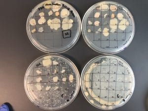
This is the PDA patch plate I made, and where I isolated my only producer. The other producers I have were generously given to me by Elizabeth Altman.

this is an image of my producer. It appears to have both a mix of both gram + and gram – bacilli.  I’m not sure how to characterize this, it could be a mixed culture.


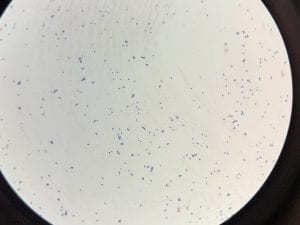
Both isolates from Elizabeth looked very similar.  They show individual gram positive bacilli on the left, and filamentous “clusters” of cells on the right.

Fun with Soil – Kara

“Where did you obtain your soil sample? Why did you choose this location?”
I obtained soil from under a tree that was located near my dorm. I chose this location because the day I collected soil it was very wet outside, so the soil under the tree was drier than most other soil. I also chose this location because it is near the dorm compost bin. I figured maybe because of the proximity to the compost there would be some interesting growth.

“Do you expect a lot of isolates? Why or why not? Have your initial observations supported this?”
I didn’t really know what to expect when I collected the soil. It was close to the compost, which likely has a lot of microbes. However, the soil under the tree is rather protected. My initial observations showed less robust growth than I expected. My CFU was only 4.5-1.2 10^6 colonies / gram of soil.
When I let the plates grow for several days there was more growth, but there appear to be only a few different types of colonies.

“What media did you choose? What dilutions? Will you need to redo any? How did you sample differ on the different media?”
I decided to use 2 “rich” medias-LB and AC, and 1 “less rich” media-PDA. I chose the 2 rich medias because I was unsure how rich my soil sample was in microbes. I chose the PDA because it was mentioned in class lecture that several microbes that produce antibiotics like to grow on PDA. I originally plated 10^0,1, and 2 dilutions of media in PBS. I had to redo a few because I waited 5 days (I was away over the weekend) to look at my plates, and they were all too dense to collect a CFU and patch colonies. I then plated more of the 10^2 and 10^3 dilutions on all 3 types of media, so individual colonies were more identifiable. My PDA plates were also covered in a thick slime, which was potentially hazardous so I threw it away. The AC and LB plates showed similar growth–mostly small, white, gray, and yellow, round colonies.

(P.S.) if I accidentally replied to people my apologies! it took me a few tries to get this is the right spot