And the name of my microbe is… – Le Minh Nguyen

What were the results of your 16S analysis?
Although I originally only got one PCR product for my L.5 isolate, I obtained two 16S RNA gene sequences from Prof. Salvo. Using BLAST, I was able to determine the identity of my two producers, P.1 and L.5. Both isolates were identified with very high confidence to belong to Pseudomonas genus (P.1: 980 base pairs, 100% query cover, 99% identity; L.5: 885 base pairs, 100% query cover, 99% identity). 

Does your gram stain agree?
Unfortunately, because I only obtained a single PCR product, I just performed a Gram-stain on L.5 isolate but not on P.1 isolate. The Gram stain of L.5 producer allowed me to identify it as a Gram-negative rod-shaped bacterium, which is consistent for Pseudomonas as they are Gram-negative bacilli (1) (picture of my L.5 isolate’s Gram-stain can be found in my “Meet my Microbes!” blog post).

a) General cellular and morphological characteristics of the genus (taxonomic classification, nutrition, cell shape, habitat).
Pseudomonas are Gram-negative rod-shaped bacteria (1). They belong to Bacteria kingdom, Proteobacteria phylum, Gammaproteobacteria class, Pseudomonadales order, and Pseudomonadaceae family (2). This genus is found in soil, water, plants, and animals but is also known to inhabit in hospitals (1). Pseudomonas aeruginosa, a species of Pseudomonas, is known to have simple nutritional requirements as it can grow in just “distilled water” but can also withstand extreme physical conditions as it can grow in jet fuel or diesel (3). Additional information on the cellular and morphological characteristics of P. aeruginosa that belongs to this genus can be found in my “Meet the ESKAPE Pathogen” blog post. 

b) Information regarding antibiotic production in this genus.
There are few cases of antibiotic-producing bacteria from the Pseudomonas genus:

One of them is mupirocin. It is an antibiotic that is used topically to treat skin infections. It is isolated from Pseudomonas fluorescens and has a broad spectrum activity against Gram-negative and Gram-positive bacteria (4). It works by inhibiting the bacterial isoleucyl-tRNA synthetase (5). 

Another example of an antibiotic production is a bioactive organometallic compound that has shown to have an antibiotic ability against microorganisms; it can be isolated from Pseudomonas aeruginosa LV strain in the presence of copper chloride (6). 

Lastly, isolated Pseudomonas viscosa shows broad antibiotic spectrum against a wide range of Gram-negative and Gram-positive bacteria that seems to have greater antibiotic ability relative to P. aeruginosa and P. fluorescens (7). 


Works Cited
1. Iglewski, Barbara H. “Pseudomonas.” Current Neurology and Neuroscience Reports., U.S. National Library of Medicine, 1 Jan. 1996.
2.“Pseudomonas.” Encyclopedia of Life, eol.org/pages/83175/overview.
3. Putty, Murali. “Pseudomonas aeruginosa.” EMLab P&K, Mar. 2007
4. Matthijs, Sandra., et al. “Antimicrobial Properties of Pseudomonas Strains Producing the Antibiotic Mupirocin.” Research in Microbiology, 7 Oct. 2014.
5. Hughes, J, and G Mellows. “Inhibition of Isoleucyl-Transfer Ribonucleic Acid Synthetase in Escherichia coli by Pseudomonic Acid.” Current Neurology and Neuroscience Reports., U.S. National Library of Medicine, 15 Oct. 1978. Biochem J, 179 (1978), pp. 305-318.
6. Gionco Barbara., et al. “New Insights about Antibiotic Production by Pseudomonas aeruginosa: A Gene Expression Analysis.” Front Chem., 15 Sep. 2017.
7.Chinn, S. H. F. “An Antibiotic-Producing Bacterium of the Genus Pseudomonas.” Canadian Journal of Microbiology, 1973. 

Extract News! – Le Minh

I was able to obtain an extract from the only isolate (LB #5) that I had. The extract appeared insoluble in methanol as I had a hard time resuspending it; the extract formed a large white clump that would not fully dissolve even after lengthy vortexing. However, I still carried out the following procedure of testing the extract with the obtained solution. I chose to test it against the Gram-negative #2 E. coli and #7 E. aerogenes as well as against the Gram-positive #4 E. raffinosus and #6 B. subtilis as my isolate was able to grow in those tester strains during the ESKAPE testing we performed before. 

In the end, my extract did display some antibiotic-producing ability (so the extract was somewhat successfully resuspended). Out of all test plates, my extract was able to show inhibition only against E. coli but not against the other Gram-negative bacterium nor the Gram-positive bacteria. This means that my extract has narrow-spectrum antibiotic activity. 

The picture below shows the zone of inhibition around my extract (it is a little bit hard to see but it is there) that is was able to produce against E. coli 

Meet My Microbes! – Le Minh Nguyen

This is of one of my very first plates from the first week of lab. I plated my soil sample on the LB + cycloheximide plate with a 10^-4 dilution. This plate was unique because it contained those brown circular colonies with a surrounding halo-fade that also caused pigmentation in the media. As I later found out, this isolate showed some antibiotic production as it inhibited the growth of some of our testers and I eventually chose it for my PCR reaction analysis.


These plates also come from the first week of lab. The plate on the left is also a plate of my soil sample, plated on PDA + cycloheximide plate with a 10^-3 dilution. This plate shows a great diversity of bacteria as presented by a variety of different colors. A lot of colonies were picked from this plate and were patched. Its master plate looks as interesting as shown by the diverse and colorful isolates that were able to grow. 

This is a PDA + cycloheximide plate from the first round of ESKAPE testing. The tester strain is a Gram-negative E. caratovora. As seen on the plate, the tester strain did not grow very well, so the ESKAPE testing was repeated with another Gram-negative E. coli. However, what is more interesting is that only a few colonies were able to grow (#1, 4, 13, 14) while the rest seemed to be contaminated as they appear as red smears. This most probably occurred due to using the same toothpick for ESKAPE testing. 

This is an LB plate, also from the first round of ESKAPE testing with a Gram-negative E. caratovora. This time the tester strain was able to grow and my isolates were tested. As shown on the plate, colonies 5, 6, and 11 showed promise to be antibiotic producers as indicated by the presence of a halo. 


These are pictures of one of my master plates obtained during the ESKAPE testing. These isolates are grown on the PDA plate and as mentioned above, the diversity of the obtained bacteria is great; they are significantly different in morphology (as indicated by the difference in color and shape) and look really cool. 

This is one of my streak plates of the bacteria that I streaked for PCR reaction. The bacteria is grown on the PDA plate and is the same one from the previous PDA plates, indicated as #1. The single colony has a circular egg-like shape and is of deep brown color. Furthermore, after a few days, the colony causes the media to absorb the bacteria’s pigment. 


These are my Gram-positive (Staph epi) and Gram-negative (P. putida) controls. They were visible on my slide with the expected coloration so I could identify the Gram identity of my bacterium sample. 

This is my stained colony #5 from LB plate that I picked to identify as it was the only sample that produced the desired PCR product. It is a Gram-negative rod-shaped bacterium. 

Meet the ESKAPE pathogens: Le Minh Nguyen

Assigned ESKAPE Pathogen: Pseudomonas aeruginosa

Why is this ESKAPE Pathogen of interest
Pseudomonas aeruginosa is a common opportunistic pathogen that can cause disease not only in plants and animals but also in humans. This ESKAPE Pathogen is of utmost importance because it is a multidrug-resistant pathogen, with very advanced antibiotic resistance mechanism, that can survive under various environmental conditions. According to the Centers for Disease Control and Prevention (CDC), it is the most common disease-causing species. P. aeruginosa affects different sites within the body, including urinary tract, skin (burn or surgical wounds), and the respiratory tract and causes severe acute and chronic infections in immunocompromised patients with cancer and patients suffering from severe burns and cystic fibrosis. It is often associated with hospital-acquired infections because there is a higher risk for infection if you have surgical wounds or burns or if you are being treated with a mechanical ventilator and other medical devices such as urinary or intravenous catheters.

General Cellular and Morphological Characteristics of the Organism 
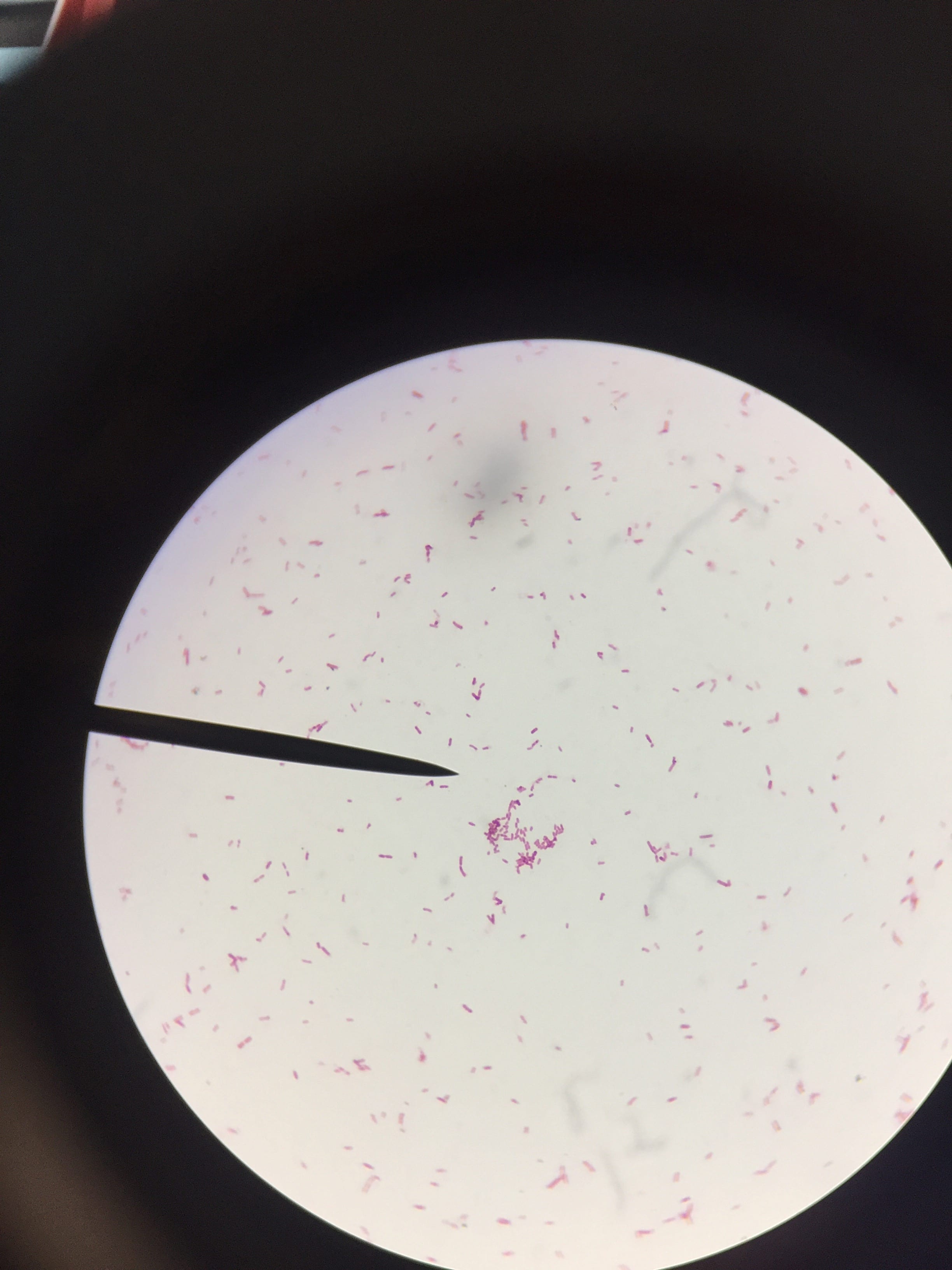
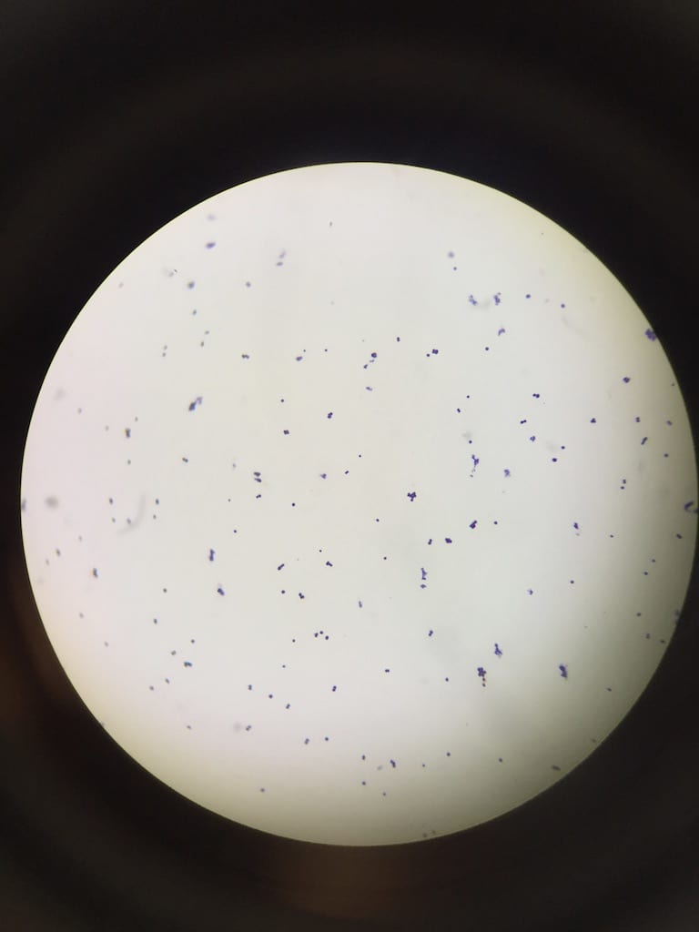
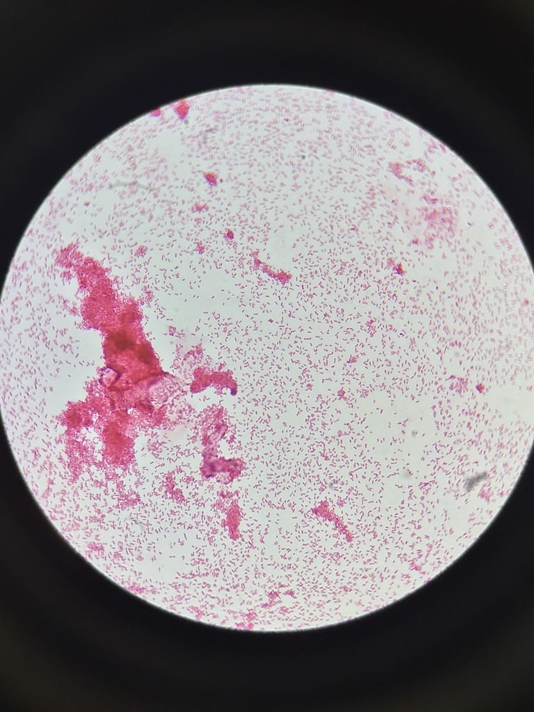
Pseudomonas aeruginosa is a Gram-negative, rod-shaped, asporogenous, and monoflagellated bacterium (Wu, 2014). Its size is about 0.5 to 0.8 µm, and it belongs to the bacterial family Pseudomonadaceae (Putty, 2007).

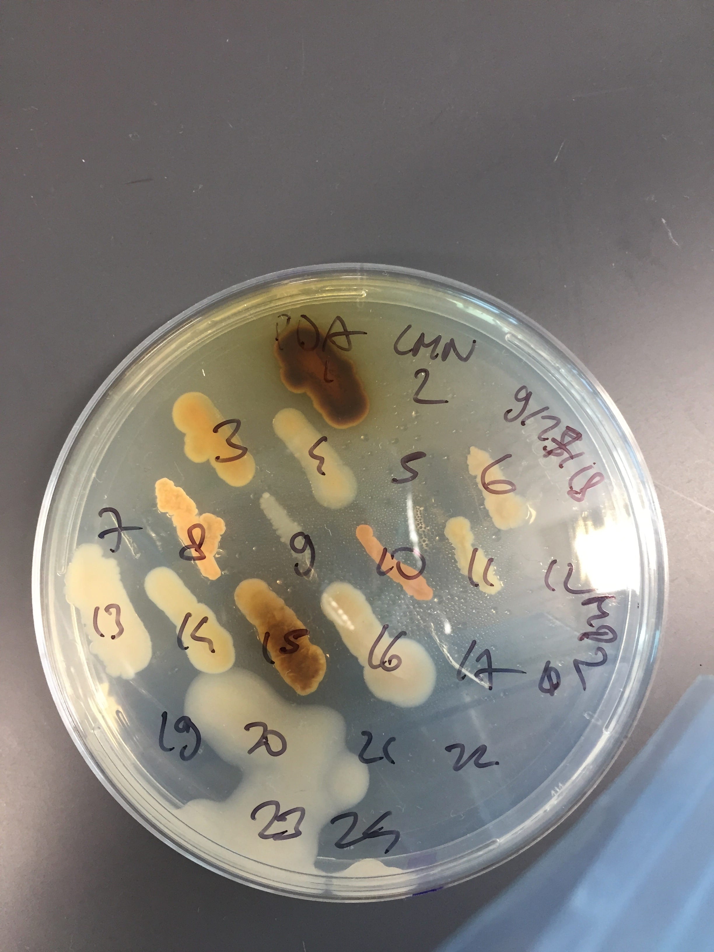
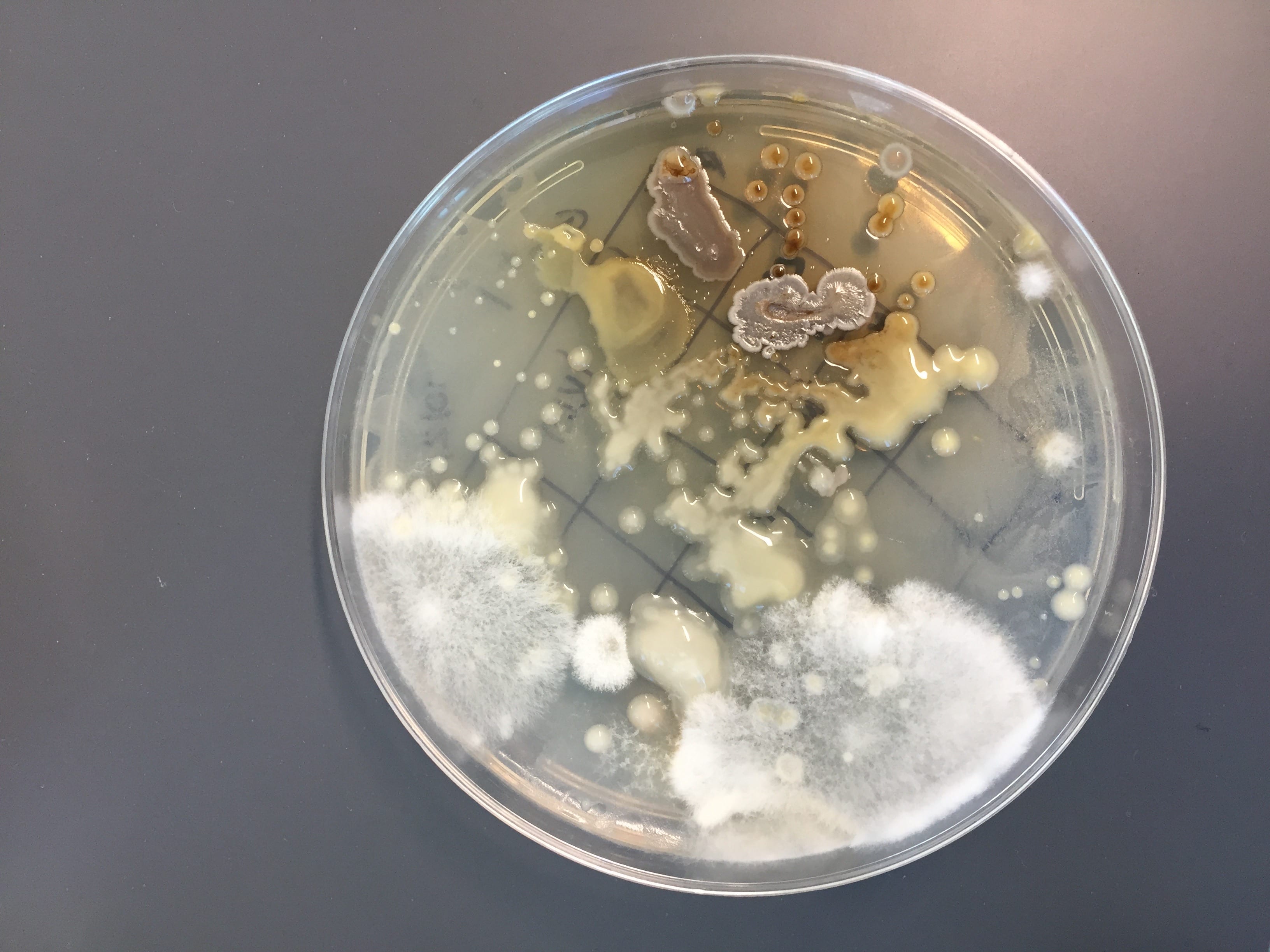
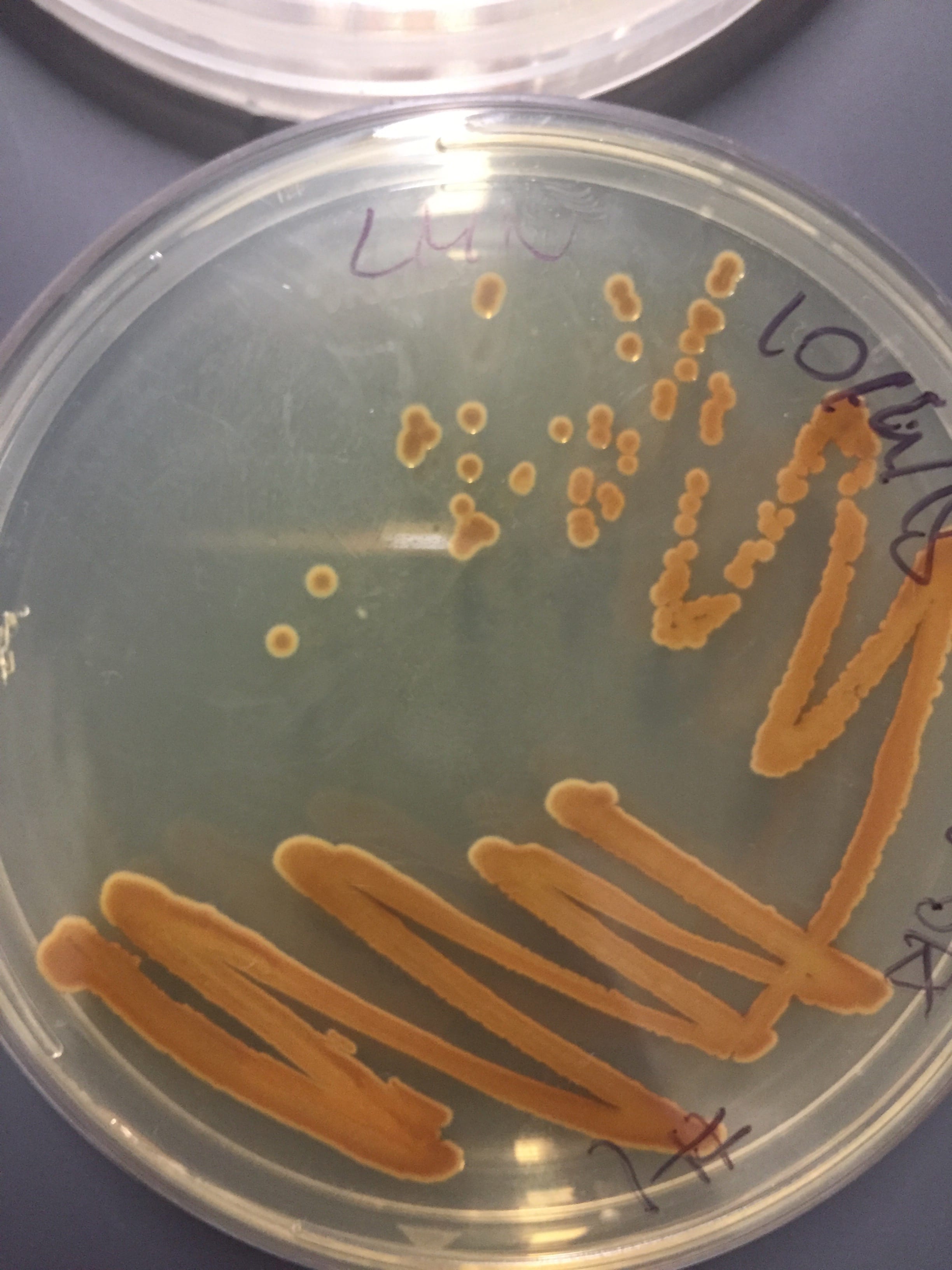
Pseudomonas aeruginosa is often identified by its pearlescent appearance and grape-like or tortilla-like odor (Putty, 2007). P. aeruginosa strains can produce one or more pigments: pyocyanin (blue-green), pyoverdine (yellow-green and fluorescent), and pyorubin (red-brown) (Wu, 2014).

Pseudomonas aeruginosa has simple nutritional requirements as it is often observed to grow in “distilled water” (Putty, 2007). It grows well at 25°C to 37°C, but it is its ability to grow at 42°C that help us differentiate it from many other Pseudomonas species (Wu, 2014).

Like most environmental bacteria, P. aerugionosa lives predominantly in slime-enclosed biofilms adherent to available surface from which it periodically releases (Putty, 2007). It is present in soil and aquatic environments. However, as mentioned before, it is tolerant of a variety of environmental conditions. It is also capable of growing in diesel and jet fuel, where it is known as a hydrocarbon utilizing microorganism (or “HUM bug”), causing microbial corrosion (Putty, 2007). Furthermore, it is resistant to high concentrations of salts and dyes, weak antiseptics, and many antibiotics (Putty, 2007).

Clinical Importance and Prevalence
The most difficult challenge when facing P. aeruginosa is its ability to rapidly develop resistance to multiple classes of antibiotics during treating an infection, ability to survive on minimal nutritional requirements and ability to tolerate a variety of physical conditions. These contribute to the organism’s capacity of persisting in hospital settings.

Data collected by the CDC National Nosocomial Infections Surveillance System from 1986 to 1998 reveals that P. aeruginosa was the fifth most frequently isolated nosocomial pathogen, accounting for 9% of all hospital-acquired infections in the United States; the second leading cause of nosocomial pneumonia (14 to 16%); third most common cause of urinary tract infections (7 to 11%); fourth most frequently isolated pathogen in surgical site infections (8%), and seventh leading contributor to bloodstream infections (2 to 6%) (Lister et al., 2009). In addition, more recent studies continue to show it is the leading cause among pediatric patients in the intensive care unit (Lister et al., 2009).

Infection
In hospital settings, infections by P. aeruginosa can be transmitted in hospitals by nursing staff, medical equipment, sinks, disinfectants, and food (Lister et al., 2009). Transmission occurs from improper hygiene, by patient contact with a contaminated reservoir or by ingestion of contaminated materials.

As described before, P. aeruginosa affects different sites within the body, including urinary tract, skin (burn or surgical wounds), and the respiratory tract and causes severe acute and chronic infections in immunocompromised patients with cancer and patients suffering from severe burns and cystic fibrosis (Wu, 2014).

Furthermore, exposure to contaminated water can also cause mild P. aeruginosa infections. For example, inadequately chlorinated hot tubs and swimming pools can cause ear infections and skin rashes. P. aeruginosa can also cause eye infections in users of contact lenses (Bennington-Castro, 2015).

Pathology
Bloodstream infections can cause:
• Fever and chills
• Body aches
• Light-headedness
• Rapid pulse and breathing
• Nausea and vomiting
• Diarrhea
• Decreased urination

Pneumonia can cause:
• Fever and chills
• Difficulty breathing
• Cough, sometimes with yellow, green, or bloody mucus

Urinary tract infections can cause:
• Strong urge to urinate frequently
• Painful urination
• Unpleasant odor in urine
• Cloudy or bloody urine

Wound infections can cause:
• Inflamed wound site
• Fluid leakage from wound

Ear infections can cause:
• Ear pain
• Hearing loss
• Dizziness and disorientation
(Bennington-Castro, 2015)

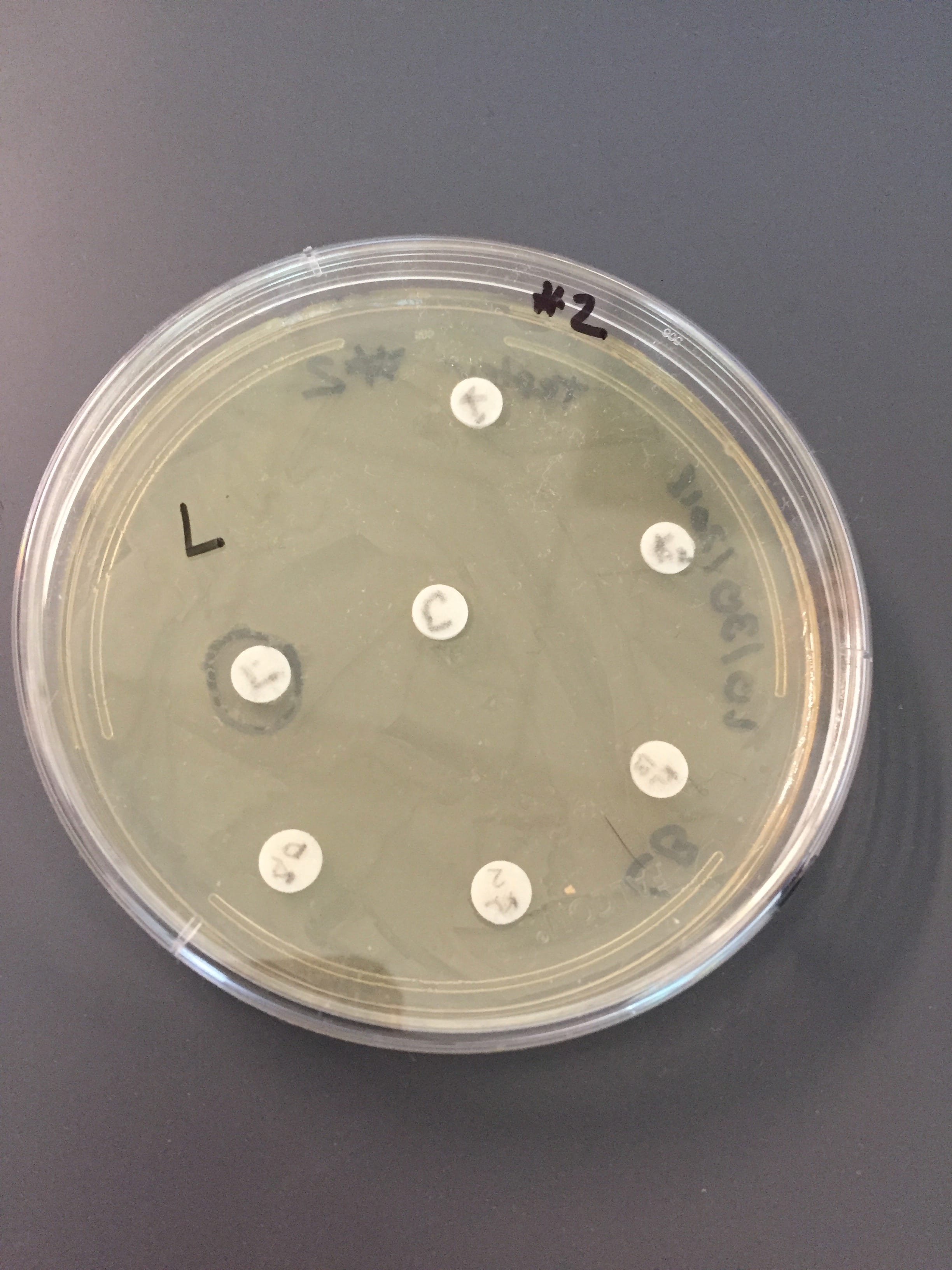
Ineffective Antibiotics
P. aeruginosa currently shows resistance to the following antibiotics: penicillin G; aminopenicillin, including those combined with beta-lactamase inhibitors; first and second generation cephalosporins; piperacillin; piperacillin and tazobactam; cefepime; ceftazidime; aminoglycosides; the quinolones; the carbapenems; colistin and fosfomycin (Hancock, 2000).


Effective Antibiotics
P. aeruginosa is most susceptible to the following antibiotics: cefepime, amikacin, ceftazidime, tobramycin, the combination of piperacillin and tazobactam, meropenem, imipenem, piperacillin, ciprofloxacin, gentamicin, and fosfomycin (Yayan et al., 2015).

Corresponding Safe Relative
The corresponding relative safe to P. aeruginosa is Pseudomonas putida. P. putida is a rod-shaped, flagellated, gram-negative bacterium that is found in most soil and water habitats where there is oxygen. It grows optimally at 25-30°C and can be easily isolated. Unlike P. aeruginosa, P. putida has a nonpathogenic nature; therefore, researchers find P. putida beneficial to research as it also happens to be very versatile and easy to handle (Marcus, 2003). For example, as P. putida assists in promoting plant development, researchers use it in bioengineering research to develop biopesticides and to the improve plant health (Espinosa-Urgel, 2000).

Works Cited
Bennington-Castro, Joseph. “What Is Pseudomonas Aeruginosa?” Stroke Center, Everyday Health, 7 Aug. 2015.
Espinosa-Urgel, Manuel, and Amparo SalidoJuan-Luis Ramos. “Genetic Analysis of Functions Involved in Adhesion of Pseudomonas Putida to Seeds.” Journal of Bacteriology, American Society for Microbiology Journals, 1 May 2000.
Hancock, R E, and D P Speert. “Antibiotic Resistance in Pseudomonas Aeruginosa: Mechanisms and Impact on Treatment.” Current Neurology and Neuroscience Reports., U.S. National Library of Medicine, Aug. 2000.
Lister, Philip D., et al. “Antibacterial-Resistant Pseudomonas Aeruginosa: Clinical Impact and Complex Regulation of Chromosomally Encoded Resistance Mechanisms.” Current Neurology and Neuroscience Reports., U.S. National Library of Medicine, Oct. 2009.
Marcus, Adam. “Versatile Soil-Dwelling Microbe Is Mapped.” GNN – Genome News Network, 10 Jan. 2003.
Putty, Murali. “Pseudomonas Aeruginosa.” EMLab P&K, Mar. 2007.
Wu, Weihui, et al. “Pseudomonas Aeruginosa.” Molecular Medical Microbiology, Academic Press, 29 Sept. 2014.
Yayan, Josef, et al. “Antibiotic Resistance of Pseudomonas Aeruginosa in Pneumonia at a Single University Hospital Center in Germany over a 10-Year Period.” PLOS ONE, Public Library of Science, 2 Oct. 2015.


Fun with Soil – Minh

Where did you obtain your soil sample?
I obtained my soil sample from the plaza that is located in between Olin, Wold, and S&E (right outside the Starbuck’s exit). The exact coordinates are 42.8175459, -73.9278173.
I dug about 2 to 3 inches below the surface next to the shrubs that are situated outside of Olin.

Why did you choose this location?
I was working late at night in my lab in Wold, so I did not want to go too far to obtain the sample. The location did not seem like a “rich” site because only some shrubs are present there but across those plants was a closed-off ground due to the presence of asbestos, so that convinced me to pick the soil from that location.

Do you expect a lot of isolates? Why or why not?
Not necessarily. As I mentioned above, the site in that area does not have a lot of diverse plants as there are mostly shrubs and grass. However, the soil might be actually quite fertile as it is the property of Union College and the school probably uses rich soil for the plants.

Have you initial observations supported this?
On the first day, 24 hours after plating, there was a lot of growth for every different media that I used at the 10^-1 dilution. Other dilutions had some growth, but there were not as many colonies as on the 10^-1 plate. In addition, after 78 hours, I could start to see a diversity of different colonies being grown as there were plenty of different colored and shaped colonies present on the plates. This suggests that the soil was much richer than I had initially assumed.

What media did you choose? What dilutions?
For the media, I chose to use LB, 10% TSA and PDA plates. As for dilutions, I spread 100 µl of each 10^-1, 10^-2, and 10^-3 dilution for every different plate.

How did you sample differ on the different media?
Every plate had something particular that made it distinct from other plates which made the plates to be very interesting. LB had some different colonies, but the colonies that struck out were evenly spread across the plate; these colonies were brown in color and had a surrounding halo of similar color around them. 10% TSA plates had some diversity, but the colonies were rather small with less diverse pigments; there was a presence of mycoides on these plates as well. PDA plates were the most diverse, in terms of shape and color; some colonies were round and filamentous, and others were colored from light yellow to dark brown or even pink. However, most importantly, every plate contained some colonies that showed inhibition as they inhibited the growth of the neighboring colonies.

Will you need to redo any?
I did not redo any of the dilutions however I did additionally plate a new dilution,10^-4, for LB and PDA plates so that it would easier for me to count the number of colonies that grew on the plates.