Meet my microbes! Sofia

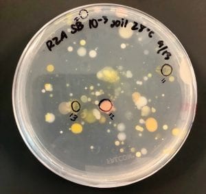
Two of my original plates with my serial dilutions from my soil sample. I plates my dilutions on R2A, LB, AC, and 10% TSA. I had the widest variety of shape, size and color on R2A. With later tests I also have the best growth, and the most inhibition on R2A, so the majority of later tests were done using R2A plates.

My second patch plate of the 15 colonies picked from my original R2A plates. Patches 4, 6, and 11 exhibited signs of inhibition. These are the isolates I used for further testing.

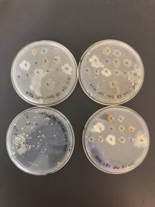
LB plates: top two are the plates used for the first test against the ESKAPE pathogens; no inhibition. Bottom left is my serial dilution plate. Bottom right is Patch plate #2.

10% TSA plates: top two are the plates used for the first test against the ESKAPE pathogens; no inhibition. Bottom left is my serial dilution plate. Bottom right is Patch plate #2. Similar growth to the R2A plates, however nothing that was patches exhibited any signs of inhibition.

AC plates: top two are the plates used for the first test against the ESKAPE pathogens; no inhibition. Bottom left is my serial dilution plate. Bottom right is Patch plate #2.

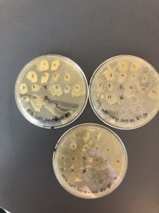
Second set of LB plates used to test against a Gm +/- strain of a “safe” ESKAPE pathogen. Lots of slimy/ gooey yellow growth, but no signs of inhibition.

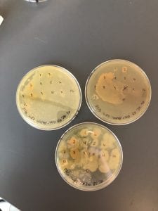
Second set of AC plates used to test against a Gm +/- strain of a “safe” ESKAPE pathogen. Also had lots of slimy/ gooey yellow and orange growth and the patch plates had very large and funky shaped patches; no signs of inhibition.

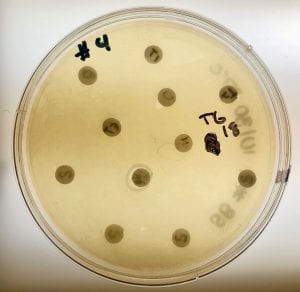
R2A plates with patches to test against Gm +/- strains of “safe”ESKAPE pathogens. There was not much inhibition against the Gm+ strain, but there was signs of inhibition against the Gm – strains. This is consistent with the results of the second ESAKPE pathogen test, in which we tested against all 8 strains. Signs of inhibition were more commonly seen against the Gm – strains.

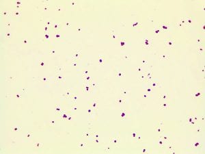
Gram stain for the Gm + control (S. epidermis)

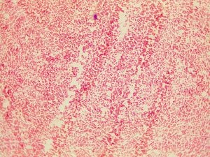
Gram stain for the Gm – control (E. coli)


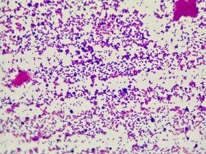
Gram stain for isolate #4; Gm -; appears to be a small cocci but further examination concludes that the bacteria is a very, very small and thick rod, that appears somewhat round.

Gram stain for isolate #4; Gm  (purple in color); small cocci, some are diplococci and there are a few  short streptococci (chains).

Gram stain for isolate #11; Gm – (pink in color); appears to be a small cocci but further examination concludes that the bacteria is a very, very small and thick rod, that appears somewhat round.

One of my plates from the extract test; My three isolates produced five different extracts, however none of them exhibited signs of inhibition. This is indicates that none of my isolates are antibiotic producers. Signs of inhibition may possibly be visible if incubation time was longer. Additionally, it is possible that something went wrong with the extract, which is why there was no visible inhibition. A repeat of this test could provide more definitive conclusions.

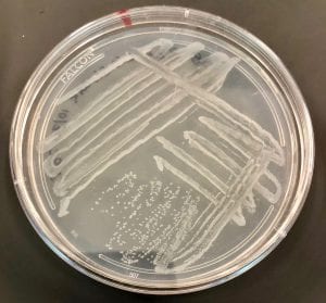
Streak plate for isolate #6 with visible single, isolated colonies.

And the name of my microbe is… Sofia

Sequence data was provided for all three of my isolates, however isolates 4 and 6 could not be identified. I was able to identify isolate number 11 though. I ran a sequence of 677 base pairs with BLAST. The closest match that came up was Klebsiella aerogenes (also known as Enterobacter aerogenes), with 98% identity and 10% query coverage (Accession number NR_102493.2).

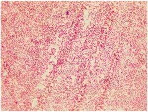
Klebsiella aerogenes is a very small, Gram negative rod. It is such a small rod, that is may sometimes appear to be a cocci at first glance. When I first looked at the Gram stain of isolate #11, I thought I was seeing cocci, however I was able to enlarge the image and what appear to be cocci, are actually very, very small rods (difficult to see in this image). Klebsiella bacteria tend to be rounder and thicker than other members of the Enterobacteriacecae family. The Gram negative pink color is consistent between my Gram stain and that of K. aerogenes.

Gram stain for isolate #11

Gram stain of Klebsiella aerogenes

Image: http://faculty.ccbcmd.edu/courses/bio141/labmanua/lab12/Entaerogenes.html

Members of the Enterobacteriaceae family, Klebseilla are facultative anaerobic, nonmotile bacteria. The Klebsiella genus consists of Gram-negative, oxidase-negative, rod-shaped bacteria with a prominent polysaccharide-based capsule (1). Klebsiella species can be found everywhere in nature, including in soil and water, on plants, and some strains are even considered to be a part of the normal flora of the gastrointestinal tracts of humans (1). One of the most well-known strains within this genus is K, pneumoniae, and ESKAPE pathogen, and a common pathogen of the human respiratory system that causes pneumonia (1). Some strains of Klebsiella have the ability to fix atmospheric nitrogen into a form that can be used by plants (2).

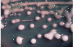
Image: https://en.wikipedia.org/wiki/Klebsiella_pneumoniae

Image: http://microbe-canvas.com/Bacteria.php?p=422

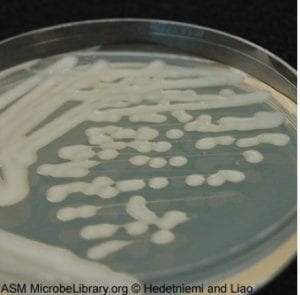
Image: https://microbewiki.kenyon.edu/index.php/Klebsiella_pneumonia

There is little research that discusses the Klebsiella genus as antibiotic producers. In fact, the majority of the literature about this genus discusses how many species within the genus have become resistant to antibiotics, and produce beta-lactamases that have the ability to break down commonly used antibiotics (4). These are often referred to as extended-spectrum beta-lactamase (ESBL) producing bacteria (4). The most common ESBLs are E. coli and K. pneumoniae. In addition to ESBL production, there are certain Klebsiella strains that are cytotoxin producers, such as K. oxytoca (5). These strains of K. oxytoca have been implicated in antibiotic-associated hemorrhagic colitis. The clinical manifestations are usually preceded by antibiotic treatment with a Beta-lactam agent (5). Cytotoxins are chemical weapons that T-cells use to destroy infected cells, protecting healthy cells. Some cytotoxins works my making holes in the cell membrane, while others turn on a program that causes the cell to self destruct (5).

Sources:

  1. Parwanu, N., K. Rogers, and A. Tikkanen. 2010. Klebsiella Bacteria Genus. Encylopedia Britanica.
  2. Buckle, J. 2014. Learn more about Klebsiella. Science Direct.
  3. 2018. Klebsiella. Wikipedia Encylopedia.
  4. Provinvial Infection Control (PIC-NL). 2011. EXTENDED-SPECTRUM BETA-LACTAMASE (ESBL) PRODUCING BACTERIA. Health.gov.nl.ca
  5. Barson, W.J., and M.J. Marcon. 2012. Etiologic Agents of Infectious Disease. Principles and Practice of Pediatric Infectious Diseases. 4th ed.