
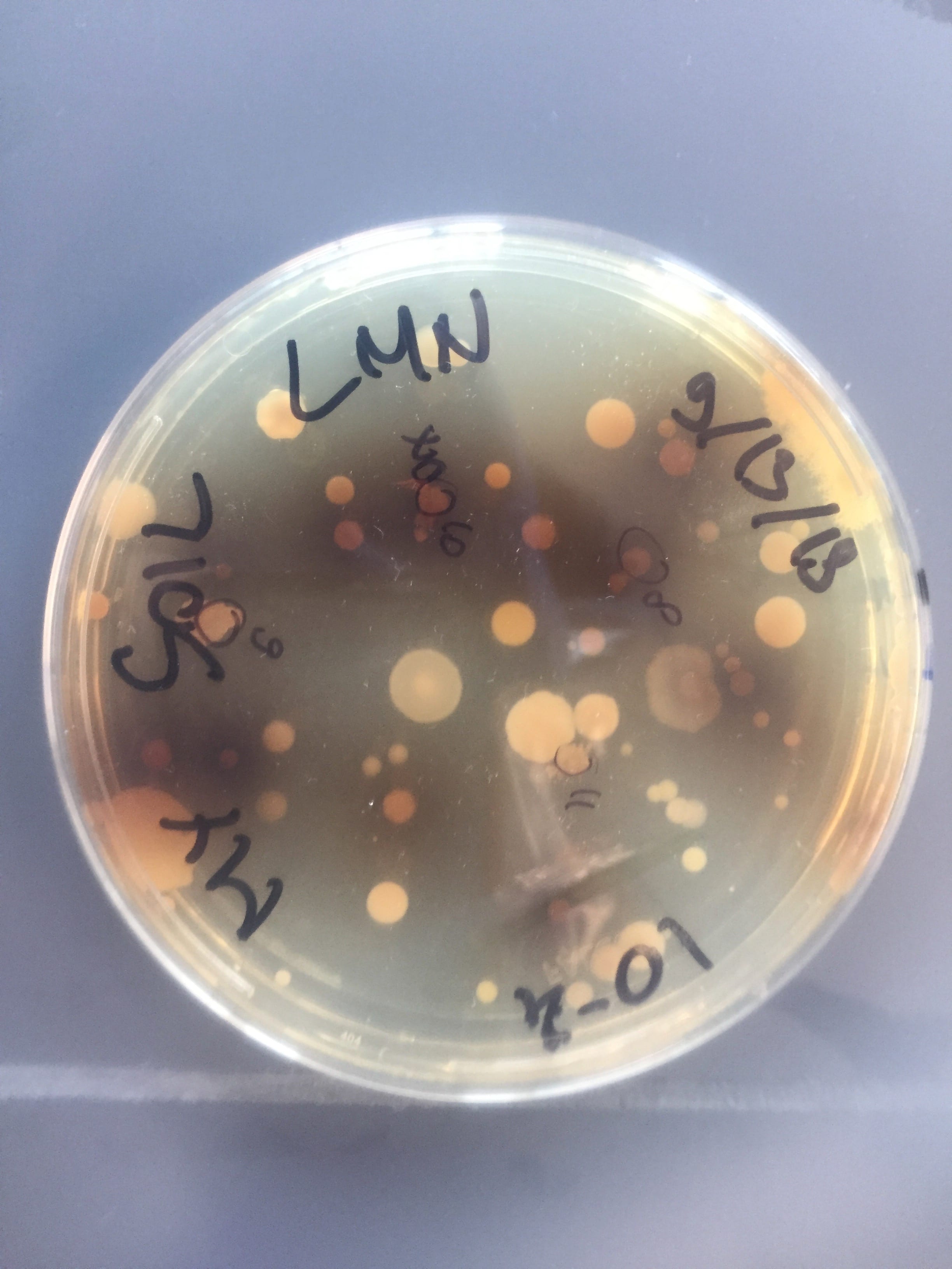
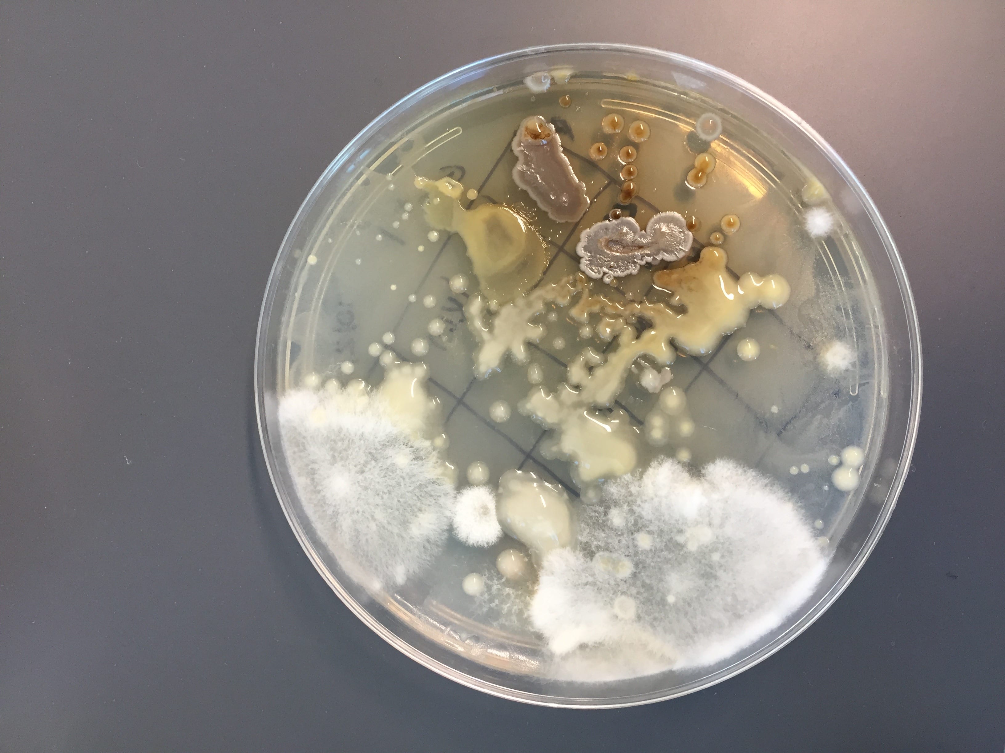
This is of one of my very first plates from the first week of lab. I plated my soil sample on the LB + cycloheximide plate with a 10^-4 dilution. This plate was unique because it contained those brown circular colonies with a surrounding halo-fade that also caused pigmentation in the media. As I later found out, this isolate showed some antibiotic production as it inhibited the growth of some of our testers and I eventually chose it for my PCR reaction analysis.


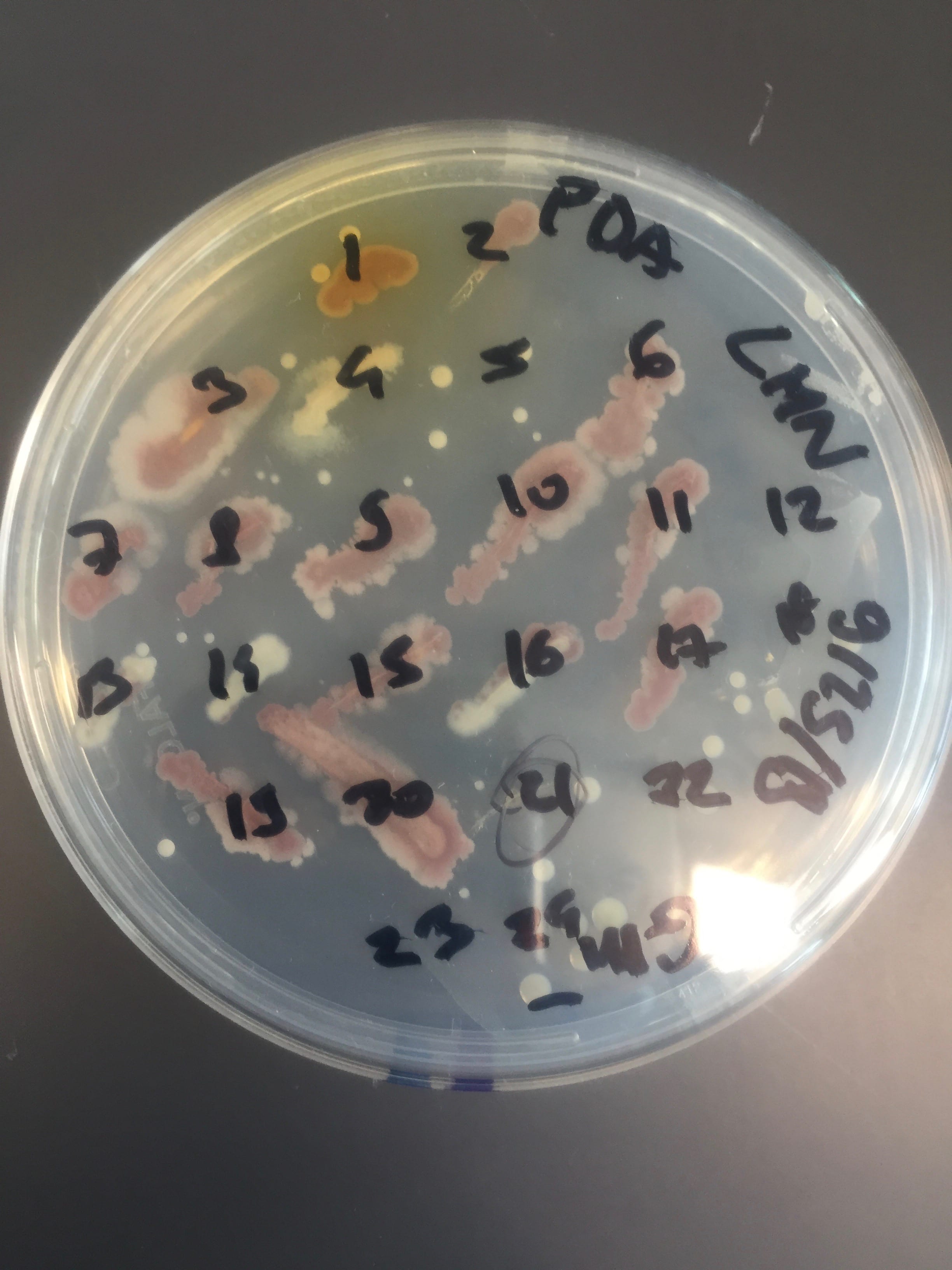
These plates also come from the first week of lab. The plate on the left is also a plate of my soil sample, plated on PDA + cycloheximide plate with a 10^-3 dilution. This plate shows a great diversity of bacteria as presented by a variety of different colors. A lot of colonies were picked from this plate and were patched. Its master plate looks as interesting as shown by the diverse and colorful isolates that were able to grow.
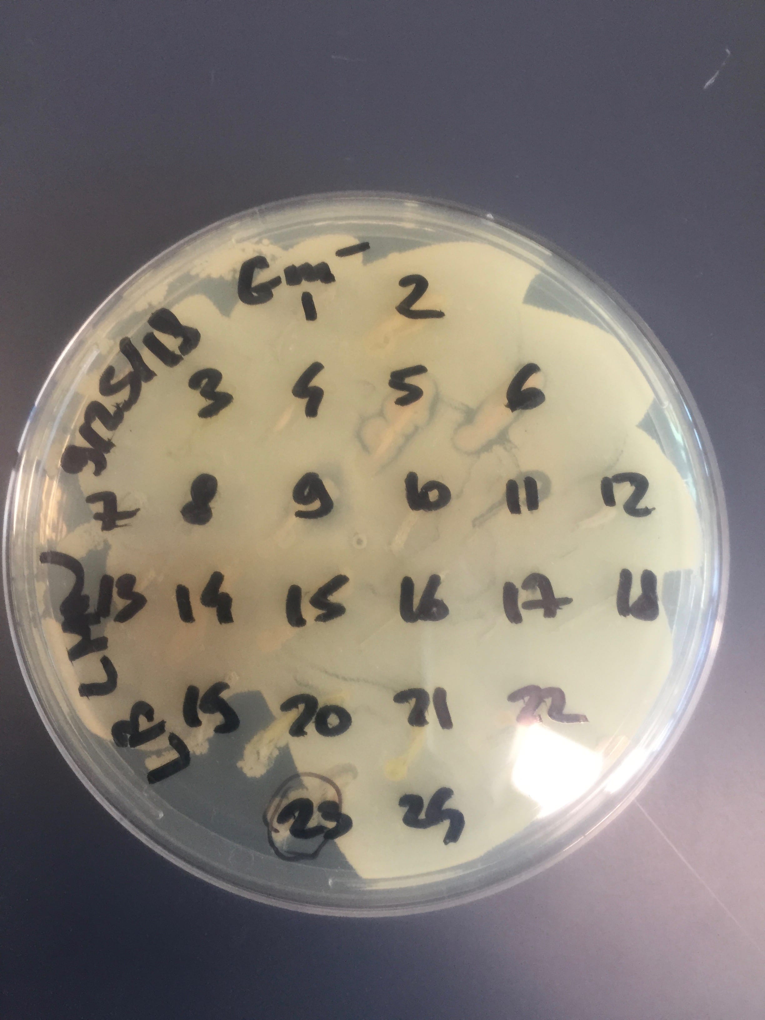
This is a PDA + cycloheximide plate from the first round of ESKAPE testing. The tester strain is a Gram-negative E. caratovora. As seen on the plate, the tester strain did not grow very well, so the ESKAPE testing was repeated with another Gram-negative E. coli. However, what is more interesting is that only a few colonies were able to grow (#1, 4, 13, 14) while the rest seemed to be contaminated as they appear as red smears. This most probably occurred due to using the same toothpick for ESKAPE testing.
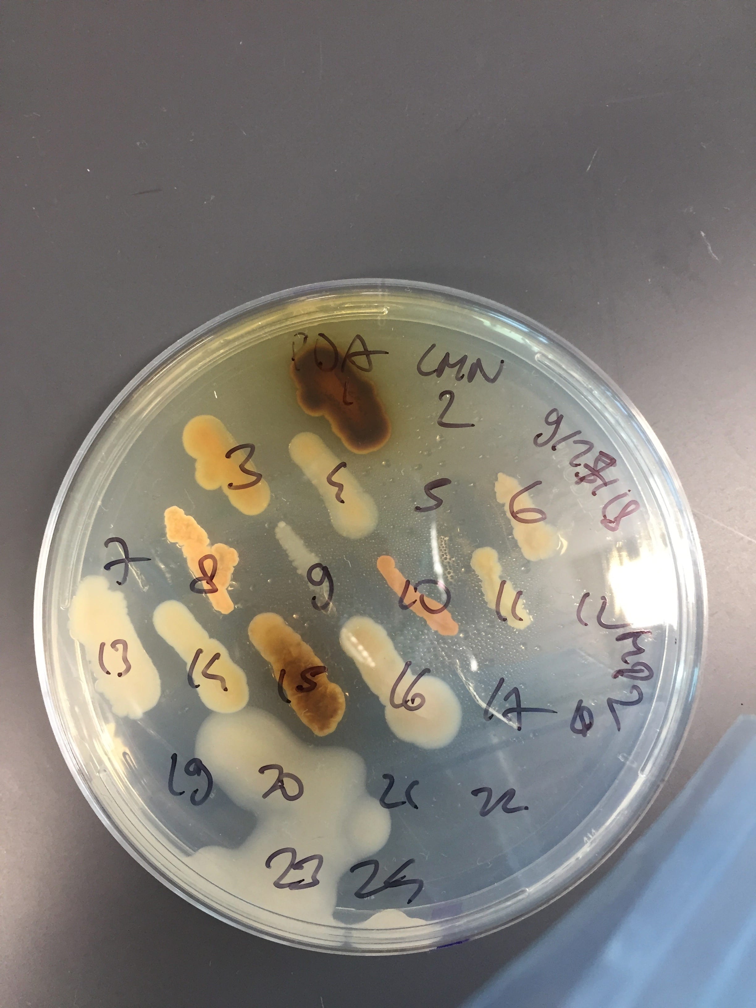
This is an LB plate, also from the first round of ESKAPE testing with a Gram-negative E. caratovora. This time the tester strain was able to grow and my isolates were tested. As shown on the plate, colonies 5, 6, and 11 showed promise to be antibiotic producers as indicated by the presence of a halo.
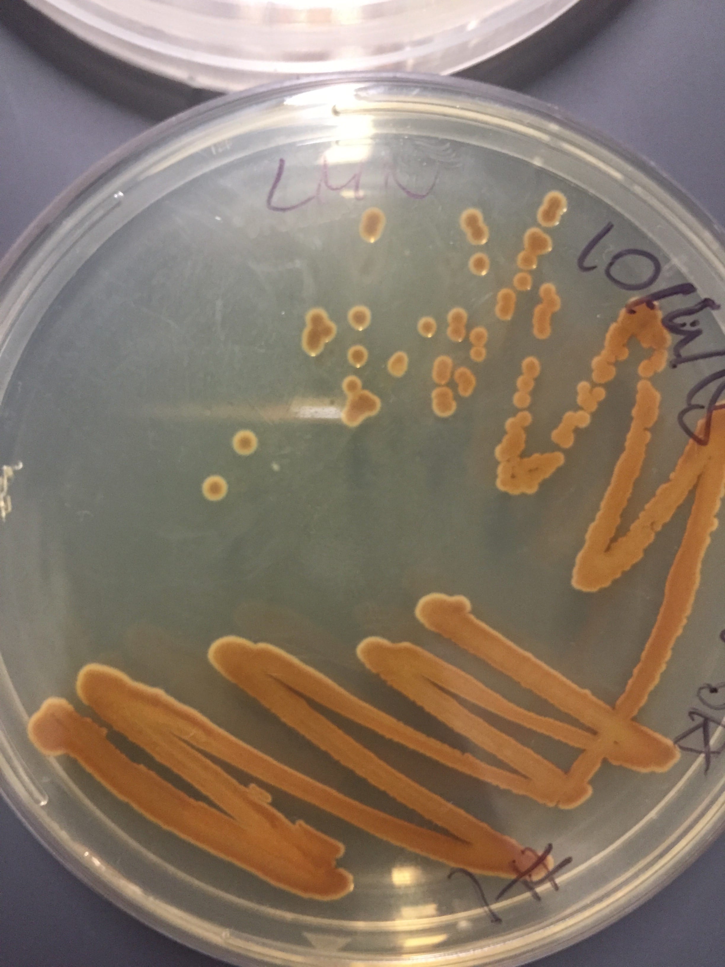
These are pictures of one of my master plates obtained during the ESKAPE testing. These isolates are grown on the PDA plate and as mentioned above, the diversity of the obtained bacteria is great; they are significantly different in morphology (as indicated by the difference in color and shape) and look really cool.
This is one of my streak plates of the bacteria that I streaked for PCR reaction. The bacteria is grown on the PDA plate and is the same one from the previous PDA plates, indicated as #1. The single colony has a circular egg-like shape and is of deep brown color. Furthermore, after a few days, the colony causes the media to absorb the bacteria’s pigment.
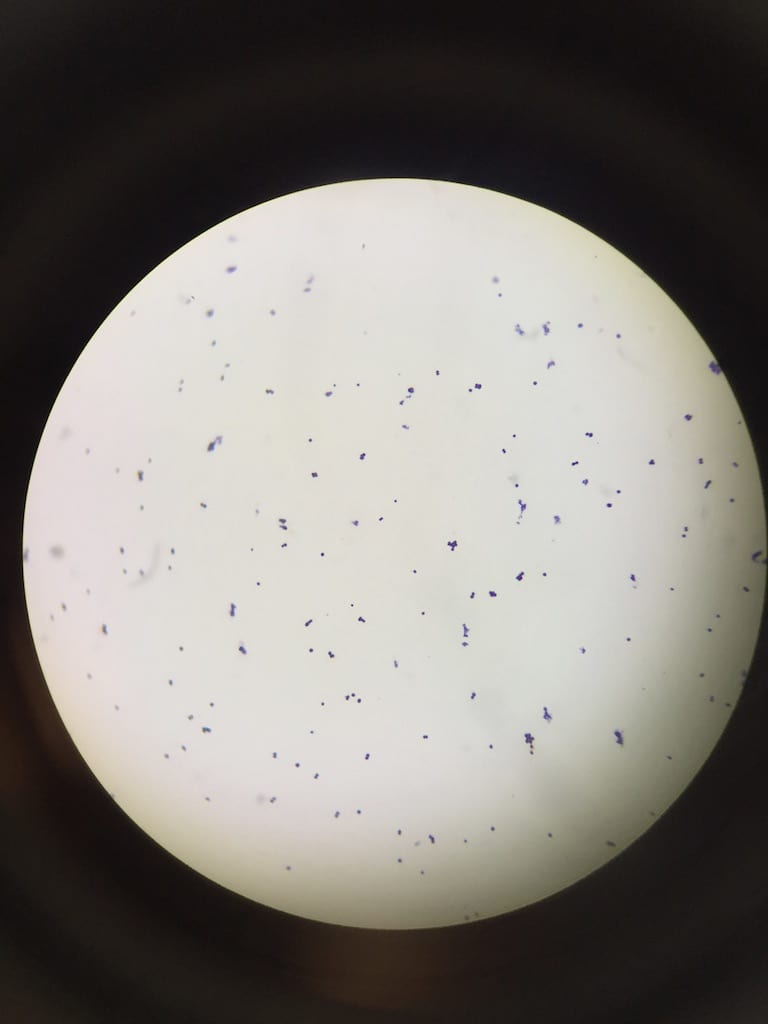
These are my Gram-positive (Staph epi) and Gram-negative (P. putida) controls. They were visible on my slide with the expected coloration so I could identify the Gram identity of my bacterium sample.
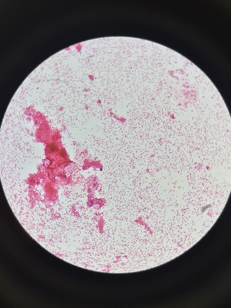
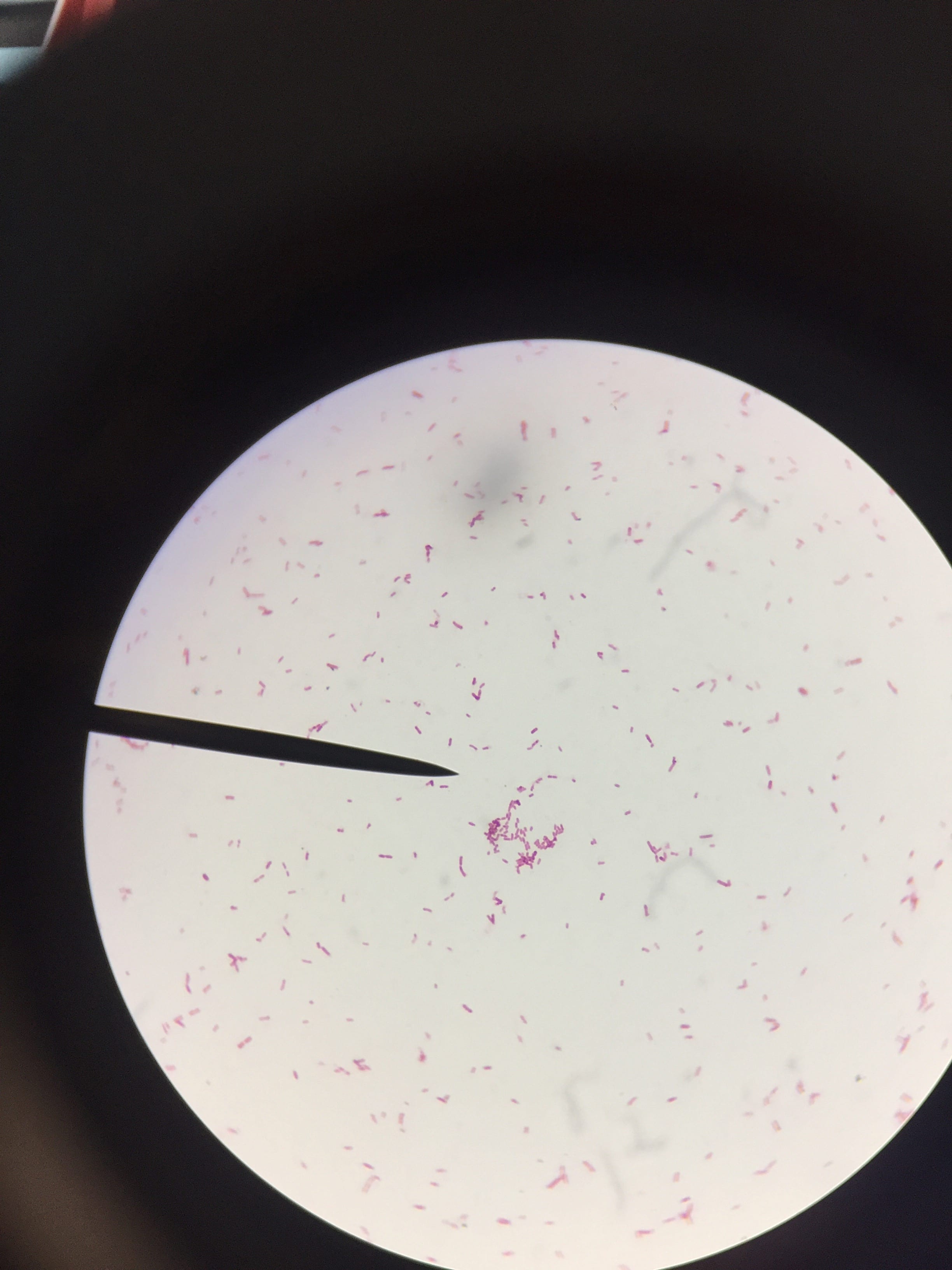
This is my stained colony #5 from LB plate that I picked to identify as it was the only sample that produced the desired PCR product. It is a Gram-negative rod-shaped bacterium.