And the name of my microbe is… – Elizabeth

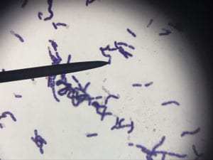
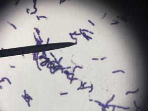
I only got sequencing results for one of my three producers, so the identity of the other two will have to remain a mystery. The one that did work yielded about 725 base pairs of reliable sequence which matched 100% to the 16S ribosomal sequence for the Streptomyces genus. This genus is made of gram positive species and my gram stain results do agree: my gram stain yielded a purple cluster of rod-shaped bacteria set up in chains (see below; image taken from the “Meet my microbes” post – please see that post for further explanation).

As far as taxonomy goes, the organism belongs to the Bacteria kingdom, Actinobacteria phylum, Actinobacteria class, Actinomycetales order, and Streptomycetaceae family (3). Streptomycesis typically live in soil, decaying plant material, and/or water (3,4). They are also known for their quite wonderful smell, which has been called “earthy” and definitely contributed to the smell of the lab that we all became so fond of (3). This genus is known for forming branching chains, as was seen during the gram stain process (4). About 80% of antibiotics used today come from species in the Streptomyces genus, including chloramphenicol, daptomycin, neomycin, streptomycin, and so many more, so production of an antibiotic from my organism is more than plausible, suggesting the experimental method used here was sufficient for our purposes (2,3). Some species also produce antifungals and antiparasitic compounds (3). One study that was done to evaluate the specific nutrient requirements to produce optimal antibiotic production from one species found that they rely on copper, magnesium, zinc, and tryptophan heavily, but excel when provided sodium nitrate, potassium phosphate, and magnesium sulfate (3).


Sources:

1. J.F. Spilsbury. Observations on the nutritional requirements of Stretomyces griseus. Transactions of the British Mycological Society. 31.3-4. 1948.
2. Rudi Emerson de Lima Procopio, et al. Antibiotics produced by Streptomyces. The Brazilian Journal of Infectious Diseases. Accessed via Science Direct. 16:5. 2012.
3. Streptomyces. Wikipedia. June 4, 2018. Accessed Nov 6, 2018. Web.
4. Streptomyces Bacterium. Encyclopedia Britannica. Accessed Nov 6, 2018. Web.

Extract News! – Elizabeth

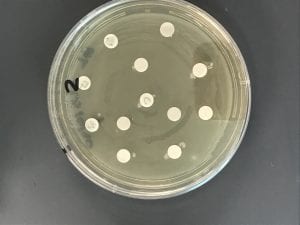
The last day of my extract procedure was completed by Professor Salvo, and thus she picked the tester strains that were used. Upon inspection of the plates, no extracts were successfully isolated; none of the disks had zones of inhibition around them. This could mean that the ethyl acetate procedure wasn’t effective for these compounds, or something else could have gone wrong with the procedure. If we had more time, I would redo the extraction with methanol instead to see if the ethyl acetate was the problem.


Example plate showing no zones of inhibition.

Meet My Microbes – Elizabeth Altman

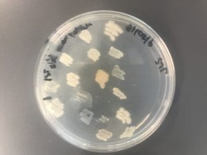
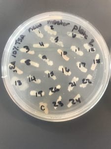
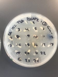
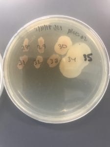
These are Professor Salvo’s initial patch plates of interesting organisms from her soil sample (Jackson’s Gardens). The first two were on LB and the last three were on TSA. I took interesting organisms from each media to get 1 set of 24 organisms on LB plates and 2 sets of 24 organisms each on TSA plates.


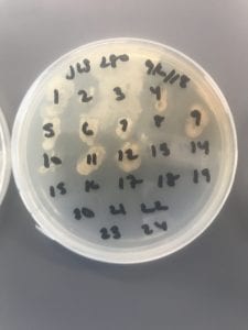
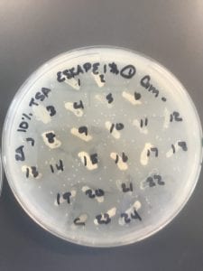
These were the LB plates from the first round of ESKAPE testing. Organisms 2 and 7 were identified as producers on the Gm- plate. These two were patched onto all the ESKAPE safe relatives by someone else.


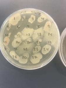
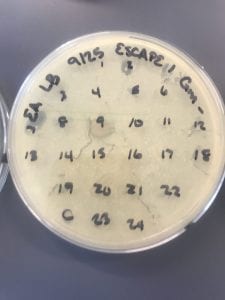
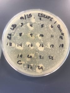
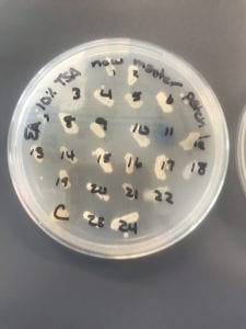
These plates are the first round of ESKAPE testing on the first set of organisms on TSA. Organisms 1, 2, 4, 8, 13, 15, 16, 20, 22, 23, and 24 were identified as producers on the Gm+ plate. I patched all 11 of these onto all ESKAPE safe relatives myself.


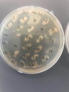
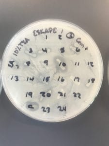
These plates are the first round of ESKAPE testing on the second set of organisms on TSA. Organisms 3 and 10 were identified as producers on the Gm+ plate. These two were patched onto all of the ESKAPE safe relatives by someone else.


This is one of the patch plates from the second round of ESKAPE testing. Only organisms 8, 16, and 20 from the original TSA first set were found to be producers at all during this round of testing. These three were put through colony PCR.


The PCR reactions with the degenerate primer worked for all three organisms, so I moved forward with all three.


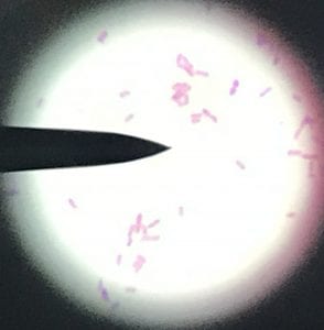
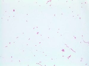
These are the positive and negative control organisms on each slide. They were visible on all slides with the expected coloration, so the stains for all unknowns were trusted. These two, along with all microscope images, were taken at 100x.


This is unknown organisms 8. It is a gram positive bacillus that seems to connect in diplobacilli chains.


Organism 16 was a very small gram negative cocci that existed in groups from two up to about twenty cells.


These are pictures of unknown organism 20. It is gram positive, but has a pretty unique appearance, looking more like rectangles than normal bacilli or cocci.

Meet the ESKAPE Pathogens – Elizabeth

Assigned ESKAPE Pathogen: Enterococcus faecium

Why is this ESKAPE Pathogen of interest:

Enterococcus faeciumis normally found in human and other mammal gastrointestinal tracts as part of the gut microbiota. However, when located elsewhere, it can cause infections. This has become a large problem in hospitals, causing about 3% of sepsis cases in NICUs­4. The emergence of VRE, or vancomycin resistant enterococci, in the early 2000s made this infection a larger issue, further compounding the growing problem of antibiotic resistance worldwide4.

General Cellular and Morphological Characteristics of the Organism (taxonomic classification, nutrition, cell shape, habitat):

This is a gram-positive aerobic bacterium that grows best on Columbia blood agar at thirty-seven degrees Celsius for twenty-four to forty-eight hours1. It typically grow as diplococci, meaning they grow in pairs of round colonies, or as short chains of round colonies and will exhibit hemolysis on some blood medias8.

As previously mentioned, they typically live in the intestines. The Enterococcus genus used to be classified as group D of the Streptococcus genus; however, genetic analysis proved too large a difference between Streptococci and Enterococci to classify them in the same genus8.

Clinical Importance and Prevalence:

Nosocomial infections are a growing problem given both antibiotic resistance and the aging of the Baby Boomer population causing an influx of ill, elderly patients in hospitals. This organism is usually resistant to penicillin (about 90% of cases in one Australian hospital) and resistance to vancomycin has hit about 50% worldwide3. Over twenty-eight percent of people with these infections in the same hospital died within 30 days3. Some research has shown that fluoroquinolone use against these infections should be reduced, as it may play a role in the development of linezolid resistance6.

Infection:

E. faecium, along with its close relative Enterococcus faecalis, cause endocarditis, urinary tract infections, prostatitis, intra-abdominal infection, and cellulitis5. These infections typically occur from lack of correct hygiene methods in hospitals, such as handwashing4.

Pathology:

Symptoms vary based on the location of the infection:

  • Endocarditis: fever, chills, fatigue
  • UTI: pain during urination, foul smelling and/or dark/cloudy urine, polyuria
  • Prostatitis: pain during urination, excessive night urination, urinary retention
  • Intra-abdominal infection: fever, chills, fatigue
  • Cellulitis: red skin, swelling, tenderness to the touch

Ineffective Antibiotics:

Strains isolated from humans have been shown to be resistant to vancomycin and linezolid (see above referenced sources). There is also evidence of strains resistant to chloramphenicol, tetracycline, ciprofloxacin, and erythromycin isolated from pigs in Malaysia5.

Effective Antibiotics:

The current recommendations include streptogramins, oxazolidinones, and Daptomycin5,6.

Corresponding Safe Relative:

The safe relative for E. faeciumis Enterococcus raffinosus. This organism has also been found to be vancomycin resistant and can cause endocarditis2. However, these infections are rare and the human immune system is normally able to withstand infection. Since the safe relative is in the same genus, it behaves similarly, but is less commonly the cause of infection in humans and less commonly resistant to antibiotics.

Sources:

  1. Culture Collections. Public Health England. Accessed October 5 2018.
  2. Dalal, Aman MD,Urban, Carl PhD, Rubin, David MD, Ahluwalia, Maneesha MD. Vancomycin-Resistant Enterococcus raffinosus Endocarditis: A Case Report and Review of Literature. Infectious Diseases in Clinical Practice. May 2008.
  3. Kelvin W. C. Leong, Louise A. Cooley, Tara L. Anderson, Sanjay S. Gautam, BelindaMcEwan, Anne Wells, Fiona Wilson, Lucy Hughson, & Ronan F. O’Toole. Emergence of Vancomycin-Resistant Enterococcus faeciumat an Australian Hospital: A Whole Genome Sequencing Analysis.Scientific Reportsvolume 8, Article number: 6274 (2018).
  4. Lakshmi Srinivasan, Jacquelyn R. Evans, in Avery’s Diseases of the Newborn (Tenth Edition), 2018
  5. Larry M. Bush, MD, Charles E. Schmidt, and Maria T. Perez, MD. Enterococcal Infections. Merck Manual. Accessed Oct 5 2018.
  6. Shiang ChietTanChun Wie ChongCindy Shuan Ju TehPeck Toung OoiKwai Lin Occurrence of virulent multidrug-resistant Enterococcus faecalisand Enterococcus faecium in the pigs, farmers and farm environments in Malaysia. PubMed. August 2018.
  7. Thomas E. Dobbs, Mukesh Patel, Ken B. Waites, Stephen A. Moser, Alan M. Stamm, Craig J. Hoesley. Nosocomial Spread of Enterococcus faeciumResistant to Vancomycin and Linezolid in a Tertiary Care Medical Center. Journal of Clinical Microbiology. June 2018.
  8. Enterococcus. Accessed Oct 5 2018.
Fun with Soil – Elizabeth

Where did you obtain your soil sample?

I got my soil from the path that walks up the right side of the football field behind Alumni Gym. The exact coordinates are Lat 42.816563, Long -73.926441. There was a layer of mulch on top that I moved out of the way and then I dug down about two inches. The soil was moist, but not wet, and dark. There were more ants in the soil than I expected once I got down into it.

Why did you choose this location?

I chose this location because I feel like it hasn’t been disturbed in a while. Most of the bushes in this area are just tended by facilities; not really replanted each year. The soil and the bacteria in it have probably been sitting long enough to have seen competition and (hopefully) some antibiotic production.

Do you expect a lot of isolates? Why or why not?

I expected the least-diluted plates to be covered, so I guess that’s one way to quantify “a lot of isolates.” However, given that its a soil sample and it was under a layer of mulch with at least some exposure to water, there’s probably a fair amount of anaerobic organisms there that we won’t grow at all, so I would expect very few of those isolates.

Have you initial observations supported this?

Yes – when I saw the plates, the ones with 1/100 dilutions were covered. They also looked like the serial dilutions were somewhat successful, as the numbers of colonies on each one decreased (based on rough estimate – I didn’t count them).

What media did you choose? What dilutions?

I chose to work with LB, 10% TSA, and R2A using dilutions of 1/100, 1/1000, and 1/10000. I used the LB as standard and then chose the 10% TSA as a “medium richness” media and the R2A as a bit of a less nutritious media. I thought this range of nutritional value would provide good comparison between plates. My dilution choices were based on our protocol from the test plates (the first lab); we weren’t seeing countable plates until the 1/10000 dilutions, so I made sure to get a 1/10000 plate for each media.

Will you need to redo any?

None of them initially seemed like they needed to be redone. They all had some growth on them and the lowest dilutions seemed countable.

How did your sample differ on the different media?

Honestly, I didn’t look at them too much. For personal reasons, my time in the lab has been limited and I haven’t been able to compare them in detail.