Meet my Microbes! -Alexa

This is a picture of my first set of spread plates. From left to right, the dilutions are 10^-2, 10^-3, and 10^-4. From top to bottom, the media used is PDA, LB, and AC.

This is a picture of my three patch plates. From top to bottom, the media is LB, AC, and PDA.

This is my patch plate on PDA. I just love the colors of the bacteria on this!

These are two of my patch plates against tester strain #6. The top plate is on AC, and colony #15 showed inhibition. The bottom plate is on LB, and #14 showed inhibition.

These are two of my patch plates against tester strain #3. The plate on the left is on AC, and colony #23 showed slight inhibition. The plate on the right is on LB, and #13 and #14 showed inhibition.

The plate on the left is the same as the LB plate against tester strain #6 as seen above. The plate on the right is my patch plate against tester strain #6 and colony #17 is showing inhibition.

The plate on the left is the same as the LB plate against tester strain #3 as seen above. The plate on the right is my patch plate on PDA against tester strain #3 and #13 showed inhibition.

These are all of my patch  plates on AC, LB, and PDA (from left to right).

This is a streak plate on PDA of two of my potential antibiotic producing bacteria. I love the pink color of colony 13!

These are streak plates of the rest of my interesting colonies. From top to bottom, there’s my colony #15 and Nikki’s colonoy #7 on AC, then my colonies #3 and #23 on PDA, then my #13 and #23 on PDA, and lastly my #13 and #14 on LB.

I patched four of my isolates against tester strains on PDA. Shown above is a patch plate against tester strain #9 and my colony #17 is showing great inhibition!

Gram stain of colony #13. It’s gram positive and rod shaped.

Gram stain of colony #14. It’s gram negative and rod shaped.

Gram stain of colony #15. It’s gram positive and coccus shaped.

Gram stain of colonoy #17. It’s gram negative and rod shaped.

And the name of my microbe is….. -Alexa

What were the results of your 16S analysis?
All four of my isolates, #13, 14, 15, and 17, did have PCR products, but I was only able to obtain the 16S rRNA gene sequence for #13, 14, and 15. When we looked at the PCR products on a gel, #17 did produce a band, but it was faint which could explain why it wasn’t able to be sequenced. BLAST showed that #13 has a 99% match with the Bacillus genus. Isolate #14 had a 99% match with the Pseudomonas genus, and #15 had a 99% match with the Staphylococcus genus.

Does your gram stain agree?
My gram stain agreed for all three of my isolates. The gram stain for isolate #13 showed it was a gram-positive, rod shaped bacteria. This matches the description for the bacteria in the Bacillus genus (1). The gram stain for isolate #14 showed it was a gram-negative, rod shaped bacteria. This also matches the description for bacteria in the Pseudomonas genus (2). The gram stain for #15 showed it was a gram-positive, coccus shaped bacteria which clumped together in random numbers. This matches the description for bacteria in the Staphylococcus genus (3).

a) General cellular and morphological characteristics of the genus (taxonomic classification, nutrition, cell shape, habitat).
I further researched the characteristics of the Pseudomonas genus which matched my isolate #14. The taxonomic classification for this is Bacteria kingdom, Proteobacteria phylum, Gammaproteobacteria class, Pseudomonadales order, Pseudomonadacae family, and Pseudomonas genus (4). They are gram-negative, straight or slightly curved rods (2). They are typically aerobic, although sometimes nitrate can be used as an alternate electron acceptor which allows it to grow anaerobically (2). Most species can not grow under acidic conditions and do not require organic growth factors (2). This bacteria is widely distributed in nature and some species within this genus are pathogenic for humans, animals, or plants (2).

b) Information regarding antibiotic production in this genus.
Pseudomonas have been found to produce a wide variety of antibiotics including mupirocin (a topical antibiotic), fosfadecin, fosfocytosin, and xantholysins (4). Xantholysins have been discovered more recently and have shown anti-fungal activity and antibiotic activity, specifically against gram positive bacteria. Pseudomonas fluorescens has been shown to produce the antibiotics  pyrrolnitrin, pyoluteorin, and 2,4-diacetylphloroglucinol (5).  It is encouraging to know that this genus has shown antibiotic production since my isolate appeared to show antibiotic production.

References
1) Liu, Y., et. al, Proposal of nine novel species of the Bacillus cereus group, International Journal of Systematic and Evolutionary Microbiology, 2017
2) Palleroni, N. J. (2015). Pseudomonas. In Bergey’s Manual of Systematics of Archaea and Bacteria (eds W. B. Whitman, F. Rainey, P. Kämpfer, M. Trujillo, J. Chun, P. DeVos, B. Hedlund and S. Dedysh).
3) Pantucek, R., et. al, Staphylococcus edaphicus sp. nov., isolated in Antarctica, harbours mecC gene and genomic islands with suspected role in adaptation to extreme environment., Applied and Environmental Microbiology, 2017
4) Javier Pascual, Marina García-López, Cristina Carmona, Thiciana da S. Sousa, Nuria de Pedro, Bastien Cautain, Jesús Martín, Francisca Vicente, Fernando Reyes, Gerald F. Bills, Olga Genilloud,
Pseudomonas soli sp. nov., a novel producer of xantholysin congeners, Systematic and Applied Microbiology, Volume 37, Issue 6, 2014, Pages 412-416
5) Sarniguet, A., et. al, The sigma factor sigma s affects antibiotic production and biological control activity of Pseudomonas fluorescens Pf-5, Proceedings of the National Academy of Sciences, 1995

Extract News! -Alexa

I tested four of my bacteria samples (13, 14, 15, and 17) against the tester strains 1 (S.Epidermis),3 (E. Carotovora) ,6 (B. Subtillis), and 9 (P. Putida). I chose these strains because my bacteria had previously shown resistance to all of them. Out of my four bacteria, only one of them, number 15, showed inhibition against strain 6. It had a very small ring around it so it didn’t show strong inhibition. Previously, number 15 did show inhibition against tester strain number 6. Number 13 and 14 both came from my LB plates and showed inhibition against tester strains 1, 3, and 6, so it was surprising that I didn’t see an inhibition from the extracts against any of the tester strains. Number 17 was from my PDA plates and showed inhibition against strains 6 and 9, but its extract also failed to show inhibition against any of the strains. Since only one of my samples showed inhibition it could be possible that I didn’t get extracts for the other ones or maybe the concentration was so low that it didn’t make a difference. It could have also been helpful to incubate the tester strains with the extracts on them longer, or let the heavy streak plates incubate longer since it’s possible the bacteria needed a few more days to start producing an antibiotic.

Meet the ESKAPE Pathogens – Alexa and Kara

Acinetobacter baumannii  is a multi-drug resistant, pathogenic bacteria that is commonly found in hospital settings.  Being that is an emerging source of nosocomial infection, and is becoming more prevalent, Acinetobacter baumannii is of interest to the medical and scientific communities (1).

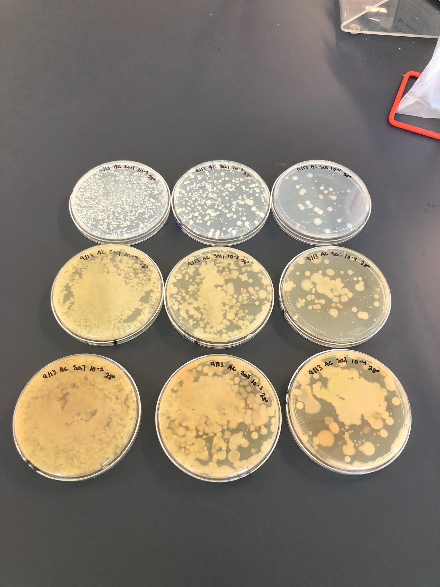
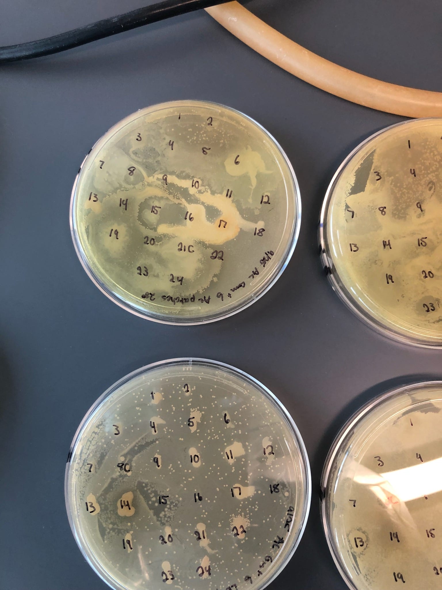
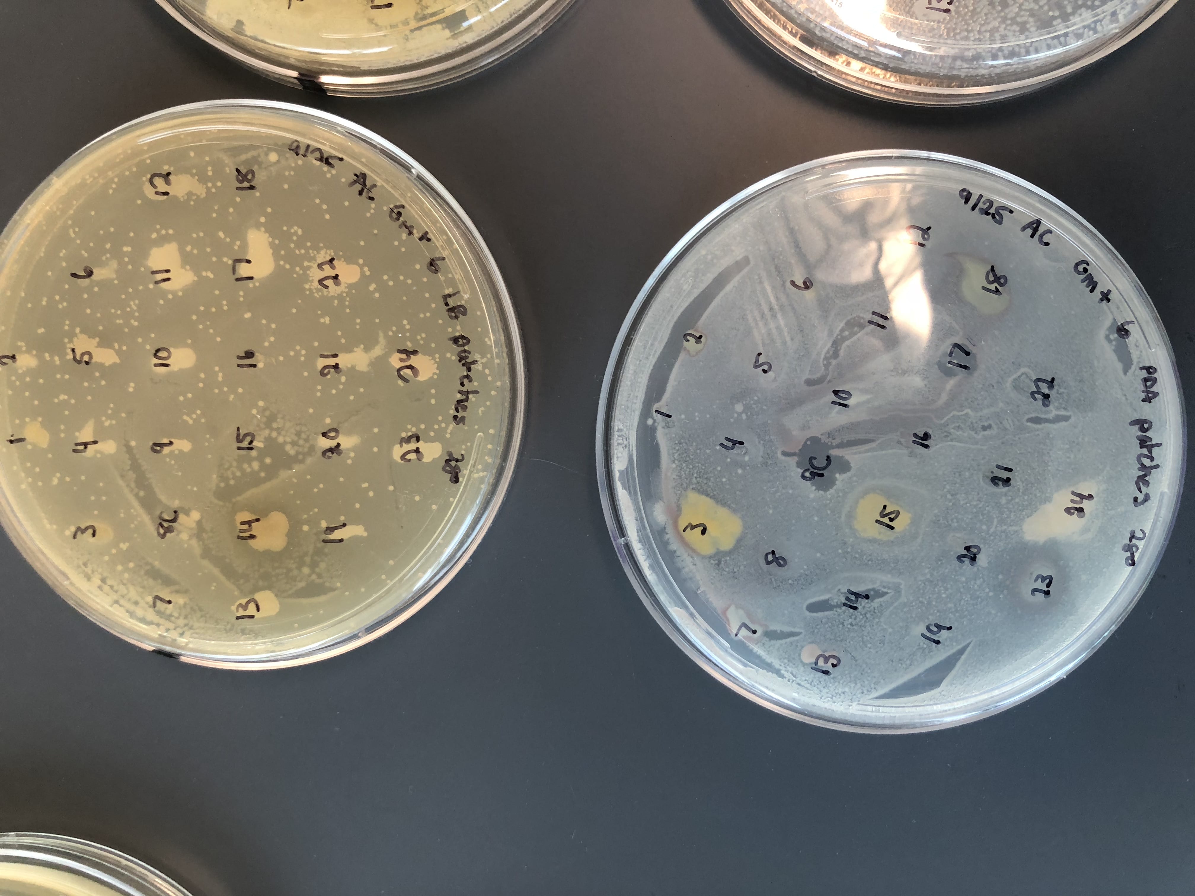
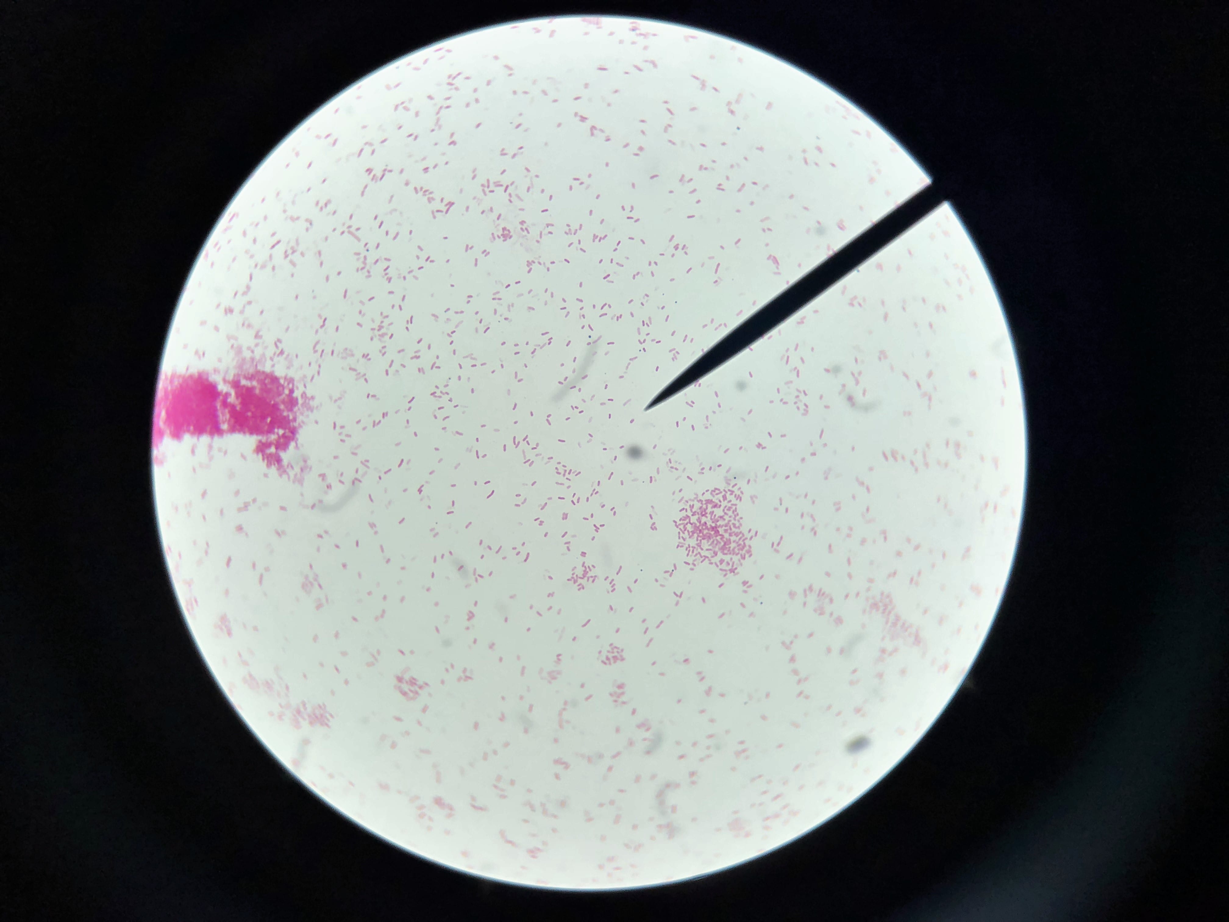
A. baumannii is gram negative, aerobic, and coccobacillus in shape (Figure 1) (3).  The genus Acinectobacter was first discovered in 1911, when a Dutch scientist isolated microorganisms from soil. Originally “Acinectobacter” was used to designate non-motile bacteria. However, today it is currently the genus for “gram-negative, strictly aerobic, nonfermenting, nonfastidious, nonmotile, catalase-positive, oxidase-negative bacteria with a DNA G+C content of 39% to 47%” (2).  Differences in the carbapenemase and gyrB genes of A. Baumannii distinguish it from its close relatives. A. baumannii’s “safe” relative is A. baylyi (2). A. baumannii, Acinetobacter genomic species 3, and Acinetobacter genomic species 13TU are extremely similar, and cannot be distinguished from one another using commercial methods.  Therefore, the 3 species are often lumped together as a complex, and treated as one (2).  Acinectobacters are commonly found in soil and water in nature, and some are part of the flora of human skin. However, virtually no individuals have tested positively for A. baumannii outside of hospital settings and combat zones, so the natural habitat of A. Baumannii is currently unknown (2). Very few individuals in China and tropical Australia have been tested positively for A. baumannii (5).  In the laboratory setting, A. Baumannii  grows well on Luria-Bertani broth (LB) (1).

As mentioned prior, A. baumannii is rarely found outside of the hospital setting.  One notable exception to this is the prevalence of A. baumannii in skin infections among soldiers and other military personnel (1).   Additionally, A. baumannii is well documented in burn units and ICUs.  Immunocompromised individuals, as well as individuals with prolonged hospital stays are at the greatest risk for infection of A. baumannii (1). Hospital-acquired pneumonia and bloodstream infections, as well as post-neurosurgical meningitis, are the diseases commonly associated with A. baumannii. In 2003 the CDC reported that 7% of ICU-acquired pneumonias were caused by A. baumannii, and between 5 and 10% of cases of ICU-acquired pneumonia were due to A. baumannii (2).  It is hypothesized that soldiers returning from Afghanistan and Iraq brought this pathogen into U.S. hospitals.  A. baumannii has thrived in this setting due to its antibiotic and antiseptic resistance (2).  Infections typically begin when an external device, such as a ventilator, is used in the treatment of a patient.  The external device provides an entry site for A. baumannii, where it can then colonize (and can form biofilms)—for this example, in the lungs (1). Figure 2 below shows how A. baumannii spreads in the hospital setting, and Figure 3 shows how it affects the human body (6). Symptoms of an infection caused by A. baumannii are hard to distinguish among already ill individuals, however, they typically include fever, chills, and cough (5).

A. baumannii is known to be resistant to cephalosporins (can degrade beta-lactam), aminoglycosides, quinolones and, lately, carbapenems (3).  When A. baumannii was first treated in the 1990’s, carbapenems were effective antibiotics, but are becoming less effective over time (3). Modified efflux pumps, porins, and lactamases enable A. baumannii to be resistant to several classes of drugs (4). The World Health Organization reports that A. baumannii is susceptible to polymyxins, but academic research has focused on new developments to combat gram-negative bacteria. Outer membrane proteins of gram-negative bacteria are being targeted experimentally as potential antibiotics (3). Additionally, bacteriophages are also being investigated as potential antibiotics, due to their ability to selectively target bacteria (1).

ESKAPE Pathogen Fig 1-27hgnn4

ESKAPE Pathogen Fig 2-18eokb8

ESKAPE Pathogen Fig 3-1t4ue2e

References:

(1)   Howard, Aoife, et al. “Acinetobacter Baumannii.” Virulence, vol. 3, no. 3, 2012, pp. 243–250., doi:10.4161/viru.19700.

(2)   Peleg, A. Y., et al. “Acinetobacter Baumannii: Emergence of a Successful Pathogen.” Clinical Microbiology Reviews, vol. 21, no. 3, 2008, pp. 538–582., doi:10.1128/cmr.00058-07.

(3)   Soojhawon, Iswarduth, et al. “Discovery of Novel Inhibitors of Multidrug-Resistant Acinetobacter Baumannii.” Bioorganic & Medicinal Chemistry, vol. 25, no. 20, 2017, pp. 5477–5482., doi:10.1016/j.bmc.2017.08.014.

(4)   Vila, Jordi, et al. “Porins, Efflux Pumps and Multidrug Resistance in Acinetobacter Baumannii.” Journal of Antimicrobial Chemotherapy, vol. 59, no. 6, 2007, pp. 1210–1215., doi:10.1093/jac/dkl509

(5)   “WPRO | Multidrug-Resistant Acinetobacter Baumannii (MDRAB).” World Health Organization, World Health Organization, 7 July 2017, www.wpro.who.int/mediacentre/factsheets/fs_20101102/en/.

(6)  Dijkshoorn, Lenie, et al., “An increasing threat in hospitals: multidrug-resistant Acinetobacter baumannii”, Nature Reviews Microbiology, vol. 5, 2007, pp. 939-951

https://doi.org/10.1038/nrmicro1789

Fun with Soil – Alexa

Where did you obtain your soil sample?
I obtained my soil sample from Peeble’s Island which is near Waterford, New York. I live in Waterford and I go to Peeble’s Island all the time when I can. I went to the beginning of one of the trails on the island, and decided to go a couple feet away from the trail and collect my soil sample there. I collected the dirt in between a tree and many smaller plants. I collected the sample about 2 inches below the surface since there was a lot of leaf litter on the top layer.

Why did you choose this location?
Since Peeble’s Island is home to many different kinds of trees, plants, and wildlife, I thought it would have very rich soil. I was also curious to see what kind of bacteria I could find in a place that I had spent so much time walking around!

Do you expect a lot of isolates? Why or why not?
I expected a lot of isolates because I expected the soil to be rich since it supported such a wide variety and large amount of plants. The soil also seemed like it would get enough oxygen to support aerobic bacteria which is the kind we are growing in the lab.

Have you initial observations supported this?
My initial observations have supported this. I was only able to determine a cfu for one plate which was on PDA and a dilution of 1:10000. All of the other plates had way too many colonies to even count, even after incubating for only 48 hours. The 1:100 dilutions were completely covered with bacteria on all the different medias I used.

What media did you choose? How did you sample differ on the different media?
I chose LB, AC, and PDA for my media. Everybody had to use LB, but I wanted to use another rich media to see how many different bacteria I could grow. I realized that since my soil was likely rich, I would need to use the weakest dilutions on LB and AC since they would support the most growth. I knew I needed to use a minimal media, so I chose PDA as my third media. I noticed there were more potential antibiotic producers on the rich media compared to the minimal media, but there was significantly more diversity on the minimal media. The most common colors on the rich media were off-white and there was occasionally a yellow colony. On the strongest dilution of PDA most of the colonies were pretty small and close to white. On the weaker dilutions there were more colors including pure white, off-white, yellow, orange, purple, dark brown, and green. There’s also fuzzy bacteria (or potentially mold) growing on the PDA plates which hasn’t grown on the rich media plates.

What dilutions?
I plated the 10-2, 10-3, and 10-4 dilutions for all of the media since I guessed the 10-1 dilution would have too much bacteria in it.

Will you need to redo any?
I am redoing the 10-4 dilution on PDA since it was my most interesting plate. I am also plating a 10-5 dilution on LB and AC since I couldn’t get a cfu on any of those plates.