Extract News! Troy

Of all the fabulous bacteria that I had, there were three strains that were worthy of testing.  Coincidentally, all three were grown on R2A – a reason for which may be that there were fewer resources in this substrate.  The extracts were gathered on Thursday, October 25, by collecting the organic layer of our previously suspended colony plates.  I extracted three different ‘antibiotics’ from my three different strains, and prepared them to be dried.  On October 30, the extracts (which were not completely dried yet) were prepared to be tested against different bacteria strains.  They were suspended in methanol, and this solution was used to create antibiotic plates that were dropped onto the different tester bacteria plates.  I chose to test against 4 strains – 3, 4, 6, and 7 -, as these strains presented two GM(+) and two GM(-) bacteria strains.  On November 1, the results were observed, and of my three extracts I had antibiotic activity against both GM(+) and GM(-) bacteria.  Strain 1A – a GM(-) bacillus – produced antibiotics that inhibited growth on bacteria strain 3 – a GM(-) bacteria.  Strain 2B – another GM(-) bacillus – produced antibiotics that inhibited bacteria strain 7 – a GM(-) bacteria.  Strain 3A – a GM(-) rod-shaped bacteria – produced antibiotics that inhibited bacteria strain 4 – a GM(+) bacteria.

I was very happy to see that I had AB production from all three of my strains.  I was interested that the two bacilli strains produced AB’s that inhibited the GM(-) testers while the rod-shaped strains produced AB’s that inhibited the GM(+) tester strain (strain 4).  I believe that these strains could be further observed to be potential drugs against bacteria, as they target a wide range of GM(+) and GM(-) bacteria.


Extract News! Kara

I tested 3 extracts from my 3 producers, one of which was isolated on PDA, while the other 2 were isolated on 10% TSA. I found that all three of my extracts inhibited strain #1 which was staph epi, but did not inhibit the other strains.  I picked the strains a bit arbitrarily, because one of my producers had inhibited all of the strains, so I chose 2 gram negative, and 2 gram positive.  I chose strains 1 (Staph epi), 2 (E. coli) , 5 (A. baylyi) and 6 (B. subtillis).  I made sure A. baylyi was one of the strains I looked at because my ESKAPE pathogen was A. baumanni (which is the pathogenic relative of A. baylyi), so I was curious as to whether or not my extracts would inhibit it (sadly they did not).

Overall, I was happy to see that my extracts demonstrated some antibiotic activity, indicating that my extraction was successful.  Unfortunately, staph epi is very “easy” to inhibit, indicating my extracts probably would not work against most microbes.

Meet my microbes! – Tommy

Here is a photo of my PDA patch plate. PDA plates gave me a lot of mold over the course of the term, but also displayed some really interesting and unique microbe morphologies. Unfortunately, none of these inhibited the ESKAPE tester strains – neither Gram positive nor Gram negative. But they look awesome!


Gram negative control.


Gram positive control. Cocci.


Streak plate of isolate 18. This is the isolate whose extraction strongly inhibited a Gram positive tester strain and a Gram negative tester strain. It was my best producer!


Unknown 18. Gram positive. Appear to be diplococci that form some longer chains as well and like to aggregate. This is what my best antibiotic producer looks like up close!


Streak plate of isolate 11


Isolate number 11. Gram positive rods. Looks like they might be spore formers.

Extract news! – Tommy

On October 25th, we performed an organic extraction of our isolates. I had two isolates that I attempted extractions on. After letting the extraction vials dry, a residue was present which was weighed and subsequently resuspended in methanol. Not all of the residue dissolved, but 20 microliters of this resuspended methanol/extract solution was put on little circles to put on tester strains to see if they inhibited tester strains. If the tester strains were inhibited, it would mean we were successful in extracting an antibiotic from the bacteria!

We tested our extractions on two Gram positive and two Gram negative tester strains. Both of my isolates had inhibited at least one Gram positive and one Gram negative tester strain. To test extractions, I chose testers 1, 3, 4, and 5 (Staphylococcus epidermidis, Erwinia carotovura, Enterococcus raffinosus, and Actinetobacter baylyi, respectively). Testers 1 (Gram positive) and 5 (Gram negative) were previously inhibited by isolate number 11 and testers 3 (Gram negative) and 4 (Gram positive) were previously inhibited by isolate number 18.

Below are pictures of the results of testing the extracts on testers 3 and 4 (Erwinia carotovura and Enterococcus raffinosus). Extract from isolate number 18 inhibited both testers as seen by the clearing of growth of the tester bacteria around the 18 extract circle. This is interesting because I chose these two testers based on inhibition that I saw from isolate number 11, not isolate 18, but extract 18 showed inhibition while isolate 11 did not. However, extract 18 also inhibited testers 1 and 5 (Staphylococcus epidermidis and Actinetobacter baylyi), albeit not as robustly. Extract 11 did not show inhibition of any tester strains. The fact that extract 18 inhibited tester strains indicates that an antibiotic was successfully extracted!

Tester 3 – Erwinia carotovura – with organic extractions. The extraction from my isolate 18 shows a clearing of growth around it, indicating inhibition of the tester.


Tester 4 – Enterococcus raffinosus – with organic extractions. The extraction from my isolate 18 shows a small inhibition of tester growth around it.

Extract News- Kelsey

I tested two extracts. One extract was an isolate of mine on 10% TSA and the other extract was the same isolate on R2A. I tested the extracts on two Gram negative strains, E. carotova and E. coli. I also tested the extracts on two Gram positive strains, E. raffinosis and B. subtilis. When testing the extracts, I found no resistance observed to any of the strains. When the two isolates were tested, they had observed resistance against E. raffinosis and B. subtilis. If I were to further test the extracts, I could try different media or the same media again. I could also leave the plates out for longer to see if any antibiotic activity would show over time. However, at this point, I conclude that the isolates did not secrete any antibiotic activity into the media on which they were plated.

Extract News! – Le Minh

I was able to obtain an extract from the only isolate (LB #5) that I had. The extract appeared insoluble in methanol as I had a hard time resuspending it; the extract formed a large white clump that would not fully dissolve even after lengthy vortexing. However, I still carried out the following procedure of testing the extract with the obtained solution. I chose to test it against the Gram-negative #2 E. coli and #7 E. aerogenes as well as against the Gram-positive #4 E. raffinosus and #6 B. subtilis as my isolate was able to grow in those tester strains during the ESKAPE testing we performed before. 

In the end, my extract did display some antibiotic-producing ability (so the extract was somewhat successfully resuspended). Out of all test plates, my extract was able to show inhibition only against E. coli but not against the other Gram-negative bacterium nor the Gram-positive bacteria. This means that my extract has narrow-spectrum antibiotic activity. 

The picture below shows the zone of inhibition around my extract (it is a little bit hard to see but it is there) that is was able to produce against E. coli 

Extract News! -Alexa

I tested four of my bacteria samples (13, 14, 15, and 17) against the tester strains 1 (S.Epidermis),3 (E. Carotovora) ,6 (B. Subtillis), and 9 (P. Putida). I chose these strains because my bacteria had previously shown resistance to all of them. Out of my four bacteria, only one of them, number 15, showed inhibition against strain 6. It had a very small ring around it so it didn’t show strong inhibition. Previously, number 15 did show inhibition against tester strain number 6. Number 13 and 14 both came from my LB plates and showed inhibition against tester strains 1, 3, and 6, so it was surprising that I didn’t see an inhibition from the extracts against any of the tester strains. Number 17 was from my PDA plates and showed inhibition against strains 6 and 9, but its extract also failed to show inhibition against any of the strains. Since only one of my samples showed inhibition it could be possible that I didn’t get extracts for the other ones or maybe the concentration was so low that it didn’t make a difference. It could have also been helpful to incubate the tester strains with the extracts on them longer, or let the heavy streak plates incubate longer since it’s possible the bacteria needed a few more days to start producing an antibiotic.

Extract News! – Brianna

I was able to obtain a total of 6 extracts between my 4 samples (there was a little bit of labeling issues, so I could not combine the duplicate sets). I chose to test these extracts against  #1 (Staphylococcus epideridis) and #4  (Enterococcus raffinosus) as the gram positive ESKAPE pathogens and #3 (Erwinia carotovura) and #7 (Enterobacter aerogenes) as the gram negative. These were chosen since my producers  showed inhibition against these pathogens (some showed against one or two, but not all three at once) with the exception of number 7 (which was chosen out of convenience).

Out of all the extracts, two showed inhibition against Staphylococcus epideridis, Enterococcus raffinosus and Erwinia carotovura . While the numbering/labeling was messed up from before, these were my two pigmented extracts. I believe these correspond to my samples, PDA #2 and LB #2.


Here is a table summarizing the cell free and colony inhibition tests.


Extract News! – Elizabeth

The last day of my extract procedure was completed by Professor Salvo, and thus she picked the tester strains that were used. Upon inspection of the plates, no extracts were successfully isolated; none of the disks had zones of inhibition around them. This could mean that the ethyl acetate procedure wasn’t effective for these compounds, or something else could have gone wrong with the procedure. If we had more time, I would redo the extraction with methanol instead to see if the ethyl acetate was the problem.


Example plate showing no zones of inhibition.

6. Extract News! – Nikki

Did you obtain an extract? Summarize the results of your testing and why you choose the tester strain(s) you did.

I was able to obtain an extract for each of my three isolates (#15, 20, and 23) following the extraction procedures we followed over the last few weeks in lab. I chose to use the following tester strains: #1 S. epidermidis (+), #2 E. coli (-), #6 B. subtilis (+), and #7 E. aerogenes (-). I chose these four tester strains because my isolates had shown inhibition previously of #1, #2, and #6 tester strains and then I chose #7 because it was a gram – strain to fulfill the 2 Gram + and 2 Gram – that we testing against in this stage. In my observations from the test extracts, I only saw a ring signaling potential inhibition around my #20 isolate/extract on the #1 S. epidermidis, a Gram + tester strain. There was no other antibiotic activity on the other plates/for the other extracts. I added a photo below of the #20 isolate on the #1 tester strain plate and it is the one circled and labeled “ND 20”. Note that this photo was taken on Thursday, November 1st, a day after Prof. Salvo took the plates out of the incubator and made the observations of antibiotic activity.


Meet My Microbes! – Brianna

Here are (some of ) my original plates from the soil sample extracts.

The first is my 10-3 dilution plated onto R2A , the second is a 10-2 dilution on PDA, and the third is 10-3 dilution on LB media. All are photographed on day three of incubation. For the most part, PDA showed the most diverse colonies with a variety of colors and shapes. From these and other dilution plates, 24 colonies (per plate type) were picked and selected to be further investigated.

Here are my master patch plates after four days of incubation, in the same order as before. A few interesting things occurred at this stage. The diversity of the colonies was much easier to identify. They went from being generic-off-white-round colonies to having slightly different pigments, shapes, and morphologies. Additionally, some of the picked colonies did not grow on the new plate (RIP), so who knows if I lost the answer to the antibiotic crisis.


Then, all of these colonies were tested for antibiotic producers. Below is my champion of my antibiotic producers, PDA #5. As you can see, a vary large halo formed around colony #5, clearing the tester ESKAPE #6, B. Subtillis.  I also had 3 other producers from other plates and colonies, but they produced only a small halo against the background.

From all the ESKAPE testing, I narrowed down my colonies to a total of 4 producers. These are the streak plates for each of these microbes. In order, they are: PDA #2, PDA #5, LB #2, and R2A #18.

These four microbes were then stained to determine if they were gram positive or negative. All images are taken under a 100X oil emersion microscope lens. For reference, here is the positive (Staphylococcus epidermidis) and negative (E. Coli) controls.


The following are of my isolates. The first image is of R2A #18. This microbe is gram positive, and appears in diploid rods. The second is LB#2, which is gram negative and appears to be a shortened more ovular rod, and tends to be in diploid clusters. The third, PDA #2,  is a gram negative cocci, and is generally diploid, but forms larger clusters as well. Lastly, PDA#5, is gram-negative, and the cells are shaped like a typical rod bacterium.


Meet My Microbes! – Le Minh Nguyen

This is of one of my very first plates from the first week of lab. I plated my soil sample on the LB + cycloheximide plate with a 10^-4 dilution. This plate was unique because it contained those brown circular colonies with a surrounding halo-fade that also caused pigmentation in the media. As I later found out, this isolate showed some antibiotic production as it inhibited the growth of some of our testers and I eventually chose it for my PCR reaction analysis.


These plates also come from the first week of lab. The plate on the left is also a plate of my soil sample, plated on PDA + cycloheximide plate with a 10^-3 dilution. This plate shows a great diversity of bacteria as presented by a variety of different colors. A lot of colonies were picked from this plate and were patched. Its master plate looks as interesting as shown by the diverse and colorful isolates that were able to grow. 

This is a PDA + cycloheximide plate from the first round of ESKAPE testing. The tester strain is a Gram-negative E. caratovora. As seen on the plate, the tester strain did not grow very well, so the ESKAPE testing was repeated with another Gram-negative E. coli. However, what is more interesting is that only a few colonies were able to grow (#1, 4, 13, 14) while the rest seemed to be contaminated as they appear as red smears. This most probably occurred due to using the same toothpick for ESKAPE testing. 

This is an LB plate, also from the first round of ESKAPE testing with a Gram-negative E. caratovora. This time the tester strain was able to grow and my isolates were tested. As shown on the plate, colonies 5, 6, and 11 showed promise to be antibiotic producers as indicated by the presence of a halo. 


These are pictures of one of my master plates obtained during the ESKAPE testing. These isolates are grown on the PDA plate and as mentioned above, the diversity of the obtained bacteria is great; they are significantly different in morphology (as indicated by the difference in color and shape) and look really cool. 

This is one of my streak plates of the bacteria that I streaked for PCR reaction. The bacteria is grown on the PDA plate and is the same one from the previous PDA plates, indicated as #1. The single colony has a circular egg-like shape and is of deep brown color. Furthermore, after a few days, the colony causes the media to absorb the bacteria’s pigment. 


These are my Gram-positive (Staph epi) and Gram-negative (P. putida) controls. They were visible on my slide with the expected coloration so I could identify the Gram identity of my bacterium sample. 

This is my stained colony #5 from LB plate that I picked to identify as it was the only sample that produced the desired PCR product. It is a Gram-negative rod-shaped bacterium. 

Meet my microbes – Kara

This is the PDA patch plate I made, and where I isolated my only producer. The other producers I have were generously given to me by Elizabeth Altman.

this is an image of my producer. It appears to have both a mix of both gram + and gram – bacilli.  I’m not sure how to characterize this, it could be a mixed culture.


Both isolates from Elizabeth looked very similar.  They show individual gram positive bacilli on the left, and filamentous “clusters” of cells on the right.

Meet My Microbes- Carly Burns

These are my master patch plates from 9/17 and 9/25.  The LB plates were labeled group “a”, the R2A plates were labeled group “b”, and the AC plates were labeled group “c”.

These are the patches from 9/25 from group a and b tested against the Gm+ tester strain E. raffinosus and the Gm- tester strain E. carotovora.  No patches showed possible antibiotic production against them.

One set of these plates are patches from group c made on 9/25 against the Gm+ tester strain E. raffinosus and the Gm- strain E. carotovora.  None of these patches showed possible antibiotic production so a new patch plate with different colonies from the original plate were made and tested against E. raffinosus and E. carotovora on 9/27.  These patches also showed no possible antibiotic production.

Since no patches showed possible antibiotic production, all of those plates were tossed in the trash and I took two possible producers from Elizabeth and made this streak plate on 9/27 for further tests.  After testing these two bacteria against all the tester strains I found that only #10 was a possible producer so I took this bacteria and two bacteria from Tommy and performed a PCR reaction with them.  These PCR reactions were then run via gel electrophoresis and this showed that only the two cultures I got from Tommy contained the band we were looking for so I sent these two out to be sequenced.

I performed the Gram stain test on the two cultures and one of them ended up being Gm+ and the cells had a deep purple color and were rod shaped (above).  This is a picture of one culture under 100x magnification.

The Gram stain test for the other culture showed that the cells were Gm- and the cells had a pink color and were oval shaped (above).  This is also a picture under 100x magnification.


Meet My Microbes – Elizabeth Altman

These are Professor Salvo’s initial patch plates of interesting organisms from her soil sample (Jackson’s Gardens). The first two were on LB and the last three were on TSA. I took interesting organisms from each media to get 1 set of 24 organisms on LB plates and 2 sets of 24 organisms each on TSA plates.


These were the LB plates from the first round of ESKAPE testing. Organisms 2 and 7 were identified as producers on the Gm- plate. These two were patched onto all the ESKAPE safe relatives by someone else.


These plates are the first round of ESKAPE testing on the first set of organisms on TSA. Organisms 1, 2, 4, 8, 13, 15, 16, 20, 22, 23, and 24 were identified as producers on the Gm+ plate. I patched all 11 of these onto all ESKAPE safe relatives myself.


These plates are the first round of ESKAPE testing on the second set of organisms on TSA. Organisms 3 and 10 were identified as producers on the Gm+ plate. These two were patched onto all of the ESKAPE safe relatives by someone else.


This is one of the patch plates from the second round of ESKAPE testing. Only organisms 8, 16, and 20 from the original TSA first set were found to be producers at all during this round of testing. These three were put through colony PCR.


The PCR reactions with the degenerate primer worked for all three organisms, so I moved forward with all three.


These are the positive and negative control organisms on each slide. They were visible on all slides with the expected coloration, so the stains for all unknowns were trusted. These two, along with all microscope images, were taken at 100x.


This is unknown organisms 8. It is a gram positive bacillus that seems to connect in diplobacilli chains.


Organism 16 was a very small gram negative cocci that existed in groups from two up to about twenty cells.


These are pictures of unknown organism 20. It is gram positive, but has a pretty unique appearance, looking more like rectangles than normal bacilli or cocci.

Meet My Microbes- Kelsey


This is a picture of where I got my soil sample! There were lots of decomposing elements to this soil including rusty beer cans, paper, organic matter, etc. The soil was most likely in the A Horizon.


These are two of my streak plates with the four producers that I found in lab. (slightly blurry due to the plastic bag).


These are my R2A plates, where I found most of my producers. You can see the mycoides growing on the 10^-2 and 10^-3 dilution plates. The second photo is a close up of the mycoides on a light box at a dilution factor of 10^-2.


A close up of my R2A plate with a 10^-4 dilution. This photo was taken on the light box.


These are three examples of patch plates. These were the first set of patch plates that we did. These photos were all taken on the light box. In both the LB and AC media plates, you can see an overgrowth of mycoides. The mycoides overtook the patches in this section and rendered them unusable. I found minimum mycoides growth in the R2A plates throughout my experiment.


These are two pictures of my Gm+ bacteria. I know it is Gm+ due to the purple color. My bacteria are most likely bacillus because they are rods and spore forming. I know that they are spore formers due to the central pink spore between the purple ends of the rods. These photos were taken at 100x.


This is another photo of my bacteria. You can see a large aggregation of colonies on the right side of the slide. This photo was taken at 1000x.


These are pictures at 1ooox of my Gram positive control (S. epi).


These are pictures at 1000x of my Gram negative control (P. putida).

Meet the ESKAPE Pathogens – Tommy

Assigned ESKAPE Pathogen

Enterococcus faecium

Why is this ESKAPE Pathogen of interest (in brief)

Enterococcus faecium can exist commensally within human or animal gastrointestinal tracts but can also be pathogenic in that it can cause neonatal meningitis and endocarditis (1). There have been strains of Enterococcus faecium identified as resistant to vancomycin – coined Vancomycin-Resistant E. faecium (VRE). Enterococcus faecium has also show tolerance to handwash alcohols in hospitals (2). Since alcohol-based disinfectants are used to control hospital infections worldwide, it is frightening that the multidrug-resistant Enterococcus faecium displays tolerance to commonly used hand rub alcohol solutions. VRE can survive on inanimate surfaces for weeks – including medical equipment – which explains how most VRE infections are acquired nosocomially (3).

General Cellular and Morphological Characteristics of the Organism (taxonomic classification, nutrition, cell shape, habitat)

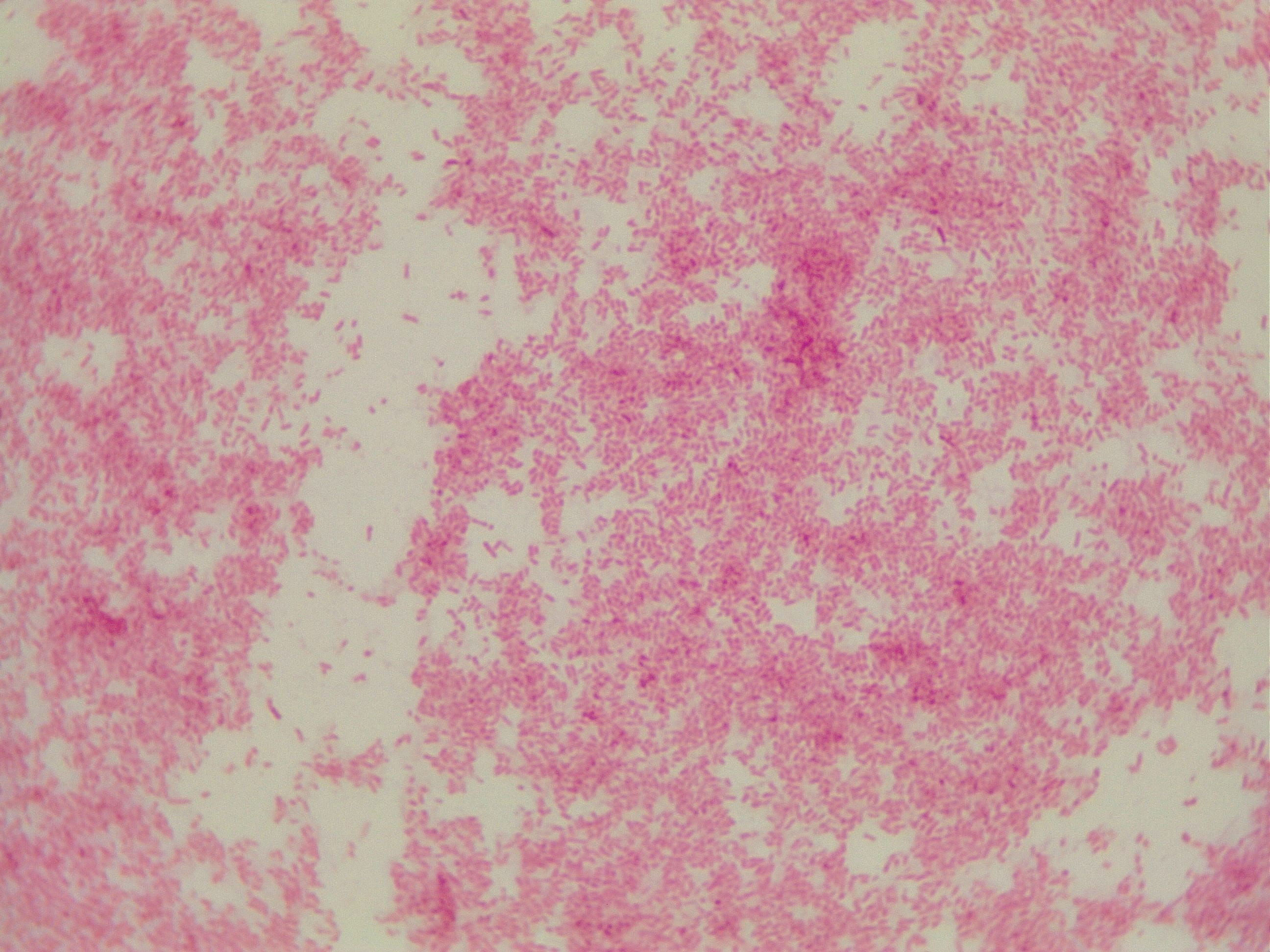
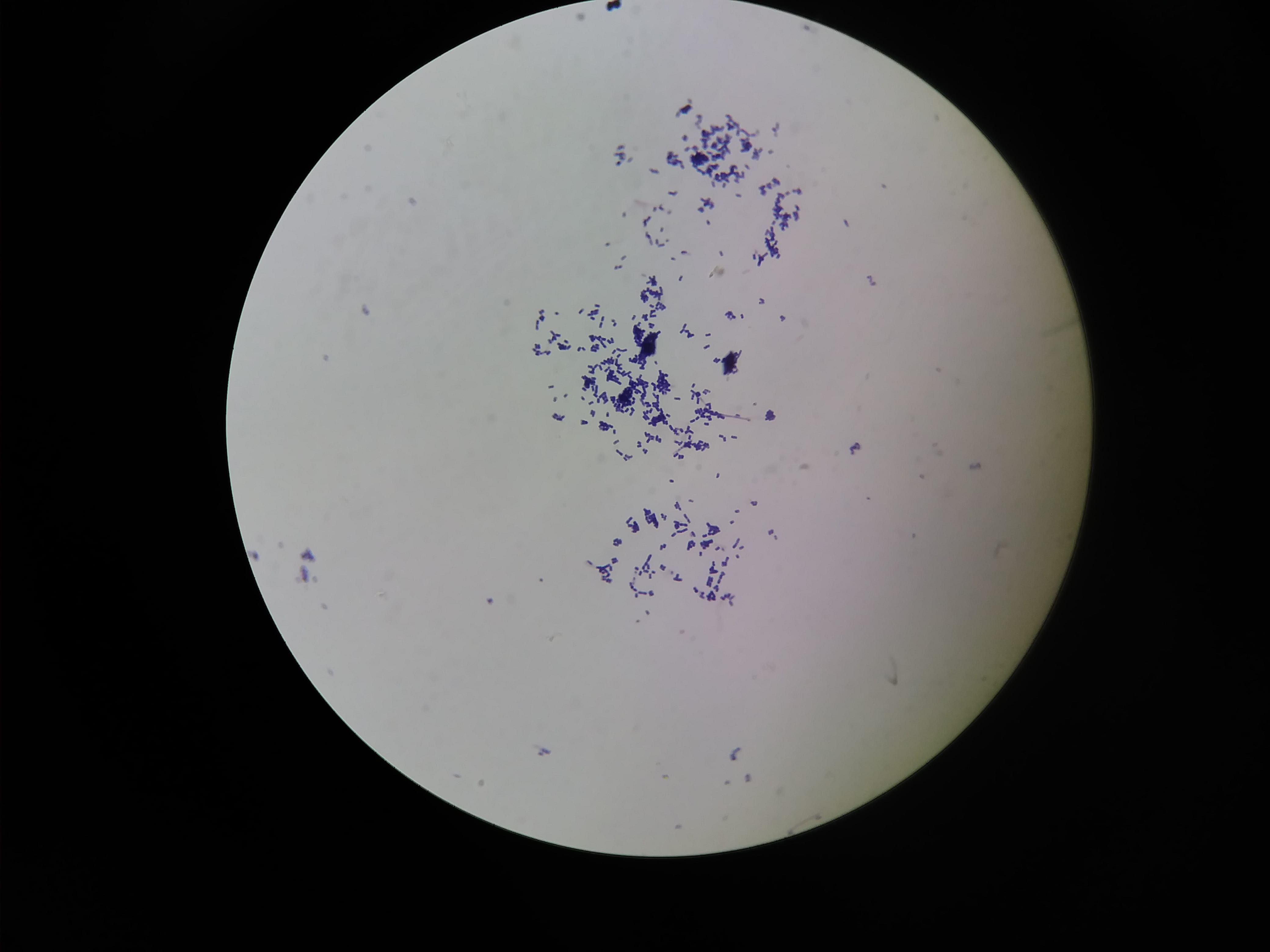
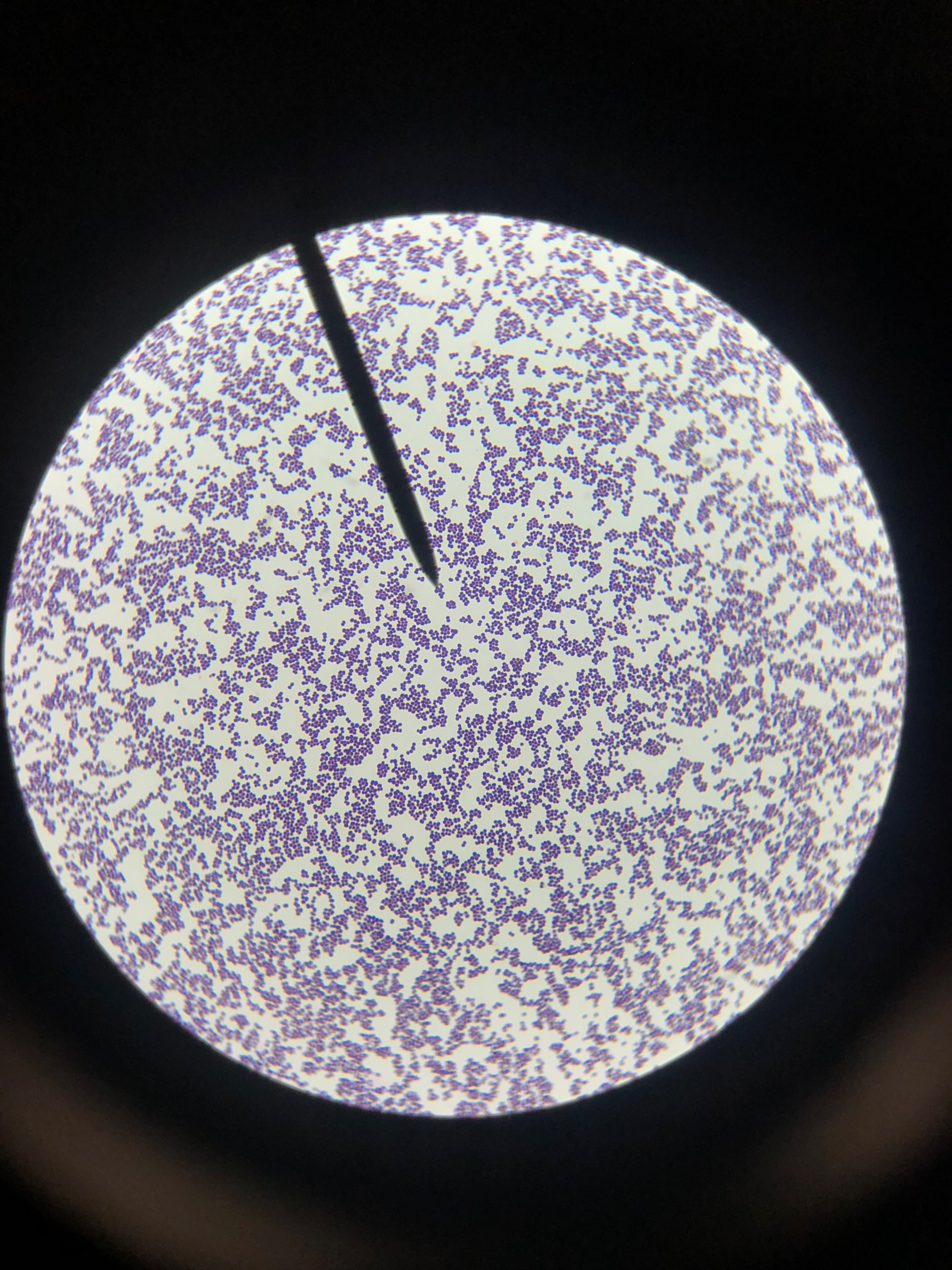
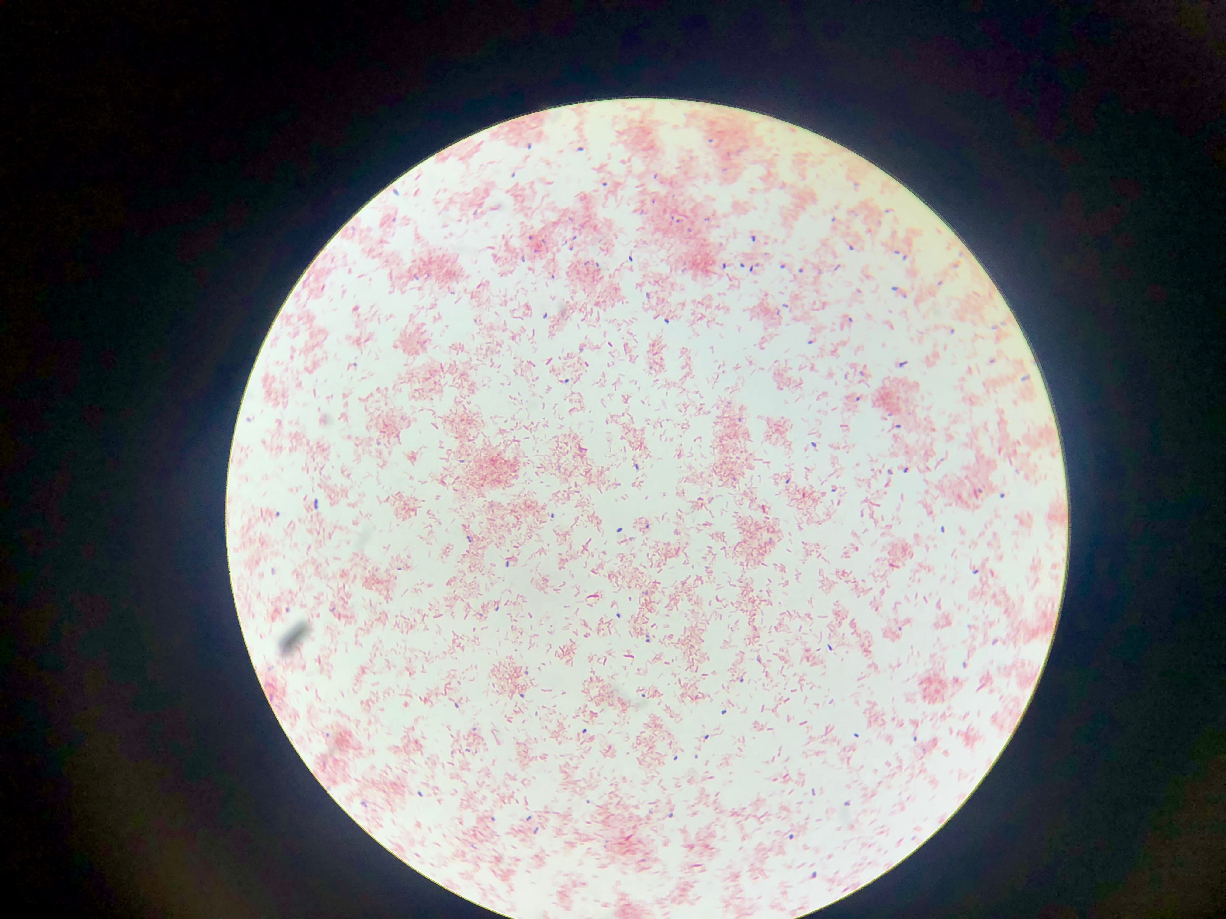
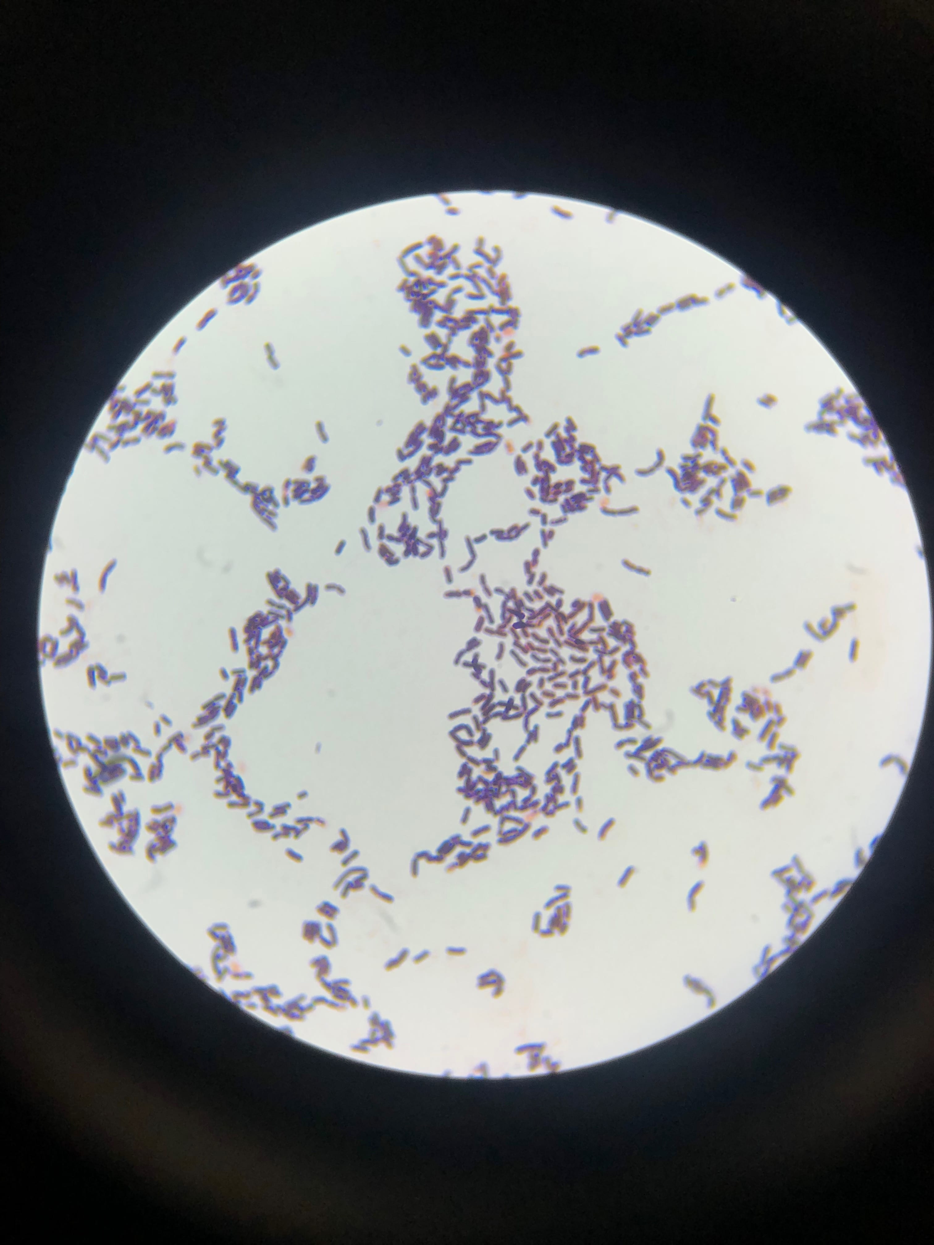
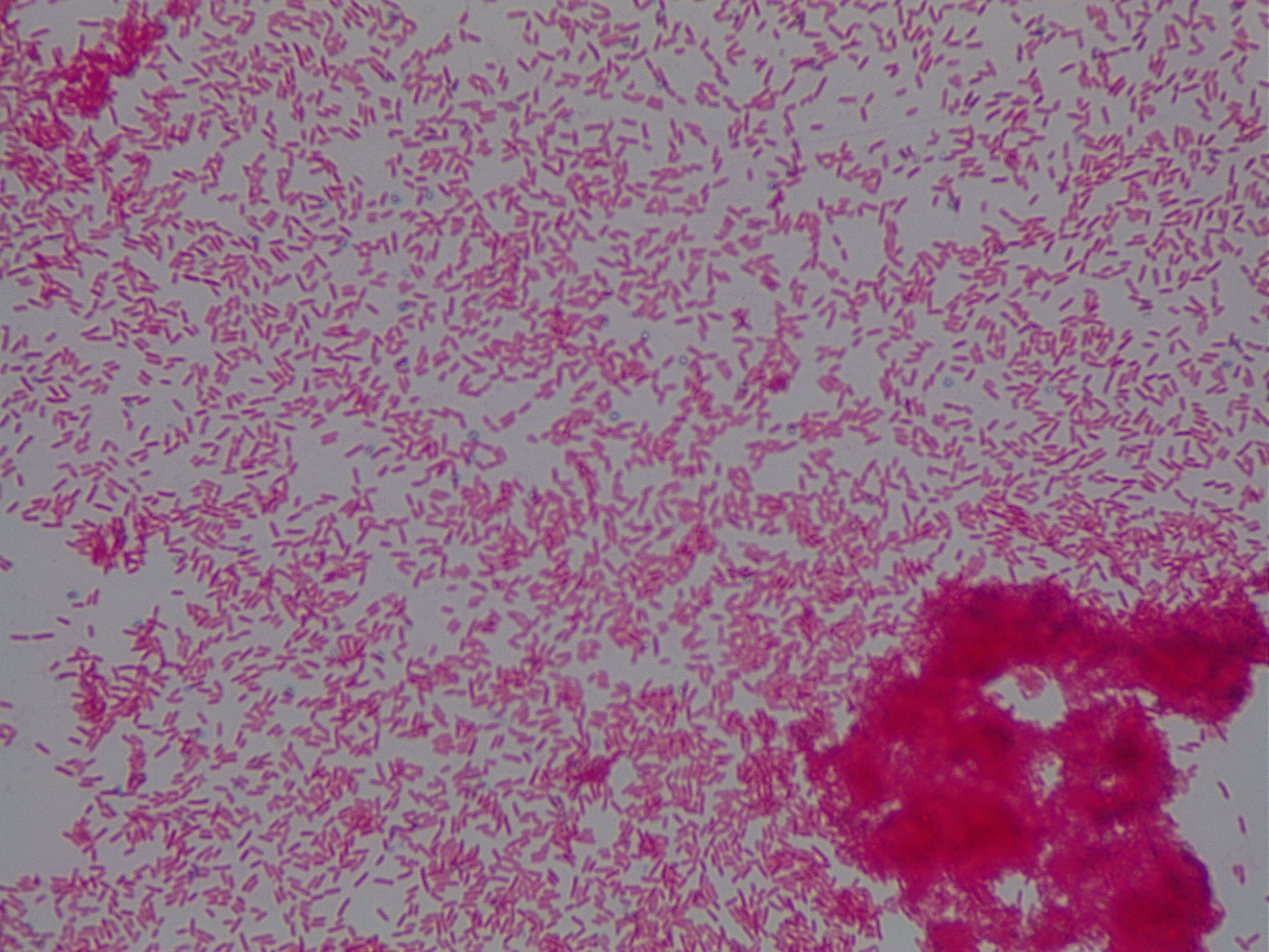
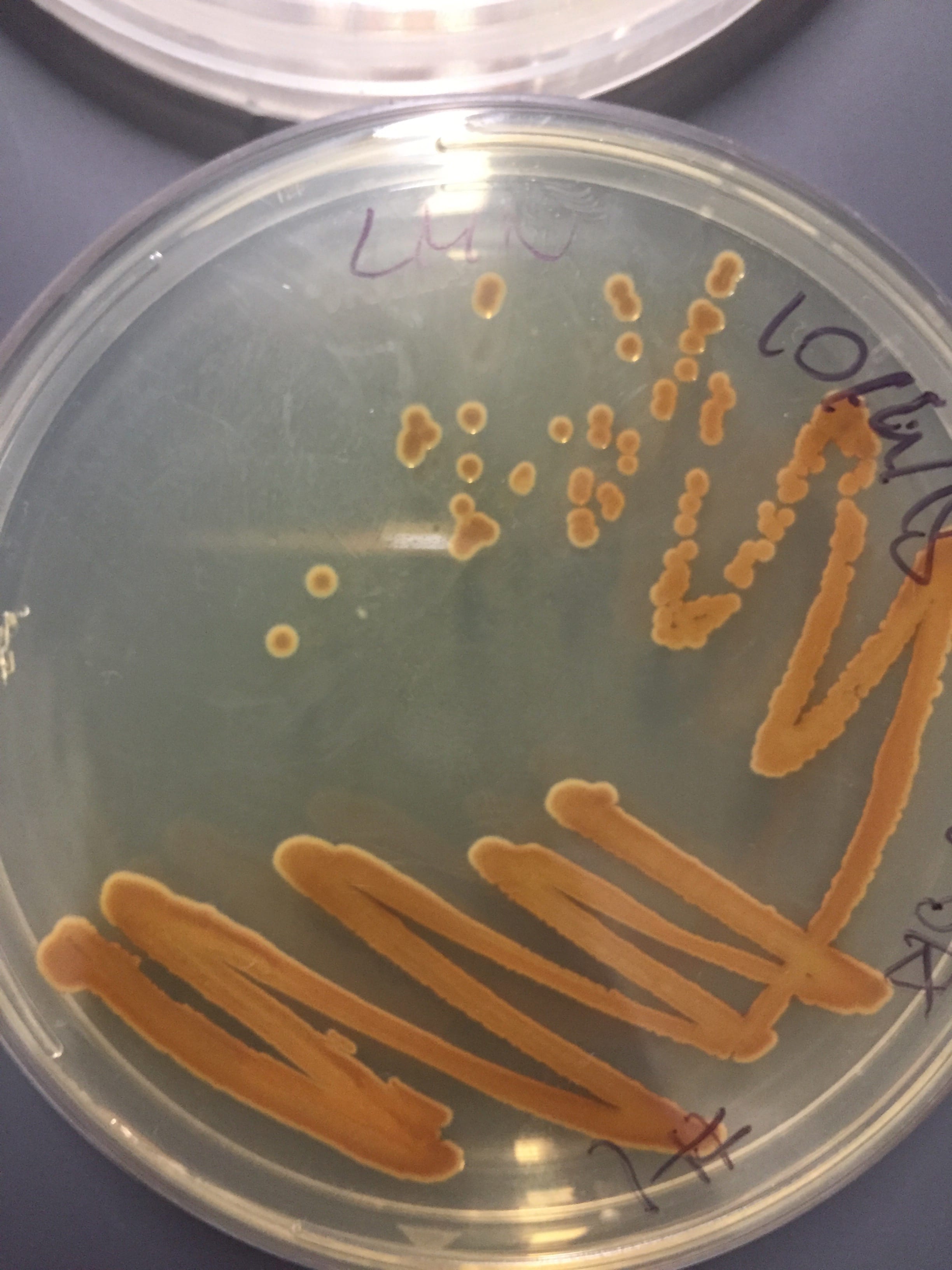
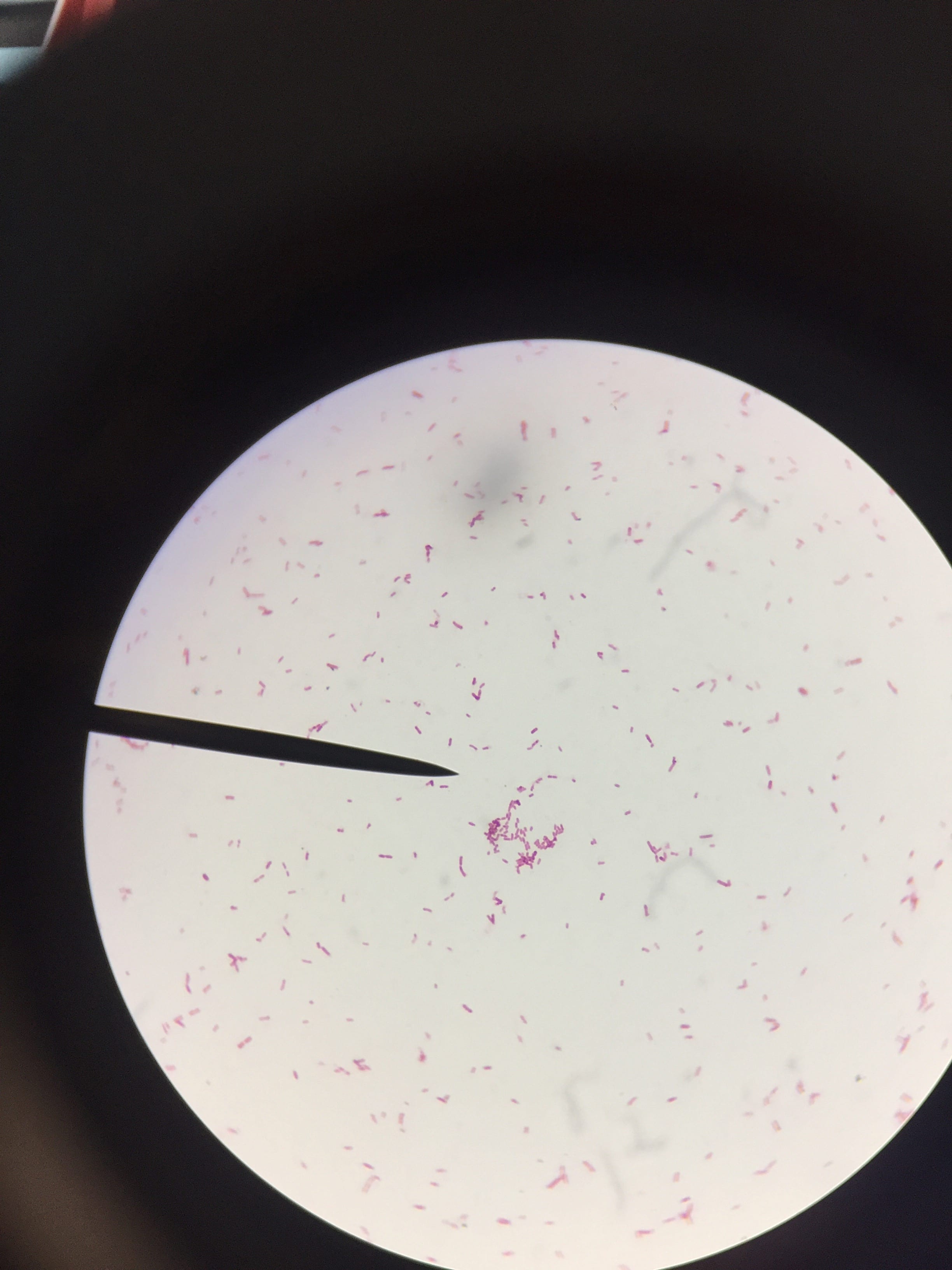
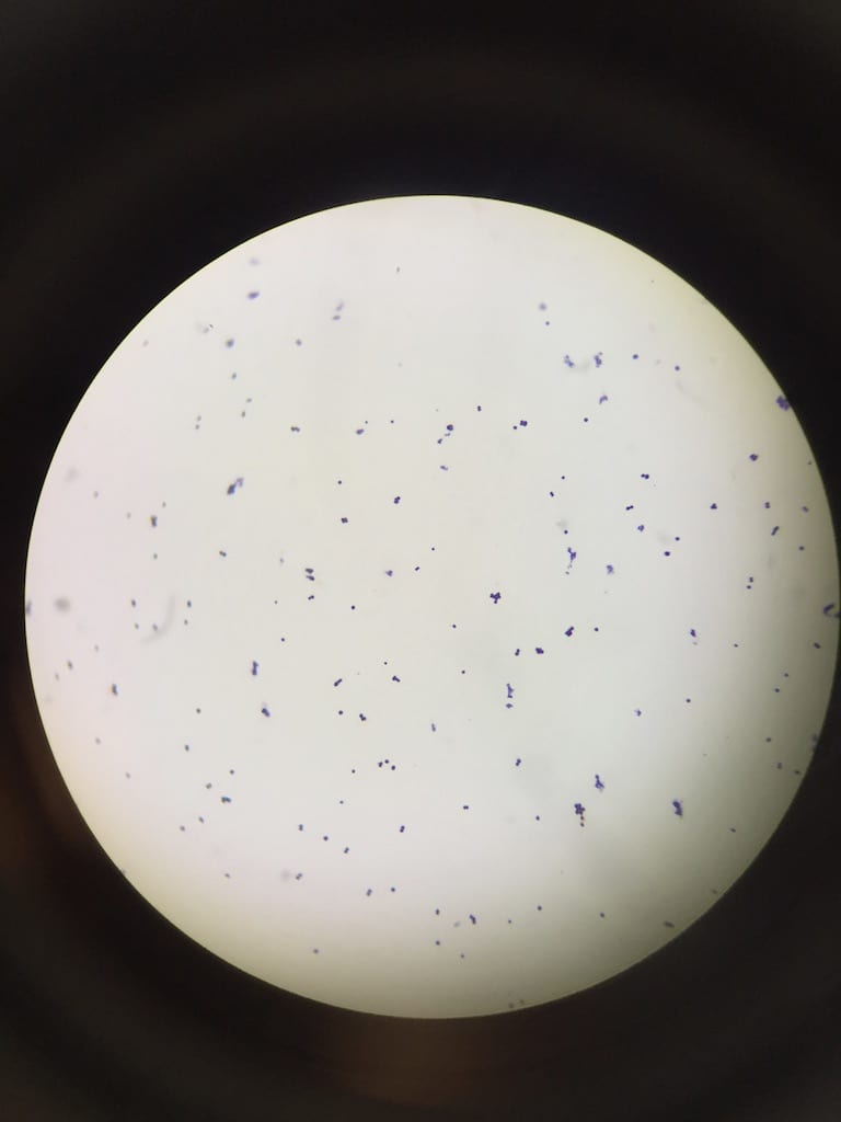
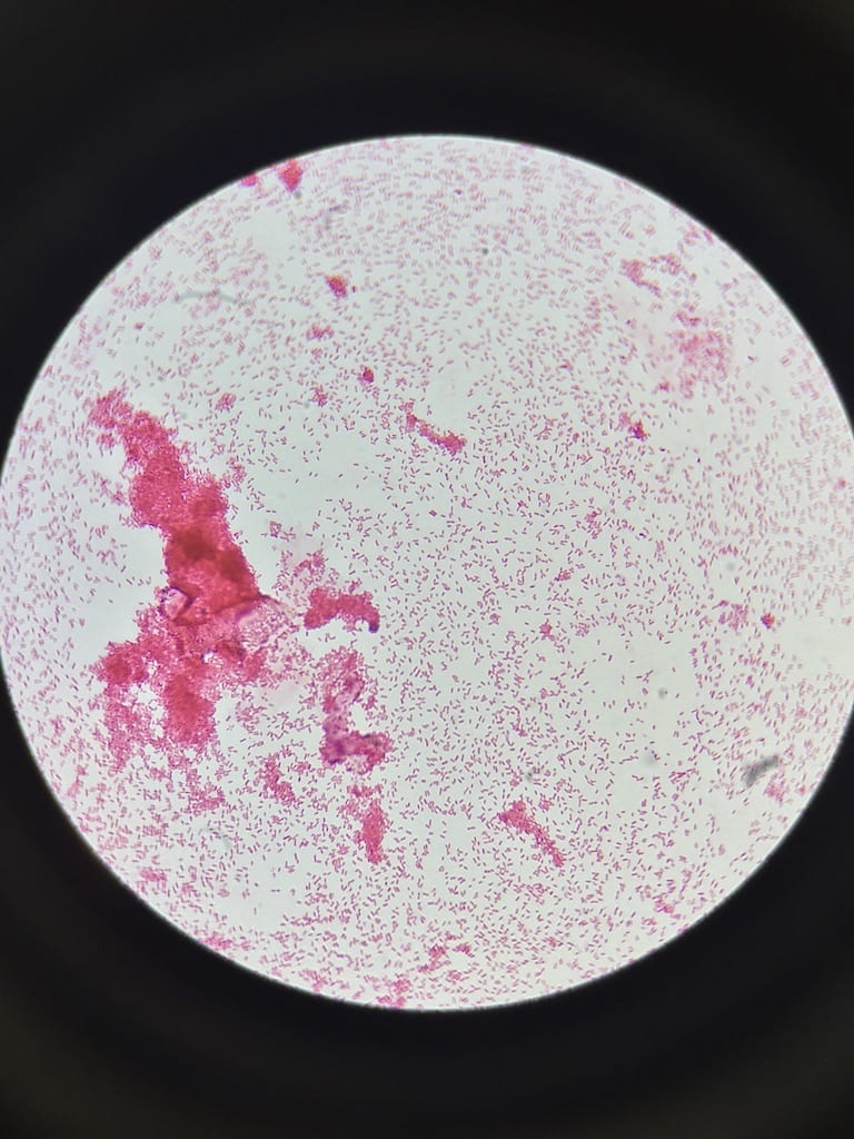
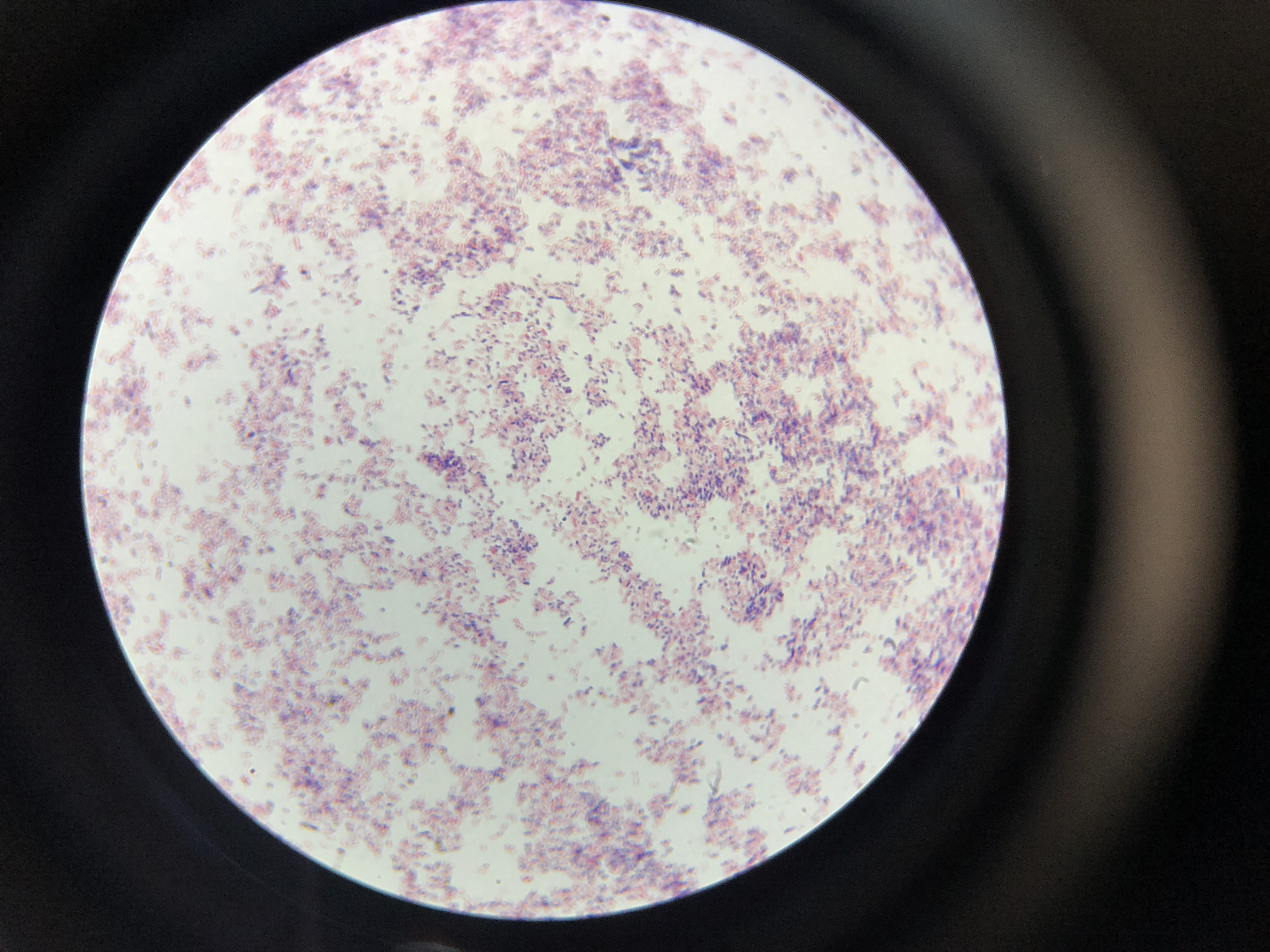
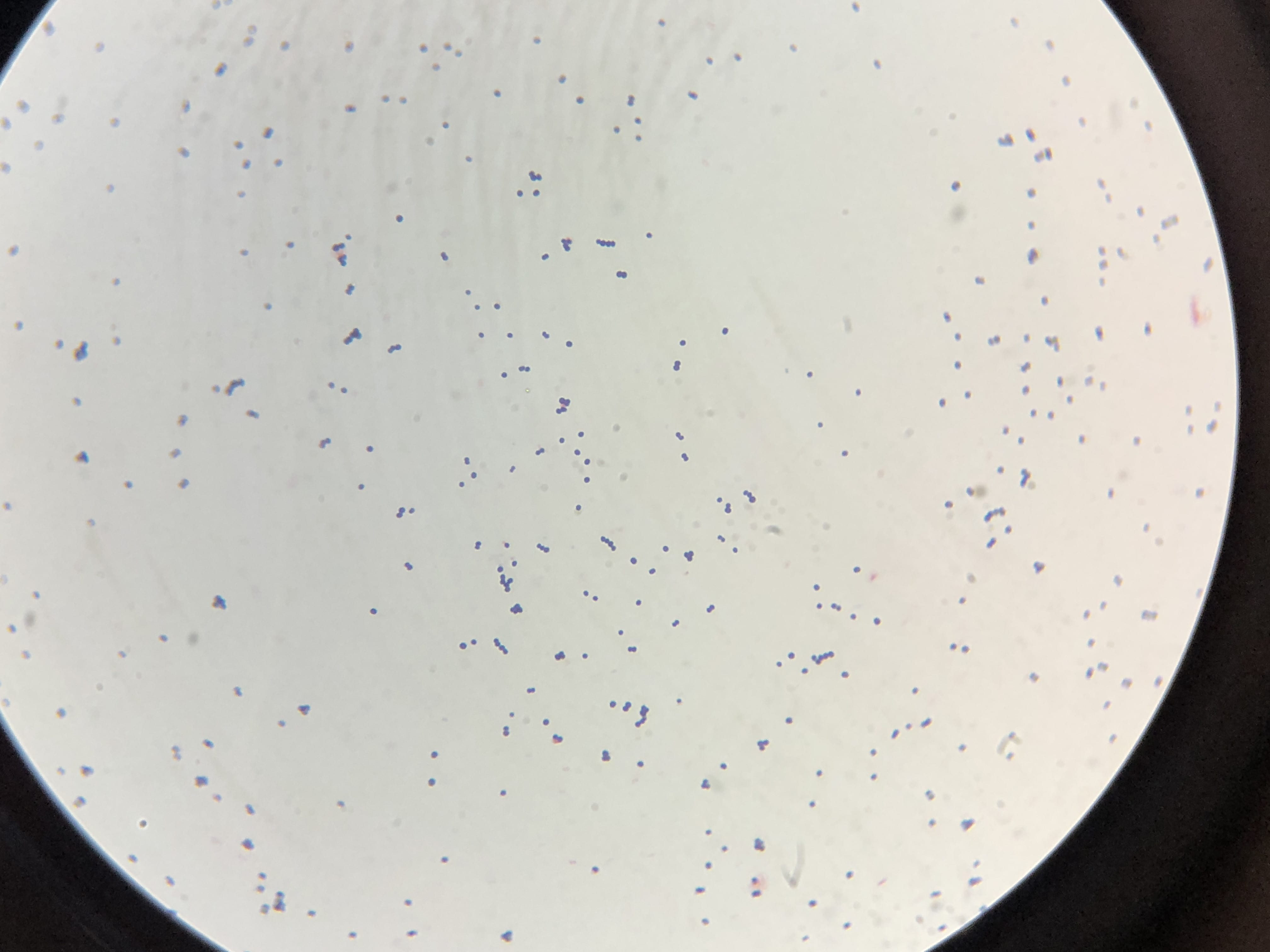
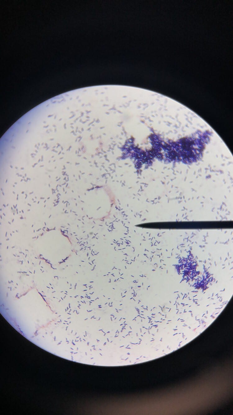
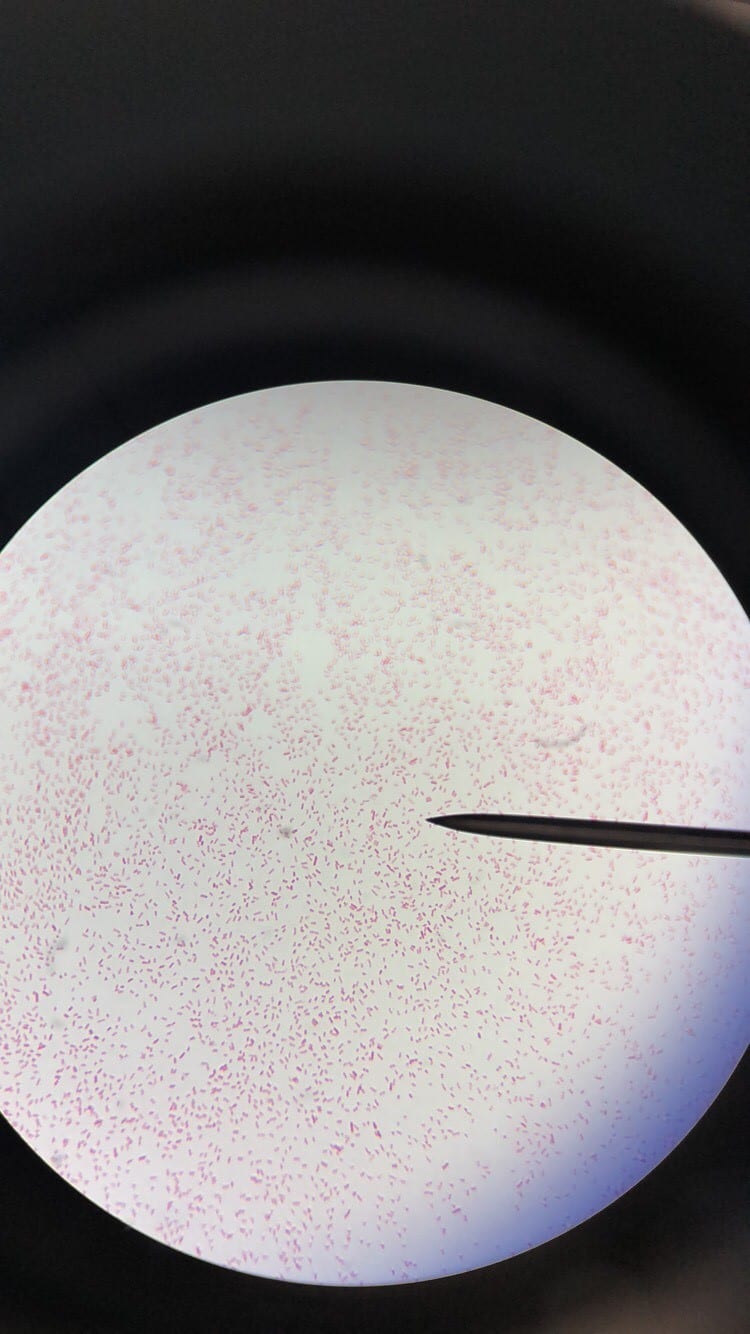
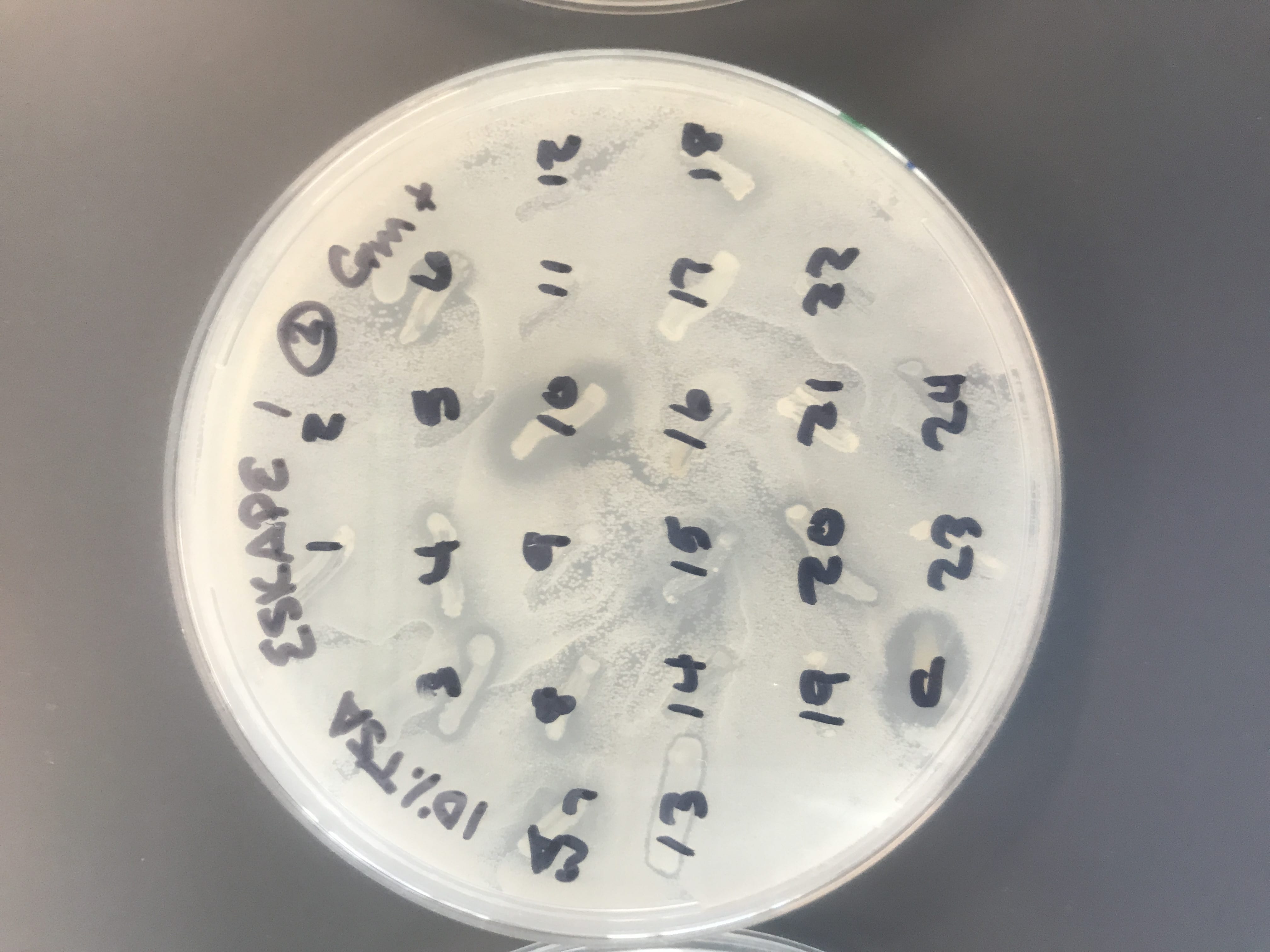
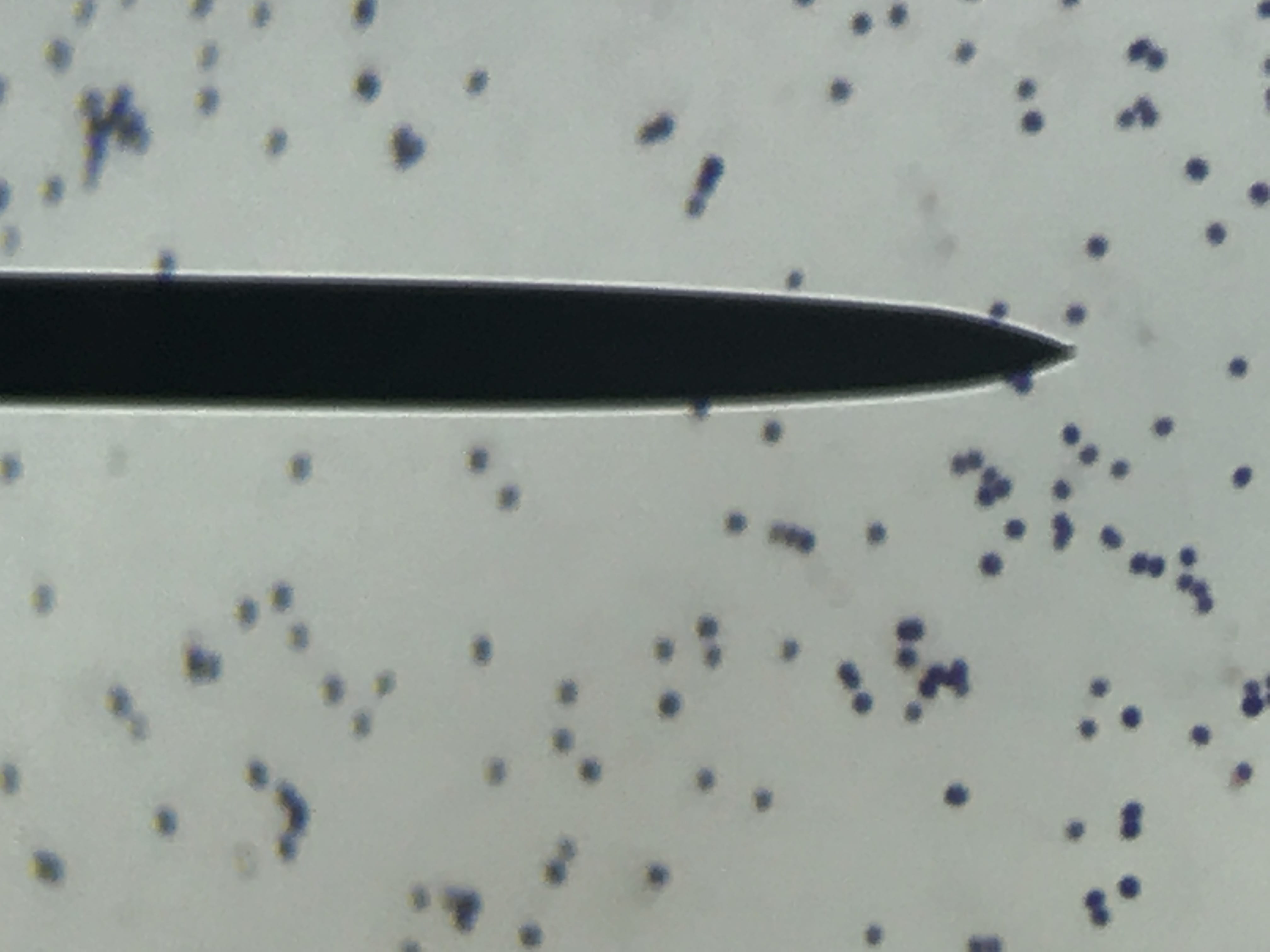
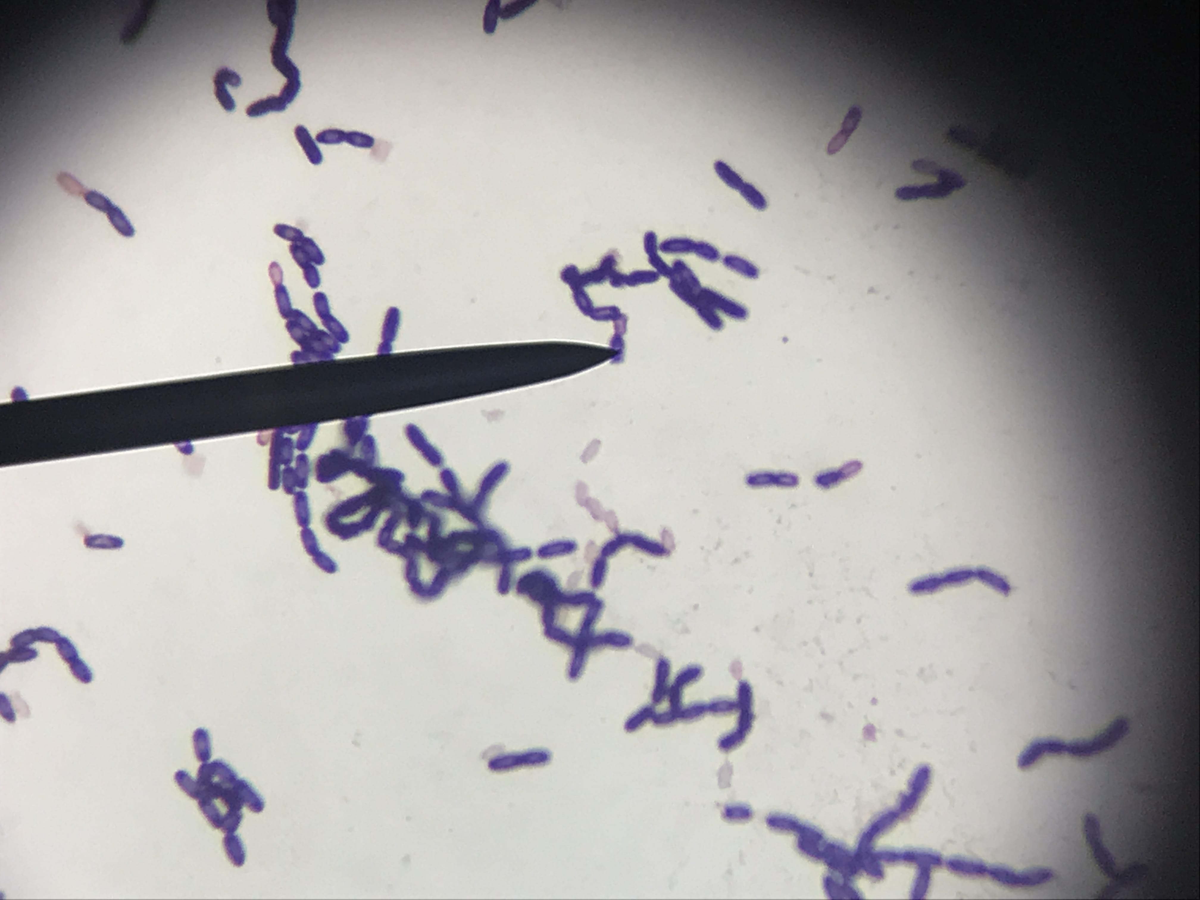
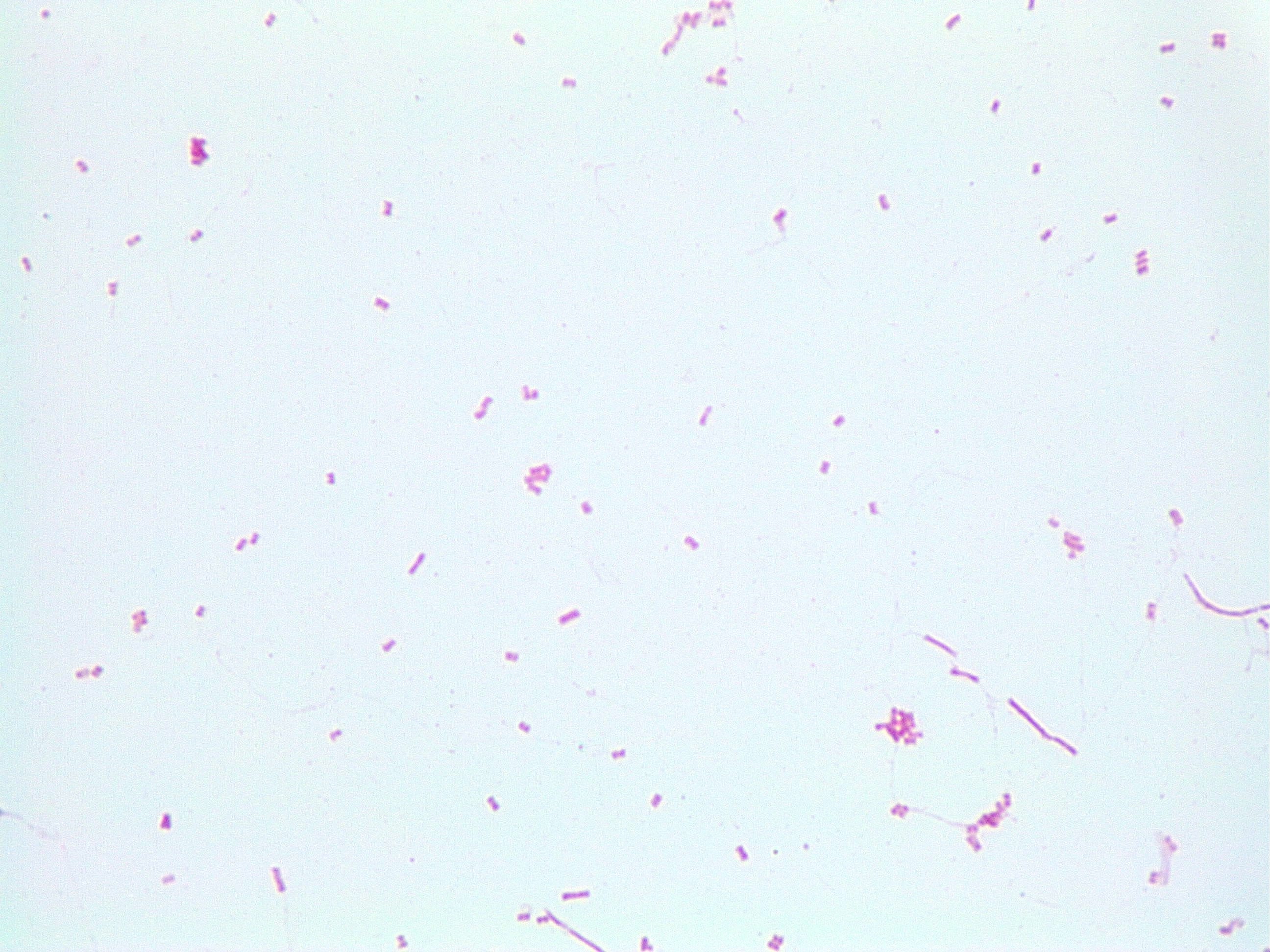
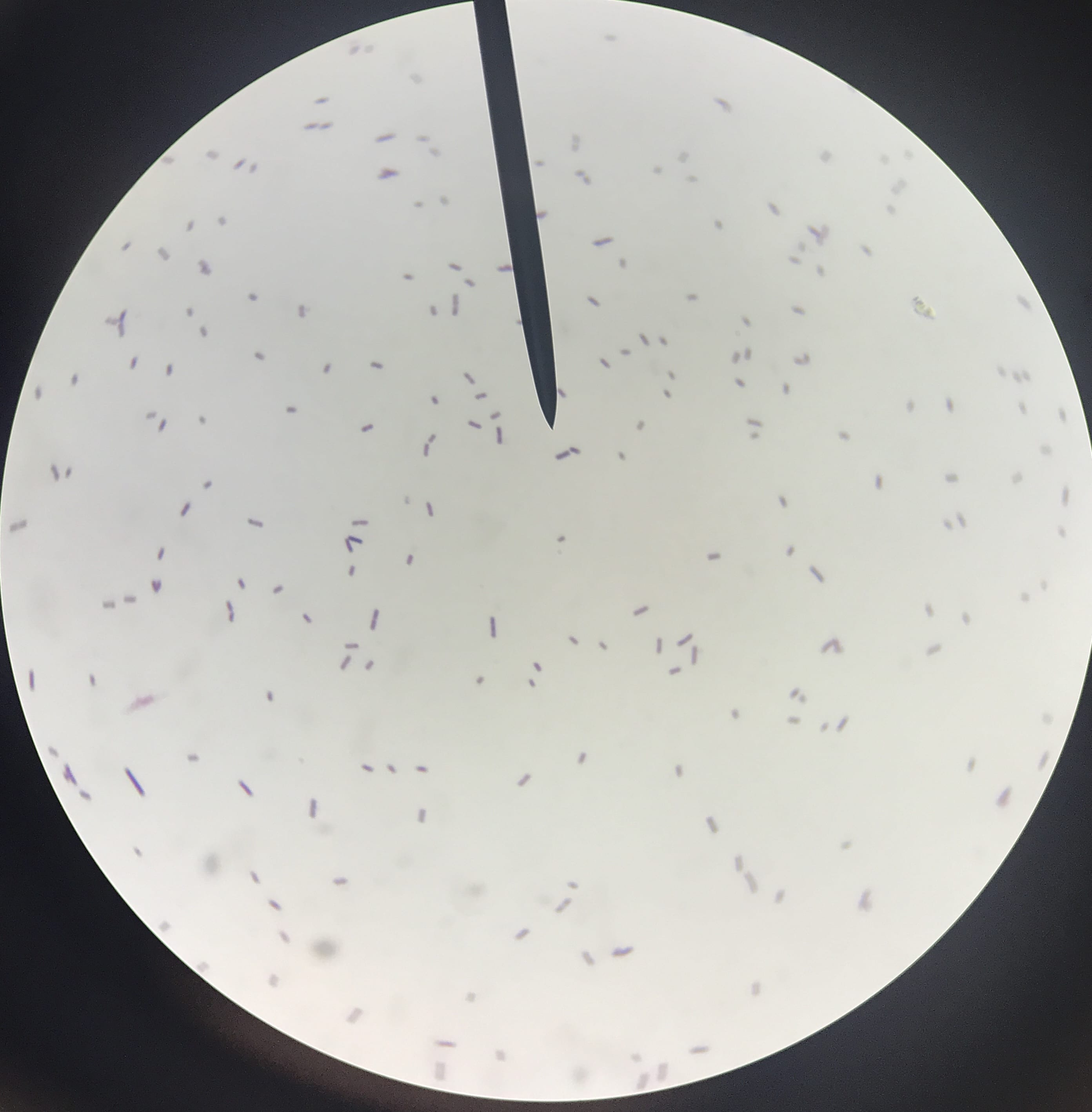
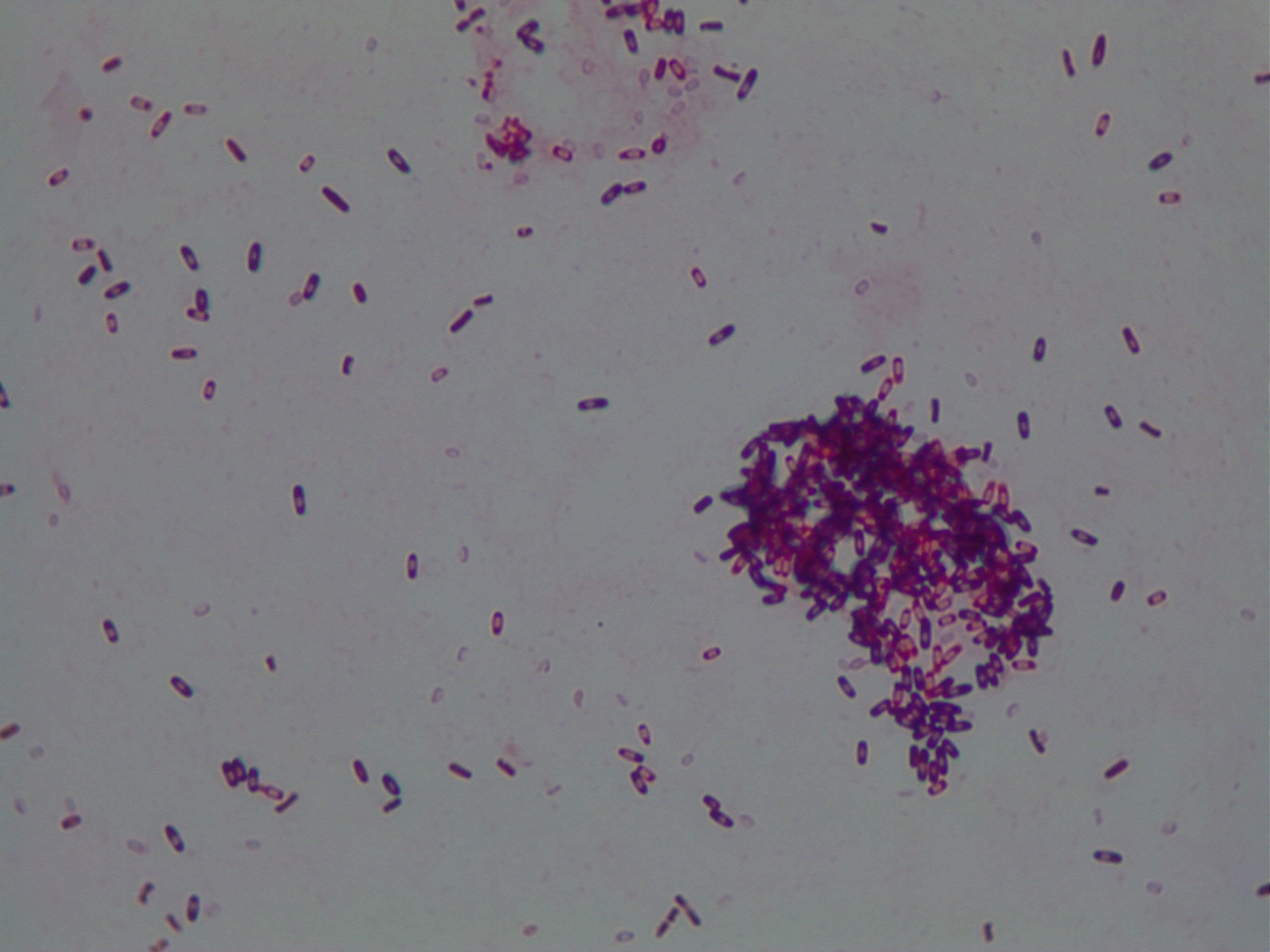
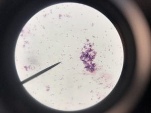
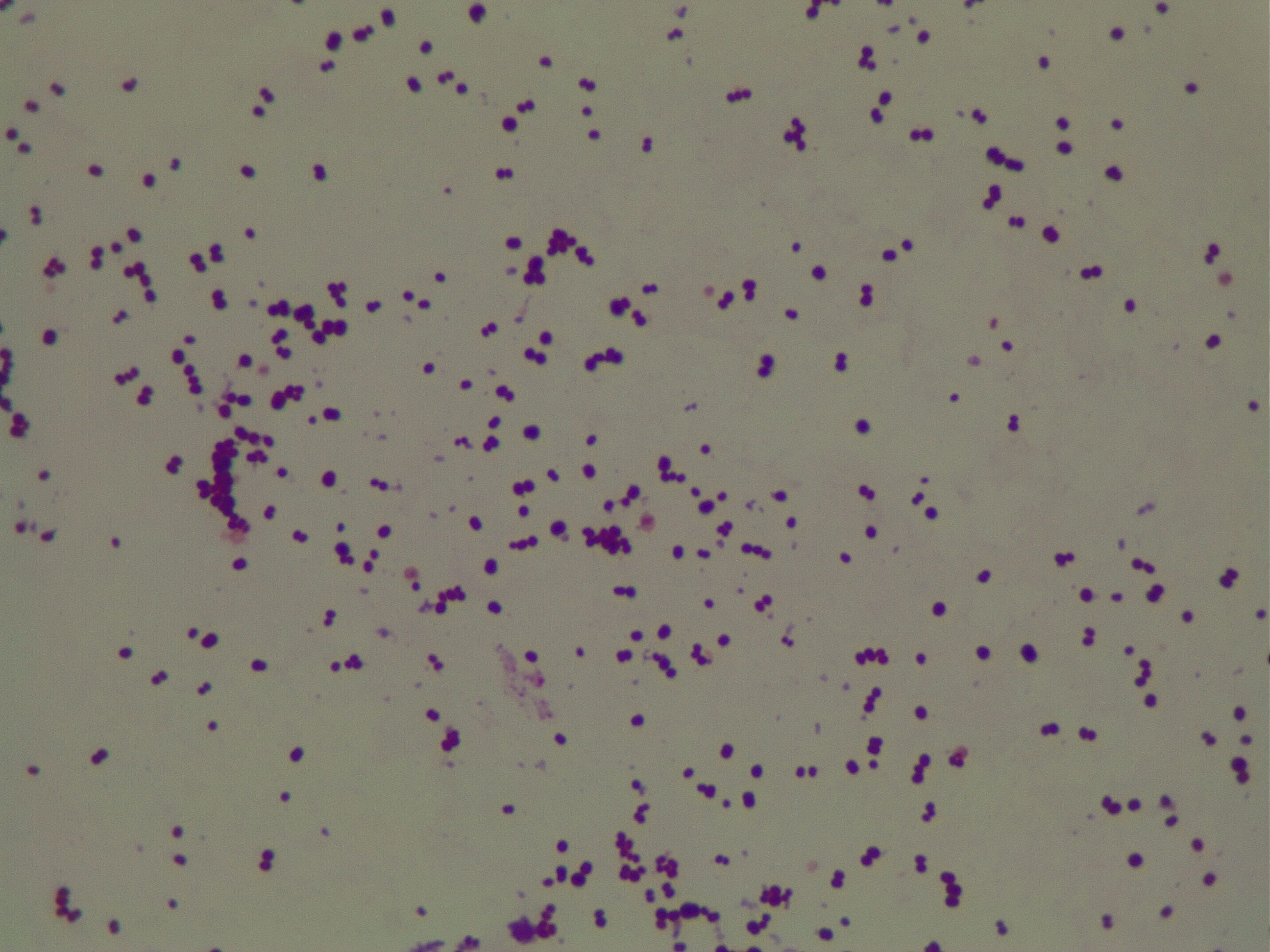
Enterococcus faecium belongs to Domain: Bacteria, Class: Bacilli, Order: Lactobacillales, Family: Enterococcaceae, Genus: Enterococcus and Species: faecium. This ESKAPE pathogen are Gram positive cocci (spherical in shape) that form short to medium length chains. They can also exist in pairs or single cells. E. faecium are also Facultative anaerobes that are ovoid in shape (1). They do not have cytochrome enzymes and are catalase negative (1). Enterococci are found in the feces of most healthy adults – where there are more faecalis than faecium although both are present. Lower percentages were found in oral cavities in healthy students (1). These bacilli tend to live in the gastrointestinal tract of humans and animals and live in feces and sewage. They are able to withstand harsh environmental conditions including high temperatures, periods of drying, and some antiseptics (1).

Clinical Importance and Prevalence

E. faecium’s high level of inherent and acquired resistance (especially VRE’s) along with the pathogen’s ability to survive on surfaces in hospitals makes it an important bacterium to study clinically. VRE constitute about 43% of all Enterococci isolates (4) making it a severe threat across the world, especially in hospitals that rely upon vancomycin or related antibiotics to combat infection. The bacterium has shown tolerance to hand-rub alcohols, making it a greater threat in hospitals (2). Additionally, this pathogen affects largely older adults with comorbidity, leading to heightened mortality rates (3). Furthermore, Enterococci harbor transferable genetic elements, meaning that resistant genes can be passed to both Gram positive and negative bacteria by conjugation systems with plasmids and transposons (4). This potential of E. faecium to pass on its multidrug resistance to other bacteria in a horizontal fashion is particularly frightening.

Infection (How does the infection occur and where is it localized?), Pathology (What disease is caused? What are the symptoms?)

E. faecium is known to cause urinary tract infections, intraabdominal, pelvic and wound infections, superinfections, and bacteremias (often with other organisms) (5). Lower urinary tract infections including cystitis, prostatitis, and epididymitis are seen in older men (5). Enterococci can also cause infection in the abdominal lining in conjunction with liver cirrhosis or in patients with chronic peritoneal dialysis (5). This ESKAPE pathogen is the third most common organism seen in nosocomial infections (3). Another notable infection of E. faecium is endocarditis. Bacteremia and endocarditis are common infections of Enterococci. The mortality associated with these infections is likely partly due to the demographic of patients who present: older adults with multiple underlying diseases such as diabetes. Synergistic, bactericidal attack is required for treatment of endocarditis (5). Multidrug resistant Enterococci are arising, including Vancomycin Resistant Enterococcus (VRE). Enterococcal surface proteins are virulence factors that contribute to infection in humans (5).

Ineffective Antibiotics (Antibiotics to which the organism has acquired resistance)

E. faecium has shown greater antibiotic-resistance than E. faecalis (3). More than half of its pathogenic isolates express resistance to vancomycin, ampicillin, and high levels of aminoglycosides (3). Additionally, the pathogen has shown resistance to some handwash alcohols (2). Enterococci also exhibit inherent and acquired resistance to cephalosporins, clindamycin, tetracycline, and penicillins. A mutation in the domain V of the 23 S rRNA of E. faecium appears responsible for linezolid resistance (5). Additionally, resistance to quinupristin-dalfopristin may be due to enzyme modification, and drug efflux (5).

Effective Antibiotics (Antibiotics known to inhibit the organism)

VRE can be successfully treated with sultamicillin (5). For most Enterococcal infections, single-drug therapies with penicillin, ampicillin, or vancomycin is adequate although resistance against these antibiotics has been observed (3). Currently, linezolid, daptomycin, tigecycline and the streptogramins (quinupristin/dalfopristin) have shown activity against VRE’s (3).

Corresponding Safe Relative

Enterococcus faecium’s safe relative is Enterococcus raffinosus (6). This non-faecialis and non-faecium enterococcus has rarely been connected to human infections (6). One of the first and only reported cases of E. raffinosus infection was endocarditis in an 85 year old man described in 2009 by Antonio Mastroianni in Le Infezioni in Medicina (6).

References

(1) Murray BE. (1990). The life and times of the Enterococcus. Clinical Microbiology Review. 3(1):46-65.

(2) Pidot SJ., Gao W., Buultjens A.H., Monk IR., Guerillot R., Carter GP., Lee JY., Lam, M., Grayson L., Ballard SA., Mahony AA., Grabsch EA., Kotsanas D., Korman TM., Coombs GW., Robinson JO., Silva A., Seemann T., Howden BP., Johnson PD., Stinear TP. (2018). Increasing tolerance of hospital Enterococcus faecium to handwash alcohols. Science Translational Medicine. 10(452).

(3) Dobbs TE., Patel M., Waites KB., Moser SA., Stamm AM., Hoesley CJ. (2006). Nosocomial Spread of Enterococcus faecium Resistant to Vancomycin and Linezolid in a Tertiary Care Medical Center. Journal of Clinical Microbiology.

(4) Olawale KO., Fadiora SO., Taiwo SS. (2011). Prevalence of Hospital-Acquired Enterococci Infections in Two Primary-Care Hospitals in Osogbo, Southwestern Nigeria. African Journal of Infectious Diseases. 5(2):40-46.

(5) Higuita NA. and Huycke MM. (2014). Enterococcal Disease, Epidemiology, and Implications for Treatment. Print.

(6) Mastroianni A. (2009). Enterococcus raffinosus endocarditis. First case and literature review. Le Infezioni in Medicina. 1:14-20.

Meet the ESKAPE Pathogens: Troy Hansen

Assigned ESKAPE Pathogen

My assigned ESKAPE Pathogen was Pseudomonas Aeruginosa

Why is this ESKAPE Pathogen of interest (in brief)

Pseudomonas Aeruginosa is of interest, because it is a common infection in hospitals after surgery, burn victims, and in individuals with weakened immune systems.

General Cellular and Morphological Characteristics of the Organism (taxonomic classification, nutrition, cell shape, habitat)

Pseudomonas Aeruginosa is a gammaproteobacteria.  This means that it is a gram negative bacteria.  It is rod-shaped and does not produce spores, but it does have a flagellum that allows it to move efficiently. It thrives in temperatures between 25ºC and 37ºC, but it can also survive in temperatures up to 42ºC making them more deadly in hospitals and clinical environments. They produce pigments that are typically a greenish color, and it can grow aerobically or anaerobically under minimal nutrition – it has even been known to grow in distilled water!

Clinical Importance and Prevalence

Pseudomonas Aeruginosa is able to grow in a wide range of environments under minimal nutrition, so it becomes dangerous in hospitals as it can easily infect someone who is immunocompromised.  Typical patients who become infected are cancer patients and burn victims,

Infection (How does the infection occur and where is it localized?)

Pseudomonas Aeruginosa can occur in most areas in the body where there is some sort of a body cavity or mucus membrane.  It can also occur on the skin of burn victims, and most places excluding in the blood.

Pathology (What disease is caused? What are the symptoms?)

Bacterial infection is caused, and can lead to colony growth, tissue invasion, and eventually the spread of the bacteria into other parts of the body.  Pneumonia can also occur from the infection of Pseudomonas Aeruginosa.

Ineffective Antibiotics (Antibiotics to which the organism has acquired resistance)

The organism has acquired resistance to all antibiotics except fluoroquinolones, gentamicin, and imipenem.

Effective Antibiotics (Antibiotics known to inhibit the organism)

Same as above.

Corresponding Safe Relative

Pseudomonas Putida


Putty, Murali. “Pseudomonas Aeruginosa.” Testing Lab Analysis: Mold, Legionella, Asbestos, Environmental Microbiology, USP 797, Radon, Lead, March, 2007.

Prince, Alice S. “Pseudomonas Aeruginosa.” NeuroImage, Academic Press, 2012

Meet the ESKAPE pathogens: Brianna Cummings

  1. Assigned ESKAPE Pathogen

Staphylococcus Aureus

  1. Why is this ESKAPE Pathogen of interest (in brief)

Staphylococcus Aureus(S. aureus)is a fairly common bacteria, anywhere from 30 to 50 percent of humans have this bacteria growing on them (1, 2). Typically, S. aureus is found on human skin and mucous membranes, and is not of any concern until it crosses into the bloodstream or internal tissues (2). Infections typically are spread in clinical and hospital settings, but can also occur in non-clinical environments (2).  The major concern regarding S. aureusis a particular strain, methicillin-resistantS. aureus, or MRSA, which is a highly difficult form of S. aureus to treat due to resistance and has a very high morbidity rate (3).

  1. General Cellular and Morphological Characteristics of the Organism (taxonomic classification, nutrition, cell shape, habitat)

Staphylococcus aureusis a Gram+bacteria with a cocci shape (round, spherical shape) and is a member of the Staphylococcaceae family (2). Interestingly, they form clusters of about 1µm in diameter, which can look like grapes when stained (2). However, unstained, S. aureus present as golden colonies (2).

The optimal growing temperature for S. aureusis 37C, while it will grow between 7 and 48C and can survive in temperatures of below -20C (4). Unlike many other bacterium, S. aureus is resistant to high salt content(4). Additionally, S. aureus has the ability to grow under aerobic and anaerobic conditions, although aerobic conditions are preferred(4). Typical medias for colony growth include, blood agar. Tryptic soy agar, and heart infusion agar (5).

As mentioned earlier, S. aureus is typically found in the mucous membranes and on the skin of humans, and are found particularly in the nasal passage (5). However, S. aureuscan also be found in food, and due to its ability to survive in harsh conditions S. aureus is a source of food poisoning (4).

  1. Clinical Importance and Prevalence

S. aureus infections have stabilized in numbers since the 1990s, with about 10-30 cases per 100,000 people per year (1). However, the number of MRSA diagnosis has been rapidly increasing over the same time period (1). Both community and health care related epidemics have contributed to the increasing number of MRSA outbreaks (1). Newer more intense infection control procedures have helped to reduce the number of outbreaks in recent years (1).

  1. Infection (How does the infection occur and where is it localized?)

Infection is most common in people at the “extremes of life,” i.e. infants and the older populations (1). Additionally, humans working in the health professions, hospitalized people, and individuals that are immunocompromised are at a greater risk of contracting the infection (2). S. aureusis transmitted via direct contact or fomites (objects that have the bacteria on them such as clothing, tools, furniture) from person to person (2). Infections can occur in virtually every part of the body, however are most common in skin and soft tissues.

  1. Pathology (What disease is caused? What are the symptoms?)

Many different diseases can be the result of  S. aureus. These include, endocarditis, osteomyelitis, septic arthritis, gastroenteritis, meningitis, toxic shock syndrome, urinary tract infections, pneumonia, but the most common being skin and soft tissue infections (2). Depending on the strain and location of the infection as well as the resulting disease, symptoms vary greatly. However, for ease of answering the question, the symptoms for some of the diseases are as follows:

Skin infections (abscess and cellulitis)  / MRSA :  Swollen, painful bumps that are warm and full of pus, and associated with a fever. Untreated, these lead to deeper more painful infections and can burrow into the skin (6).

Osteomyelitis: fever/chills, swelling of the infected limb with redness, and eventually stiffness and inability to maneuver infected limb. Diagnosis involves blood tests, and potentially a bone scan (7).

Pneumonia: Difficulty breathing, fever, chills, cough. This is diagnosed via chest x-ray and usually involves treatment with antibiotics and hospitalization (8). (Ironic because S. aureus pneumonia is usually transmitted in hospital settings)

  1. Ineffective Antibiotics (Antibiotics to which the organism has acquired resistance)

Depending on the strain, S. aureus is resistant to a variety of antibiotics (9). The emergence of antibiotic strains began in the 1950s with penicillin resistance (9). Then, in the 1960s, the first strains of methicillin resistant S. aureusemerged (9). And more recently, vancomycin resistant strains emerged (9). Thus, S. aureuscan be resistant to the majority of antibiotics, and discovery of new treatments is ever so important.

  1. Effective Antibiotics (Antibiotics known to inhibit the organism)

Treatment of S. aureusis usually done with penicillinase-resistant beta-lactams (5). Penicillin is the typical antibiotic of choice for sensitive strains (2). However, due to the many different strains ofS. aureus, characterization of the bacteria may be necessary before treatment. For example, the MRSA strains are resistant to beta lactams and are typically treated with vancomycin. Additionally, other treatments may coincide with the antibiotics such as fluid replacement.

  1. Corresponding Safe Relative

Staphylococcus epidermidis

Reference:

  1. Tong, S. Y. C., Davis, J. S., Eichenberger, E., Holland, T. L., & Fowler, V. G. (2015). Staphylococcus aureus Infections: Epidemiology, Pathophysiology, Clinical Manifestations, and Management.Clinical Microbiology Reviews28(3), 603–661. http://doi.org/10.1128/CMR.00134-14
  2. Taylor TA, Unakal CG. Staphylococcus Aureus. [Updated 2017 Oct 9]. In: StatPearls [Internet]. Treasure Island (FL): StatPearls Publishing; 2018 Jan-.Available from: https://www.ncbi.nlm.nih.gov/books/NBK441868/
  3. Green, B. N., Johnson, C. D., Egan, J. T., Rosenthal, M., Griffith, E. A., & Evans, M. W. (2012). Methicillin-resistantStaphylococcus aureus: an overview for manual therapists. Journal of Chiropractic Medicine11(1), 64–76. http://doi.org/10.1016/j.jcm.2011.12.001
  4. https://www.foodstandards.gov.au/publications/Documents/Staphylococcus%20aureus.pdf
  5. https://www.ncbi.nlm.nih.gov/books/NBK8448/
  6. Chambers, H. F., & DeLeo, F. R. (2009). Waves of Resistance:Staphylococcus aureus in the Antibiotic Era. Nature Reviews. Microbiology7(9), 629–641. http://doi.org/10.1038/nrmicro2200
  7. https://www.mayoclinic.org/diseases-conditions/mrsa/symptoms-causes/syc-20375336
  8. https://www.healthline.com/health/osteomyelitis
  9. http://www.health.state.mn.us/divs/idepc/diseases/staph/basics.html