Mostly not seen previously in the igneous rocks
Al-silicates
Other aluminous minerals
Amphiboles
Pyroxenes
Sheet silicates
Epidote group
Oxides
Remember that all of the igneous minerals can also occur in metamorphic rocks. Those shown here are either rare or absent in igneous rocks.
Al-silicates
|
|
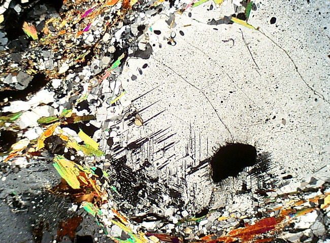
Andalusite in a muscovite–biotite schist. Note the square inclusion cloud to the bottom left, at the crystal center, and the inclusion trails that radiate from the corners of the square. This is known a “chiastolite cross”. Note the small garnet just below the square. Two {110} cleavages are visible, intersecting at approximately right angles. Andalusite has lower relief than the other Al-silicates. Birefringence is first order white to pale yellow, much like quartz and feldspars but with higher relief and very different habit. In cross-polarized light the garnet is clearly isotropic. Plane- and cross-polarized light views. Field width is 6 mm. NEIGC84-C5-2 |
|
|
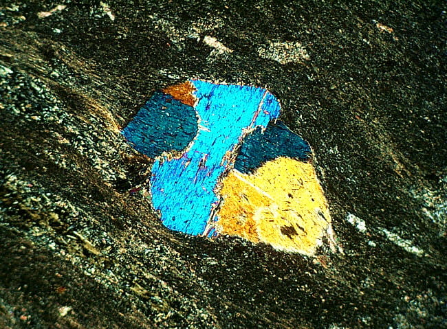
Kyanite in a muscovite–biotite schist. The four kyanite crystals are colorless, have high relief, and two have a strong cleavage parallel to their length. Relief is much higher than muscovite, which surrounds the kyanite. The kyanite crystals have interference colors up to upper 1st order, much lower than muscovite. Most sections yield slightly inclined extinction, as expected from its triclinic symmetry. Plane- and cross-polarized light views. Field width is 3 mm. TMW96-C4b |
|
|
Sillimanite fibers (variety fibrolite), in a biotite–sillimanite–muscovite schist. Sillimanite is colorless, and has relief much higher than muscovite. In medium-grade rocks sillimanite is typically of this fibrous variety. Sillimanite fibers can be included in many minerals, and can survive retrograde metamorphism in garnet and quartz. Sillimanite has birefringence up to 2nd order blue, somewhat higher than kyanite and much higher than andalusite. Extinction is parallel, as required by its orthorhombic symmetry. These fibers are so thin that individual ones scarcely have any birefringence, but thick stacks of them do. Plane- and cross-polarized light views. Field width is 0.6 mm. JBT2-XA |
|
|
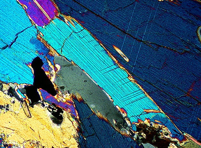
Sillimanite prisms in a biotite–garnet–cordierite schist. This granulite facies schist has coarse sillimanite prisms that can be seen in length sections, and in their diamond-shaped cross sections. The lower second order blue birefringence can be seen in the long sections. Plane- and cross-polarized light views. Field width is 3 mm. WE-1 |
|
|
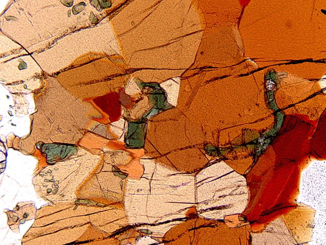
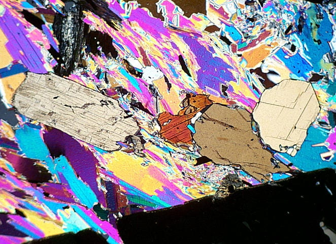
Sillimanite and andalusite together in a biotite–muscovite–andalusite schist. The large, high-relief (grayish) patches are andalusite, which are surrounded by coarse muscovite. The andalusite is partially pseudomorphed by sillimanite, which appears as high-relief, colorless diamond shapes. In cross-polarized light the andalusite mostly occupies the large gray areas, and the sillimanite domains are the vertically elongated white diamonds, scattered throughout the upper-left half of the image. Plane- and cross-polarized light views. Field width is 6 mm. JBT2-XA |


Other aluminous minerals
|
|
Cordierite in a garnet–cordierite–biotite schist. Cordierite is colorless, and relief is similar to feldspar and quartz, but it tends to be dustier than either of them. I don’t know why. Birefringence is up to 1st order white, like feldspar and quartz, and can have polysynthetic twinning (center) that somewhat resembles that in plagioclase. Note, however, that there can be THREE directions of polysynthetic twinning in cordierite, contrasting with none in quartz and typically one or two in feldspars Plane- and cross-polarized light views. Field width is 3 mm. WE-1 |
|
|
Cordierite in a garnet–cordierite–biotite schist. Enlarged view of pleochroic yellow radiation halos surrounding radioactive inclusions. In cross-polarized light the halos are typically brownish or purplish. Plane- and cross-polarized light views. Field width is 0.6 mm. WE-1 |
|
|
Staurolite in a muscovite–biotite schist. Staurolite has parallel extinction and one weak cleavage parallel to its length. It has a characteristic pale to darker golden yellow pleochroism. This example has a twin, highlighted by different color and birefringence caused by its different orientation. The birefringence of staurolite is similar to that of kyanite, upper 1st order. Plane- and cross-polarized light views. Field width is 6 mm. Gassetts Schist |
|
|
Chloritoid in a muscovite–chlorite phyllite. The pale blue to yellow pleochroism of the chloritoid, and its high relief, contrasts sharply with lower relief, pale yellow to green pleochroic chlorite. The largest chloritoid grain has prominent polysynthetic twinning, evident in parallel stripes having somewhat different birefringence and colors. The low first-order birefringence is somewhat higher than that of most chlorite, but much lower than muscovite. The polysynthetic twinning and somewhat anomalous first order interference colors of chloritoid are visible in the cross-polarized light image. Plane- and cross-polarized light views. Field width is 6 mm. IMA96-C1-1 |
 |
Chloritoid in a muscovite–chlorite phyllite. Close-up showing green chlorite between bluer and much higher relief chloritoid. Plane-polarized light view only. Field width is 1.2 mm. IMA96-C4B |
|
|
Tourmaline in a muscovite–biotite schist. Tourmaline occurs as elongate to stubby prisms having hexagonal or somewhat triangular cross sections. They are typically zoned in shades of blue, green, or brown. Tourmaline has no cleavage, though cross fractures are common, and it has a negative sign of elongation. Plane- and cross-polarized light views. Field width is 0.6 mm. East Clairindon, VT |
|
|
Tourmaline in a muscovite–biotite schist. These two images are of the same tourmaline in the image centers, rotated 90° with respect to each other. Tourmaline is unusual among common elongate minerals in having its strongest absorption when the plane of light polarization is perpendicular to the crystal length. This is the opposite of micas and most amphiboles. Both plane-polarized light. Field width is 0.6 mm. East Clairindon, VT |


Amphiboles
|
|
Actinolite in a greenstone. These actinolite grains are pale-green, and here occur as stubby crystals. These are probably pseudomorphs after augite phenocrysts in the original basalt protolith. Like most monoclinic amphiboles, actinolite has birefringence in the lower 2nd order. Twinning, possibly relict from the original augite, can be seen. Plane- and cross-polarized light views. Field width is 3 mm. NNH-3 |
|
|
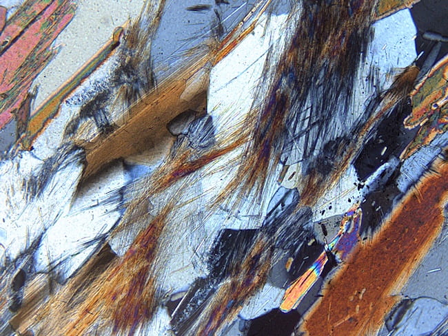
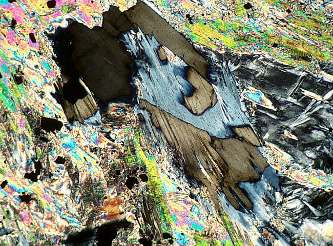
Cummingtonite in a hornblende–biotite–cummingtonite amphibolite. The cummingtonite is almost colorless, but has abundant green hornblende exsolution lamellae that are on irrational planes close to {100} and {001}. Like most monoclinic amphiboles, birefringence is in the lower second order. Plane- and cross-polarized light views. Field width is 1.2 mm. Q-603C |
 |
Cummingtonite in a hornblende–biotite–cummingtonite amphibolite. Close-up view of hornblende exsolution lamellae in cummingtonite, coming out on the two irrational planes close to {100} and {001}. Some colorless cummingtonite exsolution lamellae can be seen in hornblende to the right of the cummingtonite. Plane-polarized light view only. Field width is 0.3 mm. Q-603C
|
|
|
Gedrite in a gedrite–cordierite–biotite gneiss. Gedrite colors range from colorless to gray to green to brown. Gedrite is commonly associated with aluminous minerals like cordierite, garnet, staurolite, and aluminosilicates, as well as with other amphiboles. Anthophyllite is another, less aluminous orthoamphibole, separated from gedrite by a solvus at low temperature. The solvus is defined principally by Na and Al content, with gedrite having more of both than anthophyllite. Gedrite has lower birefringence than the monoclinic amphiboles, typically in the upper 1st order. Plane- and cross-polarized light views. Field width is 6 mm. IMA86-G2-1 |
 |
Gedrite in a gedrite–cordierite gneiss. Because gedrite is orthorhombic, crystals have extinction parallel to their lengths. The same gedrite crystals in the image immediately above is here rotated 45° counterclockwise to extinction. Cross-polarized light view only. Field width is 6 mm. W95 |
|
|
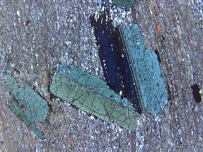
Glaucophane in a blueschist. Glaucophane is characteristically pleochroic in shades of blue and purple. This glaucophane is somewhat zoned, with pale cores and darker, more Fe3+-rich rims. As with most monoclinic amphiboles, glaucophane has birefringent colors in the lower second order. Plane- and cross-polarized light views. Field width is 1.2 mm. C15 |


Pyroxenes
|
|
Omphacite in an eclogite. Omphacite is an Na–Ca–Mg–Al pyroxene, and holds the albite component in this feldspar-free, high-pressure, low-temperature metamorphic rock. Omphacite is usually pale green, and can be very slightly pleochroic. Like most clinopyroxenes, omphacite has lower second order birefringence. Plane- and cross-polarized light views. Field width is 1.2 mm. IG16-36 |


Sheet silicates
|
|
Talc in a soapstone (metamorphosed harzbergite). Talc is colorless and resembles muscovite or colorless phlogopite, but it is much softer. In hand specimen the two are easy to tell apart because of the soapy feel of talc. Plane- and cross-polarized light views. Field width is 3 mm. 4.6.84A |
 |
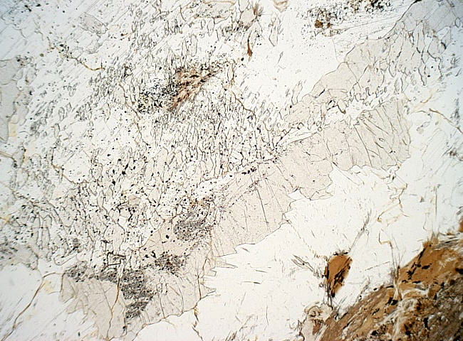
Talc in a soapstone (metamorphosed harzbergite). This talc crystal has been rotated to extinction, but there are many small areas in the central grain that are not extinct because of surface damage caused by the thin section grinding process. The micas and calcite have this surface damage effect too, but not so strongly as talc. Cross-polarized light view only. Field width is 0.3 mm. 4.6.84A |
|
|
Antigorite in a soapstone (metamorphosed harzbergite). Antigorite is a serpentine mineral that is platy, unlike fibrous chrysotile. It is typically colorless to pale green, and in some ways resembles chlorite. Antigorite commonly has anomalous lower first order birefringence, like chlorite. Unlike chlorite, however, it can have both anomalous Berlin blue and anomalous brown interference colors in different orientations. Low birefringence chlorite, in contrast, is either almost entirely anomalous blue (Fe-rich), anomalous brown (Mg-rich), or anomalous violet (intermediate composition). The anomalous colors are caused by high dispersion of the 2V. Plane- and cross-polarized light views. Field width is 1.2 mm. 4.6.84A |


Epidote group
|
|
Epidote in an epidote–amphibolite facies metamorphosed basalt. Epidote is the term for the optically negative, high birefringence Al-Fe3+ epidote, and is the most common epidote type. It has parallel extinction relative to its usual long crystal dimension (the b direction), but inclined extinction, looking down b relative to its {001} cleavage. Although commonly light yellow-green in thin section, it may be colorless. Its interference colors commonly seem anomalously bright compared to pyroxenes, a result of epidote’s high dispersion of the 2V. Plane- and cross-polarized light views. Field width is 1.2 mm. NOR-116 |
|
|
Clinozoisite in an Mg-rich epidote amphibolite facies basaltic rock. Although clinozoisite is usually colorless, here it is pleochroic colorless to pale-yellow. Clinozoisite is optically positive, in contrast to epidote, and has lower to middle first order birefringence. In cross-polarized light the birefringence is zoned, the result of variable Fe3+ content. The interference colors of clinozoisite are anomalous, ranging from anomalous first order Berlin-blue and brown for Fe-poor varieties, to anomalous first order lemon-yellow for Fe-rich ones. The anomalous interference colors are the result of strong dispersion of the 2V. Plane- and cross-polarized light views. Field width is 1.2 mm. NOR-281 |


Oxides
|
|
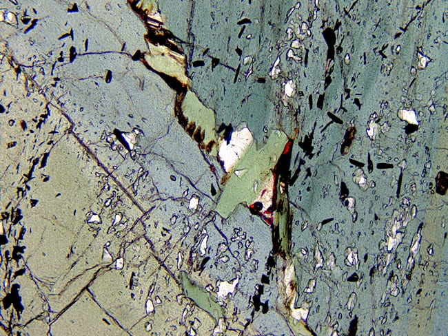
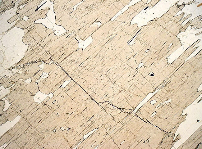
Rutile in a cordierite–gedrite gneiss. Rutile is characteristically deep yellow-brown or orange-brown, with enormously high relief. It can accept limited amounts of uranium and thorium into its structure, which can produce radiation halos that are generally weaker than those surrounding zircon, allanite, and monazite (no halos visible here). Rutile has very high birefringence, rarely discernable because of the dark mineral color, and because the high colors are mostly pastels. Birefringence is discernable on very thin edges, or thin rutile fibers. Plane- and cross-polarized light views. Field width is 1.2 mm. W95 |
|
|
Spinel in a hornblende–pyroxene granulite. Spinel in common rocks is usually an intermediate Fe-Mg solid solution, and so is usually green. It has very high relief. Spinel is isotropic, but its green color distinguishes it from colorless to pink garnet and colorless fluorite, and its high relief and color from sodalite. Sodalite and spinel are unlikely to occur in the same rock anyway. Plane- and cross-polarized light views. Field width is 1.2 mm. TMW96-A4-A |