Prograde reactions
Diffusion-controlled reactions
Metasomatic reactions
Retrograde hydration reactions
Pseudomorphs
Prograde reactions
|
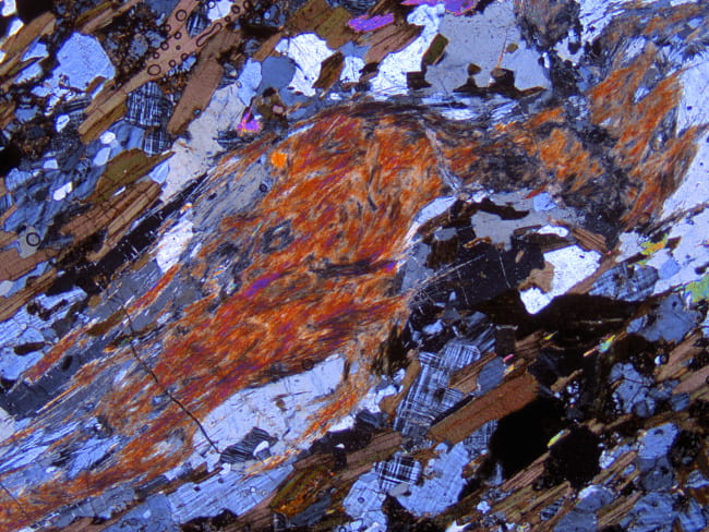
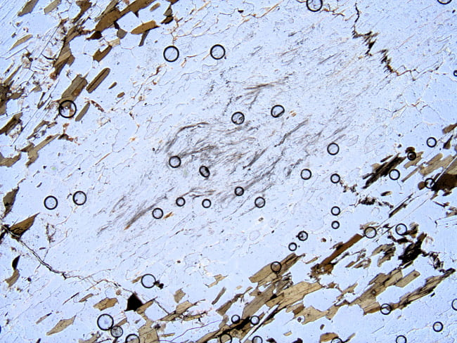
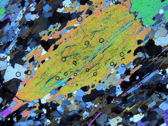
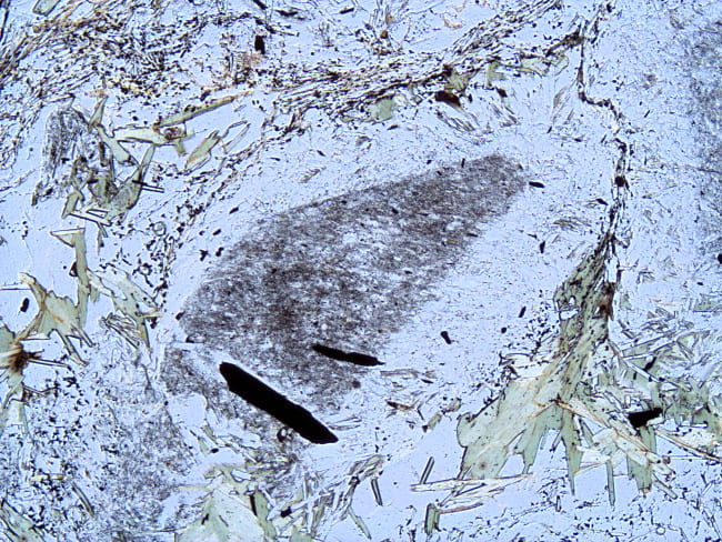
Sillimanite nodule in a biotite gneiss. The sillimanite grew by the reaction muscovite + quartz = sillimanite + K-feldspar + H2O. Small amounts of muscovite in the foliation are still present, so the reaction did not run to completion. Microcline and quartz can also be distinguished in the image, so this represents the reaction assemblage. Colorado. Plane- and cross-polarized light views. Field width is 6 mm. 22-Gn |
|
|
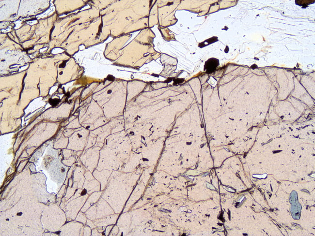
This shows a large garnet, containing inclusions of chloritoid (small blue crystal to the lower-right, other grains are other pleochroic colors). No chloritoid occurs outside the garnet, which now has an assemblage that includes kyanite and staurolite (upper-left). The garnet therefore holds evidence of an earlier metamorphic assemblage. Such evidence can allow partial reconstruction of the metamorphic history. Muscovite schist, Vermont. Plane- and cross-polarized light views. Field width is 3 mm. Gassetts Schist |
|
|
Somewhat complex sample showing the prograde replacement of andalusite (irregular, somewhat grayish-brown mineral, dark-gray birefringence) partially replaced by sillimanite (elongate diamonds having medium-gray birefringence). Andalusite transforms to sillimanite relatively easily, requiring a few broken bonds and a small rotation to the structure. The rotation can be to the left or right, so some new sillimanite domains nucleated one way, and some the other, with a few degrees difference in extinction angle between them. Much of the andalusite has also been replaced by muscovite, which are the large, colorless crystals with colorful birefringence. Muscovite schist, Maine. Plane- and cross-polarized light views. Field width is 6 mm. JBT-2-XA |
Diffusion-controlled reactions
|
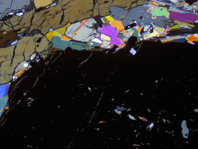
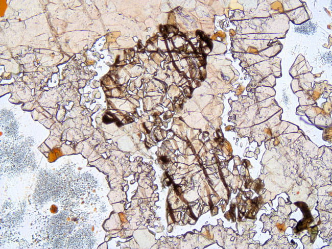
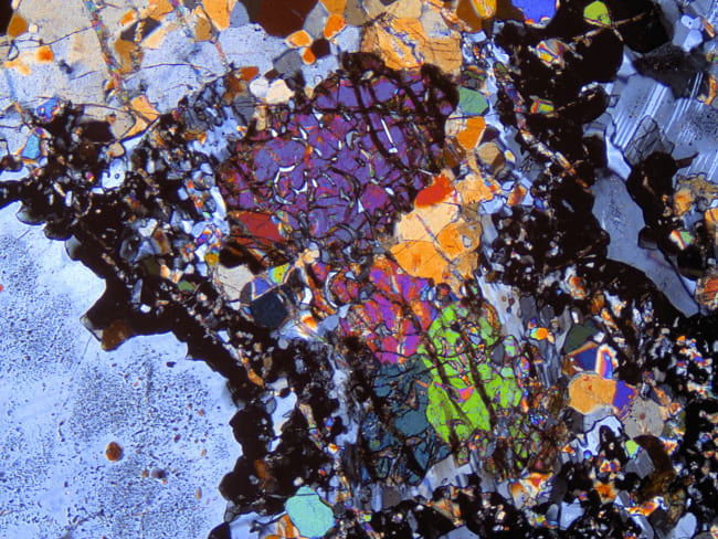
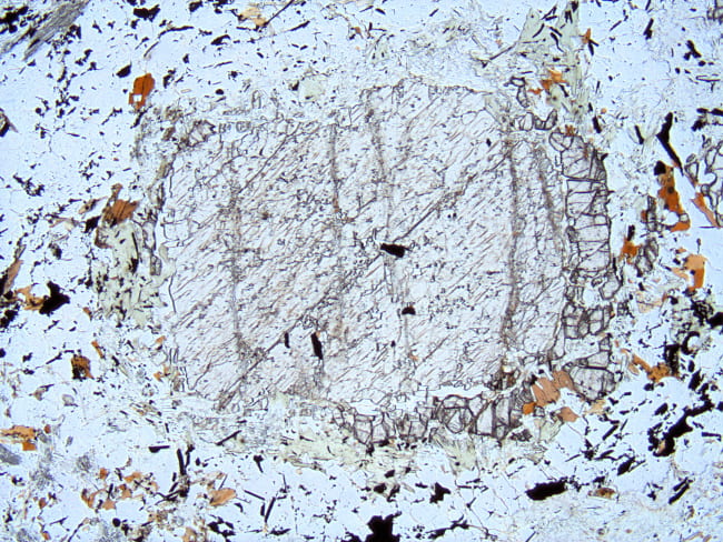
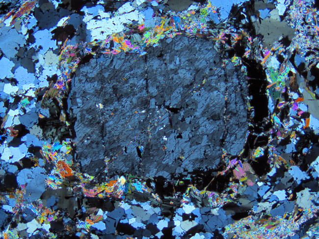
This is a polycrystalline clump of augite, surrounded by a rim of garnet, with plagioclase around that. During granulite facies metamorphism under fluid-absent conditions, a reaction grew garnet at the interface between augite, which supplied Fe, Mg, and Ca, and plagioclase, which supplied Ca, Al, and most of the Si. Garnet growth was a continuous reaction, changing the compositions of the remaining augite and plagioclase. Gabbroic anorthosite, Adirondacks, New York. Plane- and cross-polarized light views. Field width is 6 mm. I-27A |
|
|
“Corona” texture, developed between igneous plagioclase and olivine in a gabbro, almost without deformation, during granulite facies metamorphism. The reaction zone is only about 0.5 mm thick. It starts with central olivine crystals, which are overgrown by successive layers of enstatite, a plagioclase moat, garnet + augite, and finally garnet, with plagioclase on the outside. Olivine supplied Fe and Mg, which diffused outward. Plagioclase supplied Ca, Al, and Si, which diffused inward. The activity gradients established which minerals precipitated in each layer. There was some left over Fe, Mg, and Al, which precipitated as green spinel dust in the outer plagioclase. Adirondacks, New York. Plane- and cross-polarized light views. Field width is 3 mm. TMW96-A2-A |
|
|
This is a different “corona” reaction texture found around ilmenite in a different piece of the gabbro, seen immediately above. Here, the ilmenite (probably originally titanomagnetite) supplied Fe and Ti, and the surrounding plagioclase supplied Na, Ca, Al, Si, and probably K. The successive layers, starting from ilmenite, are biotite, hornblende, and garnet, with plagioclase on the outside. The red-brown colors of the hornblende and biotite indicate that they have high titanium contents, and are relatively low in Fe3+. Again, left over Fe, Mg, and Al precipitated as green spinel in the surrounding plagioclase. Adirondacks, New York. Plane- and cross-polarized light views. Field width is 3 mm. TMW96-A2-B |
Metasomatic reactions
|
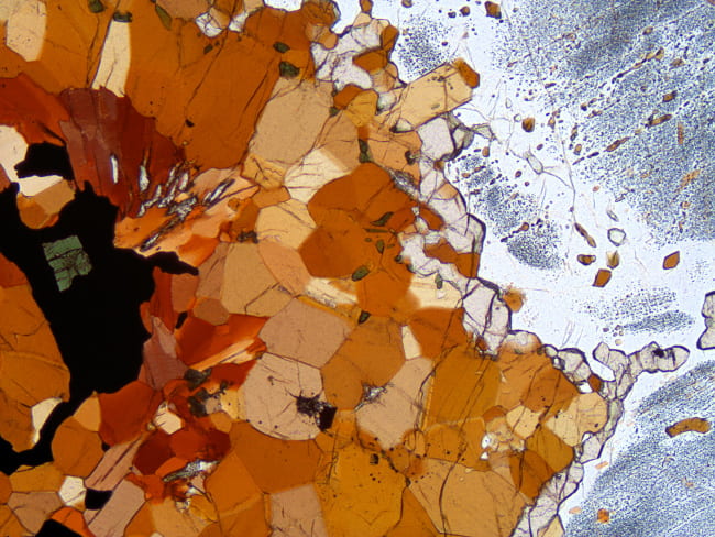
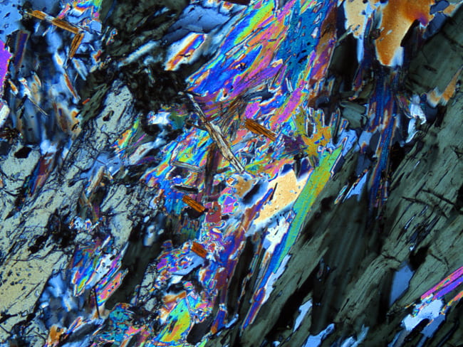
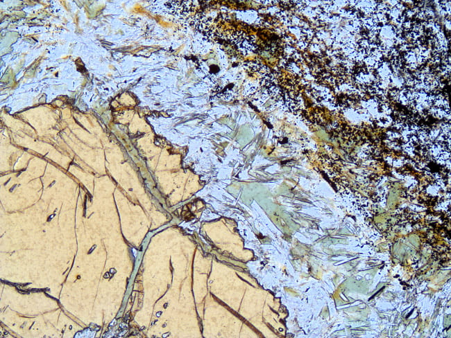
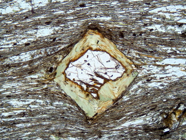
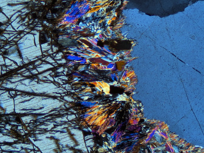
This rock is a chlorite schist, made mostly of chlorite with lesser amounts of staurolite, muscovite, quartz, ilmenite, and rutile. As can be seen, the staurolite has been largely replaced by a thick rim of muscovite, chlorite, and kyanite. This rock has no source of potassium to run the replacement reaction, requiring both K2O and H2O to have been added to this part of the rock, probably by moving metamorphic fluids. Vermont. Plane- and cross-polarized light views. Field width is 3 mm. IMA86-C1-3 |
Retrograde hydration reactions
|
This is a muscovite-biotite gneiss that has peculiar, large, lens-shaped muscovite crystals, in addition to smaller muscovite crystals in the matrix foliation. All of the larger crystals have sillimanite inside of them. This suggests that a retrograde hydration reaction occurred: K-feldspar + sillimanite + H2O = muscovite + quartz. The precursor was probably a sillimanite nodule, like that seen in the image above. In fact, that sample is from the same unit a few kilometers away. Colorado.. Plane- and cross-polarized light views. Field width is 6 mm. 19-Gn |
|
|
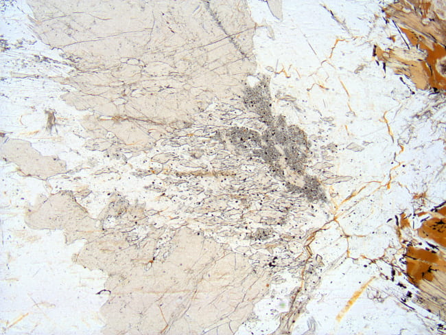
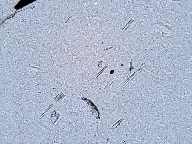
Retrograde reactions may completely destroy prograde minerals like sillimanite. However, relics of them may survive trapped inside other minerals, like garnet, muscovite, and quartz. Here, sillimanite was completely lost from the rock assemblage, but a few grains remain inside these quartz crystals, where they were protected from reaction. Massachusetts. Plane- and cross-polarized light views. Field width is 0.6 mm. 1A |
|
|
Staurolite crystal in a muscovite schist, that has been retrograded along its rim and fractures to a mixture of muscovite and chlorite. The potassium necessary for new muscovite came from biotite, which was also consumed in the retrograde reaction: staurolite + biotite + quartz + H2O = muscovite + chlorite + ilmenite. Massachusetts Plane- and cross-polarized light views. Field width is 3 mm. MDC-6A-170.5 |
|
|
Partially retrograded garnet in muscovite schist. Including calcium, an important part of the garnet solid solution, the retrograde reaction was: garnet + biotite + ilmenite + H2O = chlorite + muscovite + titanite + quartz. Apparently, the chlorite grew after foliation development. Massachusetts. Plane- and cross-polarized light views. Field width is 3 mm. NS-54A |
|
|
This sample is part of a partial melt segregation in amphibolite that formed during upper amphibolite facies metamorphism. The melt, derived from the amphibolite, was tonalitic in composition, and crystallized to an assemblage of plagioclase, quartz, hornblende, biotite, and orthopyroxene. At lower temperature, water gained access to the rock and partially retrograded the orthopyroxene margins to a mixture of cummingtonite and magnetite, by the reaction Opx + quartz + H2O + O2 = cummingtonite + magnetite. In the host amphibolite there are small mats of fine-grained, polysynthetically twinned cummingtonite that mark the former occurence of orthpyroxene in the amphibolite as well. Massachusetts. Plane- and cross-polarized light views. Field width is 3 mm. WD-H37Ab |
Pseudomorphs
|
This sample is from a schist xenolith inside of a pluton. The original schist had andalusite crystals, which then underwent contact metamorphism and transformed to sillimanite. As mentioned above, during the transformation of andalusite to sillimanite, the new crystal structure is rotated one way or the other relative to the andalusite precursor. The result is sillimanite, in the shape of the original andalusite, that is a patchwork of rougly diamond-shaped domains that have extinction angles a few degrees apart. At higher magnification, not shown here, the pseudomorphs can be seen to contain small inclusions of spinel, remarkable given the abundance of quartz in the matrix. Massachusetts. Plane- and cross-polarized light views. Field width is 6 mm. Rt. 2 Philipston |
|
|
This is a pseudomorph of chlorite and muscovite, after staurolite, where the reaction seen above went to completion. The inner part of the original staurolite had abundant graphite inclusions, whereas the outer part did not. The pseudomorph retains the pattern of graphite inclusions, showing that little deformation took place during the replacement process. Plane- and cross-polarized light views. Field width is 3 mm. NS-111A
|

